. 2002 Jun 11;99(12):8236-41.
doi: 10.1073/pnas.122686299.

FLIP switches Fas-mediated glucose signaling in human pancreatic beta cells from apoptosis to cell replication

Affiliations

FLIP switches Fas-mediated glucose signaling in human pancreatic beta cells from apoptosis to cell replication

Kathrin Maedler et al. Proc Natl Acad Sci U S A. .

Abstract

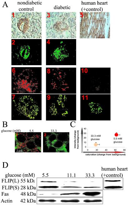
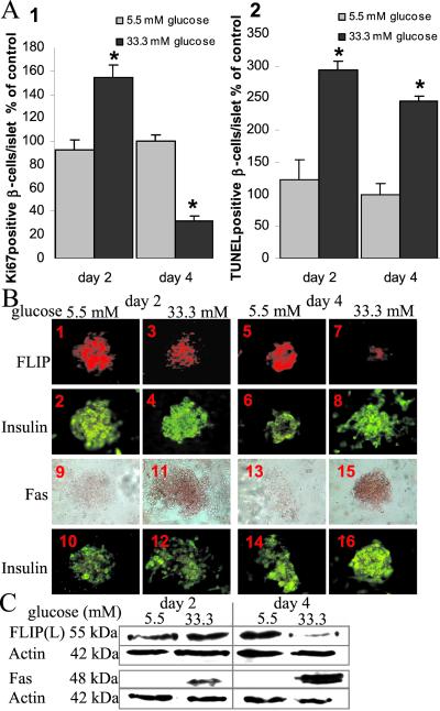
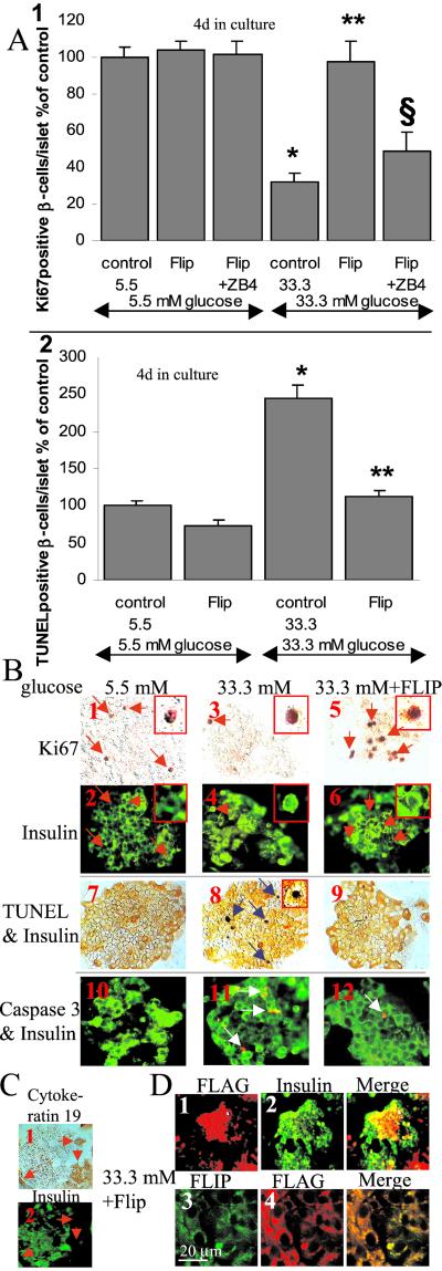
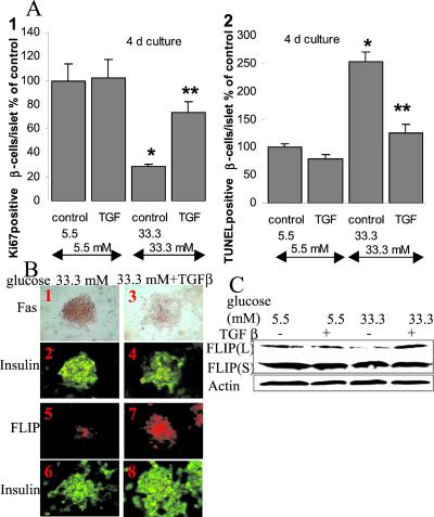
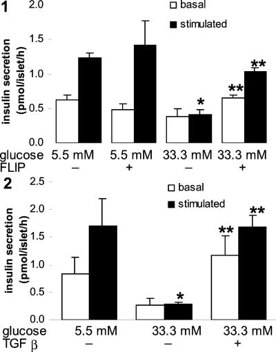
Type 2 diabetes mellitus results from an inadequate adaptation of the functional pancreatic beta cell mass in the face of insulin resistance. Changes in the concentration of glucose play an essential role in the regulation of beta cell turnover. In human islets, elevated glucose concentrations impair beta cell proliferation and induce beta cell apoptosis via up-regulation of the Fas receptor. Recently, it has been shown that the caspase-8 inhibitor FLIP may divert Fas-mediated death signals into those for cell proliferation in lymphatic cells. We observed expression of FLIP in human pancreatic beta cells of nondiabetic individuals, which was decreased in tissue sections of type 2 diabetic patients. In vitro exposure of islets from nondiabetic organ donors to high glucose levels decreased FLIP expression and increased the percentage of apoptotic terminal deoxynucleotidyltransferase-mediated UTP end labeling (TUNEL)-positive beta cells; FLIP was no longer detectable in such TUNEL-positive beta cells. Up-regulation of FLIP, by incubation with transforming growth factor beta or by transfection with an expression vector coding for FLIP, protected beta cells from glucose-induced apoptosis, restored beta cell proliferation, and improved beta cell function. The beneficial effects of FLIP overexpression were blocked by an antagonistic anti-Fas antibody, indicating their dependence on Fas receptor activation. The present data provide evidence for expression of FLIP in the human beta cell and suggest a novel approach to prevent and treat diabetes by switching Fas signaling from apoptosis to proliferation.


Figures

Figure 1
Figure 1
Expression and regulation of FLIP in human islets. (A) Double immunostaining for FLIP in brown (1, 3, 5) and insulin in green (2, 4) in tissue sections of pancreases from a nondiabetic patient (1, 2) and from a patient with type 2 diabetes (3, 4) and in a tissue section of human heart (positive control, 5). In situ hybridization for FLIP mRNA in red (6, 8, 10) double immunostained for insulin in green (7, 9, 11) in tissue sections of pancreases from a nondiabetic patient with antisense probe (6) and with sense probe (negative control; 10), and from a patient with type 2 diabetes using anti-sense probe (8) (×250-fold). (B) Double immunostaining (confocal microscope) for FLIP in red and insulin in green in human islets cultured on extracellular matrix-coated dishes and exposed for 4 days to media containing 5.5 mM glucose (1) or 33.3 mM glucose (2). (C) Intensity and degree of saturation of FLIP immunostaining relative to background in β cells identified by double staining for insulin. Results are shown as mean ± SE of human islets from 3 donors exposed for 4 days to 5.5 or 33.3 mM glucose. (D) Immunoblotting of FLIPL, FLIPS, Fas, and actin. Human islets cultured in suspension at 5.5, 11.1, or 33.3 mM glucose were analyzed after 42-h incubation. Cardiac tissue was used as a positive control for FLIP. The antibodies were blotted on the same membrane after stripping. One representative of five experiments from five donors is shown.
Figure 2
Figure 2
Time course of the effect of elevated glucose concentrations on human β cell proliferation and apoptosis in correlation with the expression of FLIP and Fas. (A) Relative number of Ki-67-positive (1) and TUNEL-positive (2) β cells per islet after 2- and 4-day culture in 5.5 or 33.3 mM glucose normalized to control incubations at 5.5 mM glucose alone for 4 days (100%; in absolute values: 1.63 Ki-67-positive β cells per islet and 0.30 TUNEL-positive β cells per islet). The mean number of islets scored from each donor was 29 for each treatment condition. Islets were isolated from six organ donors. Results are shown as mean ± SE. *, P < 0.01 relative to islets at 5.5 mM glucose. (B) Double-immunostaining for FLIP in red (1, 3, 5, 7) or Fas in brown (9, 11, 13, 15) and insulin in green (2, 4, 6, 8, 10, 12, 14, 16) in human islets cultured on extracellular matrix-coated dishes and exposed for 2 (14, 912) and 4 days (58, 1316) to media containing 5.5 mM (1, 2, 5, 6, 9, 10, 13, 14) or 33.3 mM glucose (3, 4, 7, 8, 11, 12, 15, 16) (×250). (C) Immunoblotting of FLIPL, Fas and actin in human islets cultured on extracellular matrix-coated dishes and exposed for 2 and 4 days to media containing 5.5 mM or 33.3 mM glucose.
Figure 3
Figure 3
FLIP protects human β cells from glucose-induced apoptosis and restores β cell proliferation. (A) Human islets were cultured on extracellular matrix-coated dishes for 4 days in 5.5 and 33.3 mM glucose (control) after transfection with an expression vector coding for FLIP (FLIP) with or without addition of the antagonistic Fas antibody ZB4 (FLIP + ZB4). Results are means ± SE of the relative number of Ki-67-positive (1) and TUNEL-positive β cells (2) per islet normalized to control incubations at 5.5 mM glucose alone (100%; in absolute values: 1.63 Ki-67-positive β cells per islet and 0.30 TUNEL-positive β cells per islet). The mean number of islets scored from each donor was 32 for each treatment condition. Islets were isolated from six organ donors. *, P < 0.001 relative to islets at 5.5 mM glucose; **, P < 0.01 relative to islets at 33.3 mM glucose; §, P < 0.01 relative to FLIP transfected islets at 33.3 mM glucose. (B) Islets were exposed for 4 days to media containing 5.5 mM glucose (1, 2, 7, 10) or 33.3 mM glucose (3, 4, 8, 11) or 33.3 mM glucose and after transfection with FLAG-tagged FLIP (5, 6, 9, 12). Detection of β cell proliferation with anti-Ki-67 (orange; 1, 3, 5) and with anti-insulin antibody (green; 2, 4, 6). Double-immunostaining (79) for insulin (orange) and DNA fragmentation by the TUNEL assay (black). Double-immunostaining (1012) for cleaved caspase-3 (red) and insulin (green). The red arrows mark cells stained positive for Ki-67 and for insulin; the blue arrows mark β cells nuclei stained positive for the TUNEL reaction; the white arrows mark β cells stained positive for cleaved caspase-3 (×250, with higher magnifications of Ki-67+- (16) or TUNEL+- (8) β cells in the inserts. (C) Double-immunostaining for cytokeratin 19 (1) and insulin (2) in human islets cultured for 4 days in 5.5 mM glucose. The red arrows mark ductal cells (×250). (D) Islets were transfected with FLAG-tagged FLIP and exposed for 4 days to media containing 33.3 mM glucose. Double-immunostaining for FLAG (1) and insulin (2) or FLIP (3) and FLAG (4). In 3 and 4, images are of a selected field in which essentially all cells were seen to have been transfected (FLAG positive). (×200) (1, 2); confocal microscopy (3, 4).
Figure 4
Figure 4
TGF-β protects human islets from the deleterious effects of high glucose on β cell turnover. (A) Human islets were cultured on extracellular matrix-coated dishes for 4 days in 5.5 and 33.3 mM glucose alone (control) or with TGF-β (TGF). Results are mean ± SE of the relative number of Ki-67-positive (1) and TUNEL-positive (2) β cells per islet normalized to control incubations at 5.5 mM glucose alone (100%; in absolute values: 1.04 Ki-67-positive β cells per islet and 0.31 TUNEL-positive β cells per islet). The mean number of islets scored from each donor was 23 for each treatment condition. Islets were isolated from three organ donors. *, P < 0.001 relative to islets at 5.5 mM glucose; **, P < 0.01 relative to islets at 33.3 mM glucose. (B) Double-immunostaining for Fas (brown; 1, 3) or FLIP (red; 5, 7) and insulin (green; 2, 4, 6, 8) in human islets cultured on extracellular matrix-coated dishes and exposed for 4 days to media containing 33.3 mM glucose alone (1, 2, 5, 6) or in the presence of TGF-β (3, 4, 7, 8). B originates from the same experiment as in Fig. 2B and has therefore the same controls. (×250). (C) Immunoblotting of FLIPL, FLIPS and actin. Human islets cultured in suspension at 5.5, or 33.3 mM glucose without or with 20 ng/ml of TGF-β were analyzed after 42-h incubation. The antibodies were blotted on the same membrane after stripping. One representative of three experiments from three donors is shown.
Figure 5
Figure 5
FLIP and TGF-β preserve glucose-stimulated insulin secretion in human islets exposed to high glucose for 6 days. Islets were cultured on extracellular matrix-coated dishes for 6 days in 5.5 and 33.3 mM glucose (control) or after transfection with an expression vector coding for FLIP (FLIP) (1) or with addition of 20 ng/ml TGF-β (2). Basal and stimulated insulin secretion denotes the amount secreted during a 1-h incubation at 3.3 and 16.7 mM glucose, respectively, after the 6-day culture period. Each bar represents the mean of three experiments ± SE from three separate donors. In each experiment, the data were collected from three plates per treatment. *, P < 0.01 relative to islets at 5.5 mM glucose; **, P < 0.01 relative to islets at 33.3 mM glucose.

References

    1. Bernard C, Berthault M F, Saulnier C, Ktorza A. FASEB J. 1999;13:1195–1205. - PubMed
    1. Bonner-Weir S. J Mol Endocrinol. 2000;24:297–302. - PubMed
    1. Polonsky K S, Sturis J, Bell G I. N Engl J Med. 1998;334:777–783. - PubMed
    1. Cavaghan M K, Ehrmann D A, Polonsky K S. J Clin Invest. 2000;106:329–333. - PMC - PubMed
    1. Kahn B B. Cell. 1998;92:593–596. - PubMed

Publication types

MeSH terms

LinkOut - more resources