

Review
Histopathologic findings in human cerebral aneurysms embolized with platinum coils: report of two cases and review of the literature
Affiliations
- PMID: 12063226
- PMCID: PMC7976908
Item in Clipboard
Review
Histopathologic findings in human cerebral aneurysms embolized with platinum coils: report of two cases and review of the literature
AJNR Am J Neuroradiol.
2002 Jun-Jul.
Abstract
This report describes 2-week and 20-month histopathologic findings in small aneurysms embolized with platinum coils. Electron microscopy showed the presence of endothelial cells encroaching on the platinum coils at the orifice of the aneurysm in both cases. We confirm that endothelial growth can be induced as early as 2 weeks after embolization of small human aneurysms with platinum coils, similar to previous observations in animal models and human cases.
Figures
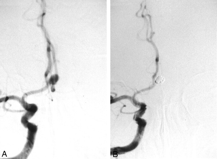
Images from the case of a 65-year-old man with an unruptured aneurysm (case 1). A, Pretreatment cerebral angiogram shows the 5-mm anterior communicating artery aneurysm. B, Post-treatment cerebral angiogram shows embolization by interlocking detachable coils (4 mm × 8 cm and 3 mm × 6 cm).
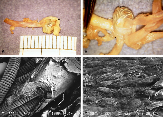
Gross pathologic findings in case 1. A, Gross outlook appearance of the thin wall of the aneurysm through which embedded coils are seen. B, Intraluminal view shows a thin transparent layer of membrane covering the whole orifice of the aneurysm and bridged over the underlying coils. C, Scanning electron microscopy shows that the coils are covered by thick neointima at the orifice of the aneurysm. Part of the neointima was removed for transmission electron microscopic study (original magnification, ×50; bar = 100 μm). D, Superficial layer of neointima with a cobblestone appearance (original magnification, ×1000; bar = 10 μm).
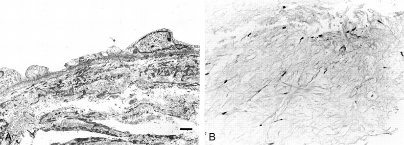
Transmission electron microscopy and light microscopy (case 1). A, Basal lamina runs contiguously along the basal endothelial cytoplasmic membrane (original magnification, ×2000; bar = 2 μm). B, Light microscopy shows cotton-like white fibrous tissue in the dome of the aneurysm (hematoxylin and eosin stain; original magnification, ×74).
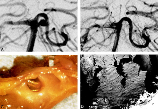
Images from the case of a 62-year-old man with subarachnoid hemorrhage (case 2). A, Right pre-embolization vertebral angiography shows 4-mm left basilar artery-superior cerebellar artery aneurysm. B, After embolization by GDC (3 mm × 4 cm and 2 mm × 4 cm), almost total obliteration was achieved. C, Gross examination shows thin membrane on coils at orifice of aneurysm. The thin fragile covering bridges the whole space across the coils at the orifice. D, Scanning electron microscopy shows neointima partially covering the coils (GDC-10 soft). A cobblestone pattern can be seen (original magnification, ×370; bar = 100 μm).
References
-
- Tenjin H, Fushiki S, Nakahara Y, et al. Effect of Guglielmi detachable coils on experimental carotid artery aneurysms in primates. Stroke 1995;26:2057–2080 - PubMed
-
- Spetzger U, Reul J, Weis J, Bertalanffy H, Thron A, Gilsbach J. Microsurgically produced bifurcation aneurysms in a rabbit model for endovascular coil embolization. J Neurosurg 1996;85:488–495 - PubMed
-
- Mizoi K, Yoshimoto T, Takahashi A, Nagamine Y. A pitfall in the surgery of a recurrent aneurysm after coil embolization and its histological observation: technical case report. Neurosurgery 1996;39:65–69 - PubMed
Publication types
MeSH terms
Substances
LinkOut - more resources
Full Text Sources
Medical