. 2002 Jun 17;21(12):2877-85.
doi: 10.1093/emboj/cdf291.

Crystal structure of auxin-binding protein 1 in complex with auxin

Affiliations

Crystal structure of auxin-binding protein 1 in complex with auxin

Eui-Jeon Woo et al. EMBO J. .

Abstract

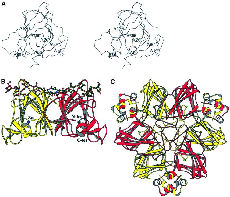
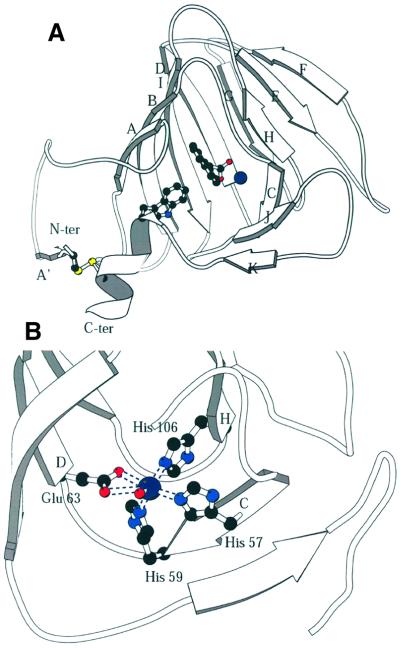
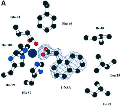
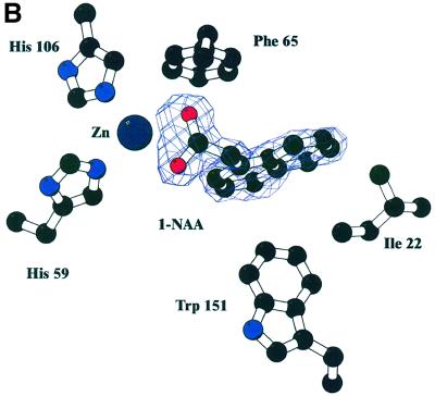
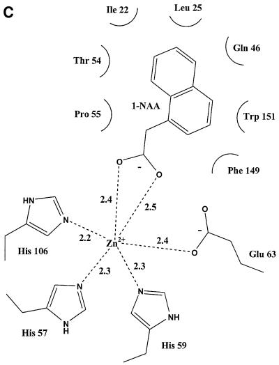
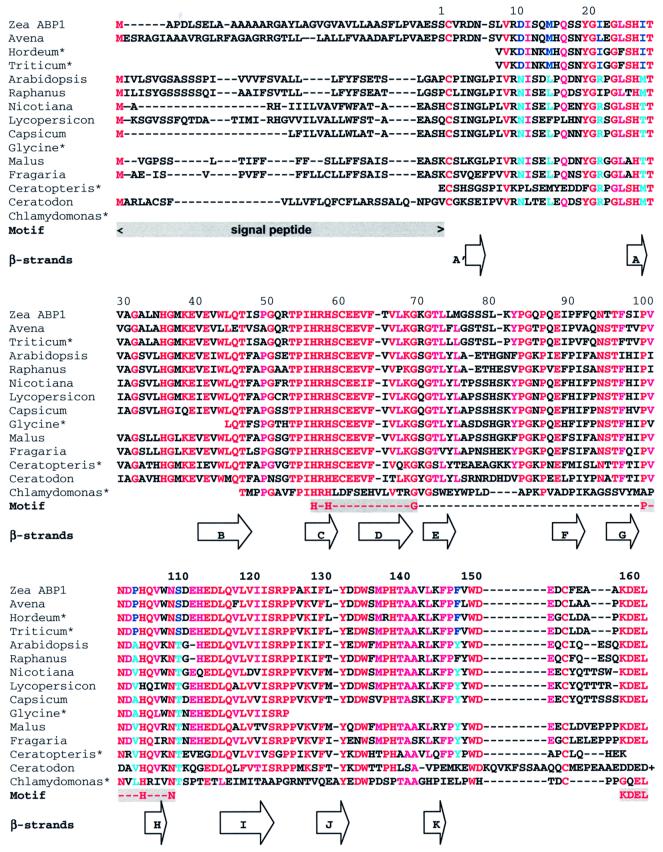
The structure of auxin-binding protein 1 (ABP1) from maize has been determined at 1.9 A resolution, revealing its auxin-binding site. The structure confirms that ABP1 belongs to the ancient and functionally diverse germin/seed storage 7S protein superfamily. The binding pocket of ABP1 is predominantly hydrophobic with a metal ion deep inside the pocket coordinated by three histidines and a glutamate. Auxin binds within this pocket, with its carboxylate binding the zinc and its aromatic ring binding hydrophobic residues including Trp151. There is a single disulfide between Cys2 and Cys155. No conformational rearrangement of ABP1 was observed when auxin bound to the protein in the crystal, but examination of the structure reveals a possible mechanism of signal transduction.


Figures

None
Fig. 1. The overall structure of maize ABP1. (A) Stereoview of an ABP1 subunit with the N- and C-terminal extensions closest to the viewer and with labels every 20 residues. (B) Schematic representation of the overall fold of the ABP1 dimer with the β-strands drawn as arrows, the α-helices as helices, and with the bound zinc ions represented as dark blue spheres, one at the centre of each subunit. The orientation of the red subunit is similar to that shown in (A). The subunits comprising the dimer are related by a vertical 2-fold rotation axis. Asn95 and the observed N-linked sugar residues [Manp(α1,6)–(Manp(α1,3))–Manp(β1,4)–GlcpNAc(β1,4)–GlcpNAc(β1,N)-Asn] are also shown. (C) Schematic representation of the germin hexamer to illustrate the relationship between the ABP1 dimer shown in (B) and the trimer of dimers in germin. The germin subunit has additional α-helical clasps that lock the three dimers together into the trimer of dimers (Woo et al., 2000a; Protein Data Bank accession code 1FI2). The subunit sequences of ABP1 and germin are 24% identical, and the r.m.s.d. for 101 equivalent α-carbon atoms is 1.85 Å. The green spheres are manganese ions, one at the active centre of each of the germin subunits. Note that (B) and (C) are not on the same scale. Figures 1, 2 and 5 were drawn using MOLSCRIPT (Kraulis, 1991).
None
Fig. 2. The subunit structure and binding site of ABP1. (A) The β-strands of the ABP1 subunit are labelled A′–K; the dimer interface is formed by strands ABIDG and the outer sheet by strands KJCHEF. This figure shows the disulfide between residues 2 and 155 that links the N- and C-terminal extensions; the sulfur atoms are the yellow spheres. Also shown is the relationship between the disulfide, the C-terminal α-helix, Trp151 (drawn as ball-and-stick model), 1-NAA (also ball and stick) and the zinc ion (dark blue sphere). The subunit is viewed from the direction of the opening to the auxin-binding pocket. (B) The zinc-binding site in detail. The protein ligands are His57 (strand C), His59 (at the end of strand C), Glu63 (at the beginning of strand D) and His106 (at the beginning of strand H). A single water molecule completes the octahedral coordination sphere. The zinc–nitrogen (His NE2) distances are between 2.2 and 2.3 Å and the zinc–oxygen distances are 2.4 Å to Glu63 OE1, 3.1 Å to Glu63 OE2 and 2.2 Å to the water molecule (shown as a small red sphere).
None
Fig. 3. Auxin binding to ABP1. (A) Omit electron density map at 1.9 Å resolution and contoured at 3σ revealing 1-NAA bound to ABP1. The view in (B) is rotated ∼60° around the x-axis compared with the view in (A), and His57, Glu63, Leu25 and Ile48 are omitted from this view for clarity. The bidentate binding of the 1-NAA carboxylate to the zinc (dark blue sphere) can be clearly seen, as can the hydrophobic environment of the naphthalene ring. (C) Simplified representation of the contacts between 1-NAA and ABP1 and between the zinc ion and ABP1 in the complex. Distances shown are in angstroms. (A) and (B) were prepared using BOBSCRIPT (Esnouf, 1997).
None
Fig. 4. Multiple alignment of ABP1 sequences. The first column gives the species from which the ABP1 originates: Zea ABP1 (Zea Mays; accession No. P13689); Avena (Avena sativa, T07797); Hordeum*, a compilation of EST entries (Hordeum vulgare, BE456042, BF259480, AL508794 and AV834303); Triticum*, a compilation of EST entries (Triticum aestivum, BE426626, BE446060, BE425494, BG274987 and BE516927); Arabidopsis (Arabidopsis thaliana, P33487); Raphanus (Raphanus sativus, AB000706); Nicotiana (Nicotiana tobacum, P33490); Lycopersicon (Lycopersicon esculentum, CAA09882); Capsicum (Capsicum annuum, CAA88361); Glycine*, an EST entry (Glycine max, BG045594); Malus (Malus x domestica, AAB47752); Fragaria (Fragaria x ananassa, CAA62956); Ceratopteris*, an EST entry (Ceratopteris richardii, BE641066); Ceratodon (Ceratodon purpureus, AAF37576); and Chlamydomonas*, a compilation of EST entries (Chlamydomonas reinhardtii, BG847560, BG860232, AV635532 and AV633592). Note that the sequence for Ceratodon extends past the KDEL terminus of other ABP1s, but the extension is not shown. The first four sequences are from monocot, the next eight from dicot plants and the last three from a fern, a moss and a green alga. The approximate positions of the β-strands identified in the text and Figure 2 are illustrated as arrows. Motif indicates the signal peptide, cupin motif and KDEL sequence. The residues for which there are clear differences between monocots and dicots are highlighted in blue. Red and cyan indicate strict and less strict conservation, respectively. The Clamydomonas ABP1 sequence may not be aligned correctly as it differs considerably from the other sequences; it is not known whether this ABP1 binds a metal. Both monocots and dicots have a glycosylation site at Asn95; dicots have an additional glycosylation site at Asn11.
None
Fig. 5. The crystal contact involving the N-terminal extensions of symmetry-related molecules in type II crystals of ABP1. The disulfide bridge and Trp151 are drawn to illustrate how this crystal contact might influence the position of the C-terminal α-helix in the protein.

References

    1. Allen F.H. and Kennard,O. (1993) 3D search and research using the Cambridge Structural Database. Chemical Design Automation News, 8, 1 and 31–37.
    1. Altschul S.F., Madden,T.L., Schaffer,A.A., Zhang,J.H., Zhang,Z., Miller,W. and Lipman,D.J. (1997) Gapped BLAST and PSI-BLAST: a new generation of protein database search programs. Nucleic Acids Res., 25, 3389–3402. - PMC - PubMed
    1. Anantharaman V., Koonin,E.V. and Aravind,L. (2001) Regulotory potential, phyletic distribution and evolution of ancient, intracellular small-molecule-binding domains. J. Mol. Biol., 307, 1271–1292. - PubMed
    1. Barbier-Brygoo H. Ephritikhine,G., Klambt,D., Maurel,C., Palme,K., Schell,J. and Guern,J. (1991) Perception of the auxin signal at the plasma membrane of tobacco mesophyll protoplasts. Plant J., 1, 83–93. - PubMed
    1. Bauly J.M. et al. (2000) Overexpression of ABP1 heightens the sensitivity of guard cells to auxin. Plant Physiol., 124, 1229–1238 - PMC - PubMed

Publication types