. 2002 Jun 17;21(12):2936-45.
doi: 10.1093/emboj/cdf289.

INK4a-deficient human diploid fibroblasts are resistant to RAS-induced senescence

Affiliations

INK4a-deficient human diploid fibroblasts are resistant to RAS-induced senescence

Sharon Brookes et al. EMBO J. .

Abstract

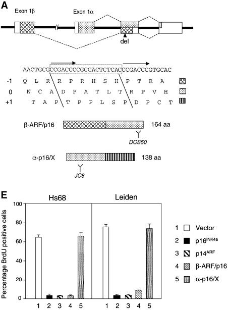
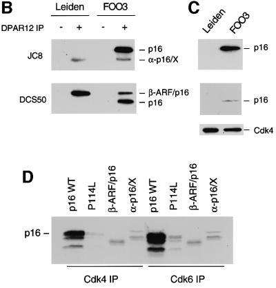
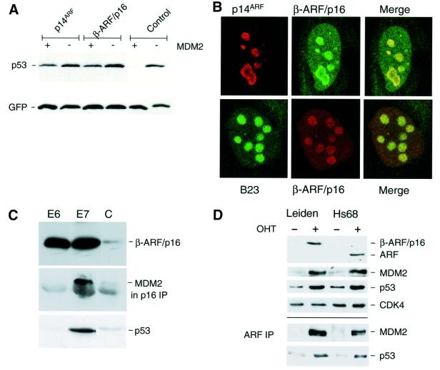
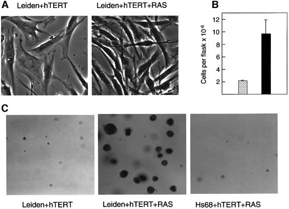
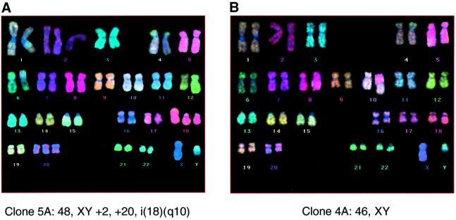
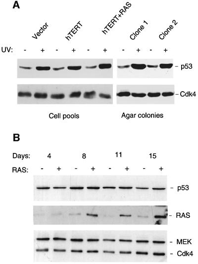
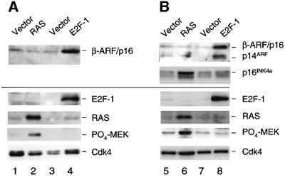
The CDKN2A tumour suppressor locus encodes two distinct proteins, p16(INK4a) and p14(ARF), both of which have been implicated in replicative senescence, the state of permanent growth arrest provoked in somatic cells by aberrant proliferative signals or by cumulative population doublings in culture. Here we describe primary fibroblasts from a member of a melanoma-prone family who is homozygous for an intragenic deletion in CDKN2A. Analyses of the resultant gene products imply that the cells are p16(INK4a) deficient but express physiologically relevant levels of a frameshift protein that retains the known functions of p14(ARF). Although they have a finite lifespan, the cells are resistant to arrest by oncogenic RAS. Indeed, ectopic expression of RAS and telomerase (hTERT) results in outgrowth of anchorage-independent colonies that have essentially diploid karyotypes and functional p53. We find that in human fibroblasts, ARF is not induced demonstrably by RAS, pointing to significant differences between the proliferative barriers implemented by the CDKN2A locus in different cell types or species.


Figures

None
Fig. 1. Leiden deletion and resultant CDKN2A fusion proteins. (A) Schematic representation of the CDKN2A locus with the exons shown as boxes. The sequences encoding p16INK4a are identified by stippling, and those encoding p14ARF by cross-hatches. The nucleotide sequence of the relevant region of exon 2 is shown, with direct repeats identified by arrows and the 19 bp Leiden deletion boxed. The encoded amino acids in the three different reading frames are shown in single letter code. The –1 (p14ARF), 0 (p16INK4a) and +1 frames are identified by the distinctive shading, and their contributions to the β-ARF/p16 and α-p16/X fusion proteins are indicated. Y defines the regions of p16INK4a recognized by the DCS50 and JC8 mAbs. (B) Lysates from the Leiden and FOO3 strains of HDFs were immunoprecipitated with either non-immune serum (– lanes) or a polyclonal antibody (DPAR12) against p16INK4a (+ lanes). After fractionation by SDS–PAGE, the samples were immunoblotted with JC8 (upper panel) and DCS50 (lower panel). The positions of wild-type p16INK4a and the β-ARF/p16 and α-p16/X fusion proteins are shown. (C) Immunoprecipitation of Cdk4 from the same Leiden and FOO3 lysates followed by immunoblotting with JC8, DCS50 and a mAb against Cdk4. (DIn vitro binding assay using unlabelled Cdk4 (lanes 1–4) and Cdk6 (lanes 5–8) mixed with [35S]methionine-labelled wild-type p16INK4a (lanes 1 and 5), the P114L variant (lanes 2 and 6), β-ARF/p16 (lanes 3 and 7) or α-p16/X (lanes 4 and 8). The mixtures were immunoprecipitated with polyclonal antisera against Cdk4 or Cdk6 and the labelled proteins were fractionated by SDS–PAGE and visualized by autoradiography. The multiple bands reflect initiation at internal methionines. (E) BrdU incorporation assays in Hs68 and Leiden cells infected with retroviruses encoding p16INK4a, p14ARF, β-ARF/p16 or α-p16/X as indicated.
None
Fig. 2. Functional appraisal of the β-ARF/p16 fusion protein. (A) U20S cells were transiently transfected with plasmids encoding p53 and green fluorescent protein, with and without human MDM2. As shown in the control lanes, MDM2 causes the marked destruction of p53 in this assay. Introduction of either wild-type p14ARF or β-ARF/p16 protected p53 from MDM2-mediated degradation. (B) NARF2 cells (U20S cells expressing p14ARF from an IPTG-inducible promoter) were transfected with a plasmid encoding β-ARF/p16 and analysed by indirect immunofluorescence. In the upper panels, p14ARF was detected with the mouse mAb 4C6/4 (red) whereas the β-ARF/p16 fusion protein was detected with a rabbit polyclonal antibody against p16INK4a (green). In the lower panels, β-ARF/p16 fusion protein was detected with the mouse mAb DCS50 (red) and the nucleolar protein B23 was visualized using a goat polyclonal antibody (green). (C) Samples (20 µg) of total protein from Leiden cells infected with retroviruses encoding the E6 or E7 proteins of HPV-16 (as indicated) or control virus (C) were fractionated by SDS–PAGE and immunoblotted with the mAbs DCS50 to detect the β-ARF/p16 fusion protein (upper panel) and DO-1 to detect p53 (lower panel). A sample (500 µg) of each lysate was immunoprecipitated with polyclonal antiserum against p16INK4a (DPAR12) and immunoblotted with mAb IF2 against human MDM2 (middle panel). (D) Control (Hs68) and Leiden HDFs expressing E2F-1 fused to the hormone-binding domain of the oestrogen receptor were treated (+) or not (–) with 4-hydroxy tamoxifen (OHT) for 24 h. Samples (20 µg) of cell lysate were fractionated by SDS–PAGE and immunoblotted with an antiserum raised against amino acids 54–75 of human p14ARF, and with mAbs against MDM2 and p53. Cdk4 served as a loading control. Samples (500 µg) of each lysate were immunoprecipitated with the same p14ARF antiserum and immunoblotted for MDM2 and p53.
None
Fig. 3. Phenotypic effects of RAS + hTERT in Leiden HDFs. (A) Pools of Leiden HDFs expressing RAS + hTERT or the respective empty vector controls were examined by phase microscopy ∼4–5 weeks post-selection. (B) The saturation densities of Leiden cells expressing hTERT alone (stippled bar) or RAS + hTERT (filled bar) were compared by counting the numbers of cells per 75 cm2 flask at 4–7 days post-confluence. The figure presents the results of four independent comparisons, and the error bars indicate the standard deviation. (C) Leiden cells expressing RAS + hTERT formed macroscopically visible colonies in soft agar (centre) whereas Leiden cells expressing hTERT alone did not (left). No significant agar growth was observed with control fibroblasts expressing RAS + hTERT (right).
None
Fig. 4. Cytogenetic analyses of anchorage-independent colonies. Representative metaphases from RAS + hTERT-expressing Leiden HDF colonies recovered from semi-solid medium and analysed by M-FISH. Clone 5A contained additional and rearranged chromosomes (A), whereas clone 4A showed a normal diploid karyotype (B).
None
Fig. 5. Induction of p53 in Leiden and FOO3 cells by UV irradiation but not RAS. (A) Anchorage-independent colonies of Leiden cells recovered from agarose (clones 1 and 2), and pools of Leiden cells infected with empty vector controls (39 PDs), expressing hTERT alone (41 PDs) or RAS + hTERT (46 PDs), were exposed to UV radiation (15 J/m2). Samples (20 µg) of total protein were then subjected to SDS–PAGE in a 10% gel and immunoblotted with a mAb against p53 (DO-1). Cdk4 served as a loading control. (B) FOO3 cells were infected with either pBABE-RAS-puro retrovirus (+ lanes) or empty vector (– lanes) and harvested at various times post-infection, as indicated, without drug selection. Samples (30 µg) were fraction ated by SDS–PAGE on a 10% gel and immunoblotted for the indicated proteins.
None
Fig. 6. Induction of CDKN2A gene products by RAS and E2F-1. Leiden (A) and FOO3 HDFs (B) were infected with retroviruses encoding RAS (lanes 2 and 6) or E2F-1 (lanes 4 and 8), or with appropriate vector controls. Because of the extensive apoptosis induced by E2F-1, the cells were analysed at 3 days post-infection, whereas the RAS-expressing cells were harvested at 11 days to maximize the effects (see Figure 6B). Samples (30 µg) were fractionated by SDS–PAGE and immunoblotted for the indicated proteins, using a 15% gel in the case of RAS, phospho-MEK and p14ARF- or p16INK4a-related proteins, and a 10% gel for E2F-1, p53 and Cdk4.

References

    1. Alcorta D.A., Xiong,Y., Phelps,D., Hannon,G., Beach,D. and Barrett,J.C. (1996) Involvement of the cyclin-dependent kinase inhibitor p16 (INK4a) in replicative senescence. Proc. Natl Acad. Sci. USA, 93, 13742–13747. - PMC - PubMed
    1. Ashcroft M. and Vousden,K.H. (1999) Regulation of p53 stability. Oncogene, 18, 7637–7643. - PubMed
    1. Bodnar A.G. et al. (1998) Extension of life-span by introduction of telomerase into normal human cells. Science, 279, 349–352. - PubMed
    1. Brown J.P., Wei,W. and Sedivy,J.M. (1997) Bypass of senescence after disruption of p21CIP1/WAF1 gene in normal diploid human fibroblasts. Science, 277, 831–834. - PubMed
    1. Bunz F., Dutriaux,A., Lengauer,C., Waldman,T., Zhou,S., Brown,J.P., Sedivy,J.M., Kinzler,K.W. and Vogelstein,B. (1998) Requirement for p53 and p21 to sustain G2 arrest after DNA damage. Science, 282, 1497–1501. - PubMed

MeSH terms