. 2002 Jun 17;21(12):3009-18.
doi: 10.1093/emboj/cdf306.

Eiger, a TNF superfamily ligand that triggers the Drosophila JNK pathway

Affiliations

Eiger, a TNF superfamily ligand that triggers the Drosophila JNK pathway

Tatsushi Igaki et al. EMBO J. .

Abstract

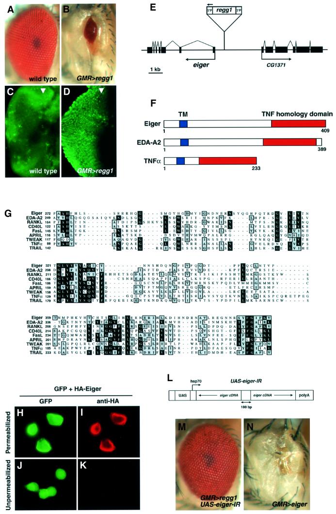
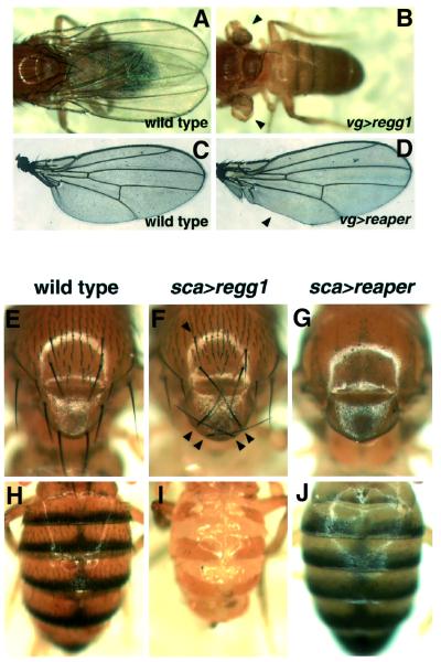
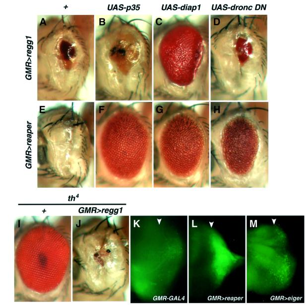
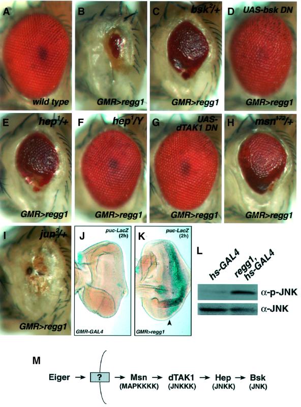
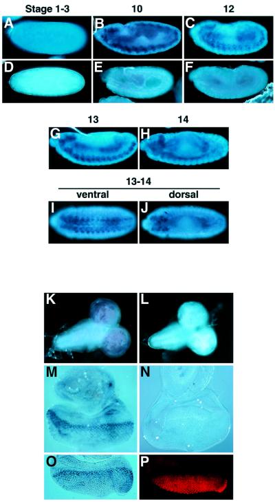
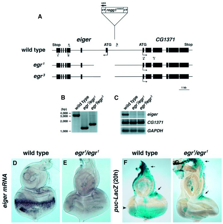
Drosophila provides a powerful genetic model for studying the in vivo regulation of cell death. In our large-scale gain-of-function screen, we identified Eiger, the first invertebrate tumor necrosis factor (TNF) superfamily ligand that can induce cell death. Eiger is a type II transmembrane protein with a C-terminal TNF homology domain. It is predominantly expressed in the nervous system. Genetic evidence shows that Eiger induces cell death by activating the Drosophila JNK pathway. Although this cell death process is blocked by Drosophila inhibitor-of-apoptosis protein 1 (DIAP1), it does not require caspase activity. We also show genetically that Eiger is a physiological ligand for the Drosophila JNK pathway. Our findings demonstrate that Eiger can initiate cell death through an IAP-sensitive cell death pathway via JNK signaling.


Figures

None
Fig. 1. Identification of Eiger, a Drosophila TNF superfamily protein. (A and B) A misexpression screen identified a GS strain, GS9830 (as Regg1), which resulted in a reduced eye when driven by a GMR-GAL4 driver. Eye phenotypes of wild type (A) and regg1GS9830/GMR-GAL4 (B) are shown. (C and D) Acridine orange staining detected numerous dying cells in third-instar larval eye discs of the regg1GS9830/GMR-GAL4 strain (D) compared with the wild-type strain (C). Many acridine orange-positive cells were observed behind the morphogenetic furrow (arrowhead), corresponding to the expression domain of the GAL4 driver. (E) A novel ORF encoding Eiger was identified from an EST clone (LP03784) containing the nucleotide sequence of CG12919, the expression of which was simulated in a GAL4-dependent manner in the Regg1GS9830 strain. (F) Schematic structures of Eiger, EDA-A2 and human TNF-α. (G) The amino acid sequence of the TNF homology domain of Eiger is aligned with the sequences of human EDA-A2, RANKL, CD40L, FasL, APRIL, TWEAK, TNF-α and TRAIL. Identical and conserved residues are denoted with blocks of black or shading, respectively. The Eiger cDNA sequence has been deposited with DDBJ/EMBL/GenBank (accession No. AB073865). (HK) Eiger is a type II membrane protein with a cytoplasmic N-terminus. S2 cells were transfected with the expression vectors for HA-Eiger (pUAS-HA-eiger) and GFP (pUAS-GFP), together with a driver plasmid, pWAGAL4 (actin-GAL4). Twenty-four hours after transfection, cells were immunostained with an anti-HA monoclonal antibody and a Cy3-labeled secondary antibody after (H and I) or prior to (J and K) fixation and permeabilization. (L) Schematic structure of the IR expression construct of eiger. Two partial sequences of eiger cDNA were cloned as a head-to-head inverted repeat that was separated by a non-palindromic 180 bp linker into the pUAST vector. (M and Neiger is the responsible gene for Regg1. The reduced-eye phenotype induced by regg1GS9830 (A) was completely suppressed by eiger-IR (M). Overexpression of exogenous Eiger caused a reduced-eye phenotype (N). Genotypes are regg1GS9830/+; GMR-GAL4/UAS-eiger-IR (M) and UAS-eiger/GMR-GAL4 (N).
None
Fig. 2. Eiger has the potential to induce severe developmental defects that are distinct from the defects caused by Reaper. Eiger or Reaper was ectopically expressed within the dorsoventral boundaries of the wing disc by a vg-GAL4 driver (B and D). Note that Eiger entirely blocked wing formation (B, arrowheads), whereas Reaper induced only a regional defect (D, arrowhead), compared with wild-type wings (A and B). Ectopic expression of Eiger in precursor cells for the external sensory organs driven by sca-GAL4 resulted in disorganization of the macrochaetae in the notum and scutellum (F, arrowheads), and a severe developmental defect of the abdomen (I), whereas Reaper induced a complete loss of bristles in these regions (G and J), compared with wild-type fly (E and H). sca>GAL4 and UAS-reaper flies were mated at 18°C, because their progeny, bearing both transgenes, died as pupae when generated at 25°C.
None
Fig. 3. Eiger activates an IAP-sensitive cell death pathway that does not require caspase activity. (A–H) Genetic interactions of Eiger or Reaper with caspase inhibitory proteins. Genotypes are as follows: regg1GS9830/+; GMR-GAL4/+ (A), regg1GS9830/GMR-GAL4; UAS-p35/+ (B), UAS-diap1/+; regg1GS9830/GMR-GAL4 (C), regg1GS9830/UAS-dronc DN; GMR-GAL4/+ (D), UAS-reaper/GMR-GAL4 (E), UAS-reaper/GMR-GAL4; UAS-p35/+ (F), UAS-diap1/+; UAS-reaper/GMR-GAL4 (G) and UAS-reaper/UAS-dronc DN; GMR-GAL4/+ (H). (I and J) Heterozygosity at the diap1 locus (th4) enhances the Eiger-induced eye phenotype. Whereas flies with a half dosage of the diap1 gene show a perfectly normal eye (I), the reduced-eye phenotype caused by Eiger overexpression (A) is strongly enhanced by the heterozygosity of th4 (J), indicating that endogenous DIAP1 negatively regulates the Eiger-stimulated death signal. Genotypes are th4/+ (I) and regg1GS9830/+; GMR-GAL4/th4 (J). (K–M) (DMe)2R staining of the eye discs shows that Eiger weakly but significantly causes caspase activation. Caspase activity was detected with rhodamine-110 fluorescence released from (DMe)2R. Genotypes are as follows: GMR-GAL4/+ (K), UAS-reaper/+; GMR-GAL4/+ (L) and UAS-eiger/+; GMR-GAL4/+ (M). Arrowheads indicate the position of the morphogenetic furrow.
None
Fig. 4. Eiger induces a reduced-eye phenotype through the activation of the JNK pathway. (A–I) regg1GS9830 genetically interacts with components of the Drosophila JNK signaling pathway. The Eiger-induced phenotype is suppressed by heterozygosity at the bsk, hep or msn locus (C, E and H, respectively). Hemizygosity at the hep locus almost completely suppresses the phenotype (F). Co-expression of a dominant-negative form of Bsk or dTAK1 completely inhibits the Eiger-induced eye phenotype (D and G). Heterozygosity at the Drosophila jun locus does not suppress the eye phenotype (I). Genotypes are as follows: wild type (A), regg1GS9830/GMR-GAL4; TM3,Sb/+ (B), regg1GS9830/bsk2; GMR-GAL4/+ (C), regg1GS9830/GMR-GAL4; UAS-bsk DN/+ (D), hep1/+; regg1GS9830/+; GMR-GAL4/+ (E), hep1/Y; regg1GS9830/+; GMR-GAL4/+ (F), regg1GS9830/UAS-dTAK1 DN; GMR-GAL4/+ (G), regg1GS9830/+; GMR-GAL4/msn172 (H) and regg1GS9830/jun2; GMR-GAL4/+ (I). (J and K) Overexpression of Eiger activates the JNK pathway. The JNK activation was monitored in GMR-GAL4/+ (J) and regg1GS9830/+; GMR-GAL4/+ (K) background eye disc by puc-LacZ expression. X-gal staining (2 h) of the eye disc shows dramatic activation of the JNK pathway in the region posterior to the morphogenetic furrow (K, arrowhead), where Eiger is overexpressed. (L) Eiger stimulates the phosphorylation of Bsk in vivo. hs-GAL4/+ or regg1GS9830/+; hs-GAL4/+ larvae were heat shocked at 37°C for 45 min and cultured at 25°C for a further 4 h, and then subjected to western blot analysis with an anti-JNK or an anti-phospho-JNK antibody. (M) A model for the JNK signaling triggered by Eiger.
None
Fig. 5. eiger is predominantly expressed in the nervous system. (AJ) Whole-mount in situ hybridization of wild-type embryos at various stages of development using eiger antisense (A–C and G–H) or sense (D–F) RNA probes. A pre-blastoderm embryo showed low levels of eiger expression (A). After germ band extension, strong staining was evident in the nervous system (B, C and G–J). (KNIn situ analysis of eiger expression in third-instar larval brains (K and L) and eye discs (M and N) using eiger antisense (K and M) or sense (L and M) RNA probes. A high level of eiger expression was evident in the brain hemispheres (K). In the eye disc, stronger staining was detected at the region posterior to the morphogenetic furrow (M). Double staining of the eye discs with the eiger RNA probe (O) and the anti-ELAV antibody (P) revealed that eiger was strongly expressed in the proliferating cells at the furrow.
None
Fig. 6. Eiger is a physiological ligand for the JNK pathway. (A) Diagram of the eiger genomic locus and location of the end points of excision mutations in egr1 and egr3 mutant alleles. Exon sequences are shown by closed boxes. (B) PCR analysis of genomic DNA from wild-type and homozygous eiger mutant adult flies using primers a and b [indicated in (A)]. The end points of genomic deletions in eiger mutants were determined by sequencing of the PCR products. (C) RT–PCR analysis of cDNA from wild-type and homozygous eiger mutant adult flies using primers c and d or e and f [indicated in (A)] to determine the expression levels of eiger and CG1371, respectively. GAPDH expression was determined as an internal control. (D and EIn situ analysis of eiger expression in the eye disc from wild-type (D) and egr1/egr1 (E) larvae. (F and G) The JNK activity was monitored in wild-type (F) and egr1/egr1 (G) background eye discs by puc-LacZ expression. Long-time (20 h) X-gal staining was able to detect the endogenous JNK activity in the region posterior to the morphogenetic furrow (F, arrowhead). The JNK activity was dramatically reduced in the eiger mutant (G). Note that the JNK activities in the disc margin, where eiger is not expressed even in the wild-type disc, are not affected in the eiger mutant (F and G).

References

    1. Abrams J.M., White,K., Fessler,L.I. and Steller,H. (1993) Programmed cell death during Drosophila embryogenesis. Development, 117, 29–43. - PubMed
    1. Adachi-Yamada T., Fujimura,K.K., Nishida,Y. and Matsumoto,K. (1999) Distortion of proximodistal information causes JNK-dependent apoptosis in Drosophila wing. Nature, 400, 166–169. - PubMed
    1. Arnett H.A., Mason,J., Marino,M., Suzuki,K., Matsushima,G.K. and Ting,J.P. (2001) TNFα promotes proliferation of oligodendrocyte progenitors and remyelination. Nature Neurosci., 4, 1116–1122. - PubMed
    1. Ashkenazi A. and Dixit,V.M. (1998) Death receptors: signaling and modulation. Science, 281, 1305–1308. - PubMed
    1. Chen P., Nordstrom,W., Gish,B. and Abrams,J.M. (1996) grim, a novel cell death gene in Drosophila. Genes Dev., 10, 1773–1782. - PubMed

Publication types

MeSH terms

Associated data