. 2002 Jun 17;21(12):3081-95.
doi: 10.1093/emboj/cdf285.

Homolog interaction during meiotic prophase I in Arabidopsis requires the SOLO DANCERS gene encoding a novel cyclin-like protein

Affiliations

Homolog interaction during meiotic prophase I in Arabidopsis requires the SOLO DANCERS gene encoding a novel cyclin-like protein

Yoshitaka Azumi et al. EMBO J. .

Abstract

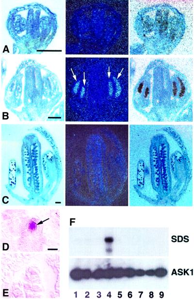
Interactions between homologs in meiotic prophase I, such as recombination and synapsis, are critical for proper homolog segregation and involve the coordination of several parallel events. However, few regulatory genes have been identified; in particular, it is not clear what roles the proteins similar to the mitotic cell cycle regulators might play during meiotic prophase I. We describe here the isolation and characterization of a new Arabidopsis mutant called solo dancers that exhibits a severe defect in homolog synapsis, recombination and bivalent formation in meiotic prophase I, subsequently resulting in seemingly random chromosome distribution and formation of abnormal meiotic products. We further demonstrate that the mutation affects a meiosis-specific gene encoding a novel protein of 578 amino acid residues with up to 31% amino acid sequence identity to known cyclins in the C-terminal portion. These results argue strongly that homolog interactions during meiotic prophase I require a novel meiosis-specific cyclin in Arabidopsis.


Figures

None
Fig. 1. Wild-type and sds mutant plant, flower and pollen development. (A) A wild-type plant with normal seedpods (arrowheads). (B) An sds mutant plant, with small seedpods (arrows). (C) An sds mutant plant with small seedpods (arrows) and two large revertant sectors that have large seedpods (arrowheads). (D) A close-up view of the top portion of a wild-type flower, showing many pollen grains (p) on the stigma and along the side of the pistil. (E) Top of an sds flower, showing anthers and the stigma (s) that lack pollen. (F) A portion of a wild-type anther with functional pollen grains that stained red. (G) Wild-type microspores. (H) A normal tetrad with four spores (numbered). (I) A portion of an sds anther, showing many abnormal pollen grains stained blue. Several pollen grains are stained red and presumably are functional (arrows); some are larger than normal. (J) Microspores from an sds anther, showing a range of sizes. (K) Six spores from a meiosis in the sds mutant. Bar = 10 mm in (A–C), 250 µm in (D) and (E), 20 µm in (F), (G), (I) and (J), and 10 µm in (H) and (K).
None
Fig. 2. Male meiosis I in wild-type and the sds mutant. Shown are images of DAPI-stained chromosomes. (A–E) Wild-type prophase I at leptotene, zygotene, pachytene, diplotene and diakinesis, respectively; note that (E) shows five brightly stained entities, representing five attached pairs of condensed homologs. (FJ) The sds mutant prophase I at similar stages; note that in (J), 10 staining bodies can be seen, indicating that the condensed homologs formed univalents. The arrows, as well as the two pairs of arrowheads, point to two strands of a partially separated chomosome, suggesting precocious separation of the arms of sister chromatids. (KN) Wild-type early metaphase I, late metaphase I, anaphase I and telophase I, respectively. Chromosomes align at the equator (K), and are elongated presumably due to the forces of the spindle (L). Homologs separate (M) and form two clusters (N). (OR) Meiosis I images from the sds mutant after prophase I. Chromosomes condense further (O), similarly to normal metaphase I chromosomes, but they did not all align at the equator. Chromosomes in (P) are elongated similarly to those in (L). Chromosomes are elongated further in (Q), suggesting that they might be pulled by the spindle as normal homologs are at anaphase I. Some chromosomes more distant from the center seem to have decondensed (R), resembling those in normal telophase I.
None
Fig. 3. Distribution of prophase I cells in wild type and the sds mutant. Chromosome spreads were examined as shown in Figure 2. The number of images at each stage is shown above the appropriate bar. The sds leptotene, zygotene, pachytene and diplotene stages were assigned on the basis on chromosome condensation and overall morphology, as shown in Figure 2. The sds zygotene and diplotene stages were not based on pairing or bivalents, respectively. The sds diakinesis images all had 10 univalents.
None
Fig. 4. Female meiosis I in wild type and the sds mutant. Shown here are DAPI-stained images of chromosome spreads from the wild type (A–C) or the sds mutant (D–I). (A) A diakinesis image showing five bivalents. (B) At metaphase I, the five bivalents are aligned in parallel. (C) At anaphase I, homologs had segregated and formed two groups of five chromosome each. (D) At diakinesis, 10 univalents can be seen. (E) A metaphase I cell with 10 univalents. (F) An anaphase I cell with one pair of chromosomes elongated (arrowheads), suggesting that they were from a bivalent. (G) A diakinesis nucleus with one bivalent (arrows) and eight univalents (arrows point to two of them). (H) A metaphase I cell with two bivalents (arrows) and six univalents. (I) An anaphase I cell with three pairs of separating chromosomes that were probably from three bivalents. Three (arrowheads) of the four univalents were on one side of the equator, probably resulting in spores that have four or six chromosomes.
None
Fig. 5. Male meiosis II in wild type and the sds mutant. (A–O) Images obtained from chromosome spreads. (A–D) Wild-type meiosis II at prophase, metaphase, anaphase and telophase, respectively. The arrows in these panels indicate the characteristic organellar band present during meiosis II. The DAPI staining patterns suggest the following events during normal meiosis II. First chromosomes condense (A) and move to the equatorial plain (B). Then sister chromatids separate (C) and move apart (D). The well-separated chromosomes form four nuclei (E). In the sds mutant, meiosis II seems to follow the normal course of events, although the distribution of initial groups of chromosomes is abnormal due to defects in meiosis I. Arrows in (F–H) and (K–N) indicate the organellar band. This band is also present in (I), but it is faint. Chromosomes condense (F and K) but they do not align into tight plates (G and L). Nevertheless, subsequent separation of sister chromatids seems normal (H, I, M and N). In some cases, even an isolated chromosome can separate into two sisters (arrowheads in H and M). The separated chromosomes then form clusters. In (I), there are two pairs of clusters with four chromosomes each, and a pair of clusters each with two chromosomes (arrowheads); in (N), one pair has four chromosomes in each cluster (arrowheads) and the other pair has six chromosomes in each cluster. These clusters then form nuclei (eight in J and six in O).
None
Fig. 6. Wild-type and sds seedlings. (A) A wild-type seedling with two evenly sized cotyledons. (BF) Abnormal seedlings from seeds of the sds mutant, with three cotyledons (B and C), two unequal cotyledons (D), one cotyledon (E), and one cotyledon and defective hypocotyl (F). All panels are at the same magnification; bar = 1 mm.
None
Fig. 7. The SDS locus and gene structure. (A) The DNA and predicted animo acid sequences of the SDS locus near the Ds insertional site. The sds mutant sequence shows an 8 bp duplication (underlined). The revertants have 9 or 6 bp insertions (bold). (B) The SDS gene structure with exons and introns, and the SDS cDNA.
None
Fig. 8. Sequence and comparison of the SDS protein. (A) The predicted SDS amino acid sequence (accession No. AJ457977). A putative destruction box with additional lysine residues is found near the N-terminus (bold). Eight putative PEST elements (underlined and marked) can be recognized. Most of the remaining portion (underlined region) is similar to cyclin cores. Asterisks are below residues that are potential sites of phosphorylation by CDKs. (B) Alignment of the cyclin-like region of SDS with representative of known Arabidopsis cyclins, based on BLAST search results and minor adjustment. The numbers to the left and at the end of the sequences are the amino acid numbers. The regions shown here (∼200 amino acids) are smaller than the cyclin cores (∼250 amino acids) of cyclins A and B (Renaudin et al., 1996) because of the lack of conservation in SDS at ends of the cyclin core. (C) Percentage identity and similarity (including conservative substitutions; number after slash) between SDS and Arabidopsis cyclin Bs, A2.1 and D1, using the regions shown in (B). Accession Nos: A2.1, Z31589; B1.1, X62279; B1.2, L27223; B2.1, Z31400; B2.2, Z31401; and D1, X83369 (Renaudin et al., 1996).
None
Fig. 9. Analysis of the SDS protein. (A) Regions in SDS and several known Arabidopsis cyclins. The regions containing putative PEST elements and the cyclin core homologous regions are shaded as indicated. The numbers indicate the beginning and end of each protein, as well as the approximate boundaries of each region. The thick line below the SDS protein represents the region included in the bait construct for the yeast two-hybrid assay. (B) Interaction of SDS with CDKs from the yeast two-hybrid assay. The Arabidopsis CycB1.1 protein and the p53 protein serve as positive and negative controls, respectively. The left panel is from a plate lacking leucine and tryptophan; the right panel is from a plate lacking, leucine, tryptophan and histidine. The cells with the SDS, CycB1.1 or p53 bait construct alone failed to grow without histidine (data not shown).
None
Fig. 10. The SDS gene expression pattern. (A–C) In situ RNA hybridization results from a radioactive probe. The left panel in each is a bright field image showing the tissues, the center panel is a dark field image, and the right panel is a composite image of both. (A) A stage 8 flower. (B) A stage 9 flower. The strong SDS expression signal corresponds to male meiotic cells inside the anthers. (C) A stage 11 flower. (D and E) In situ results from a digioxiginin-labeled probe. (D) An ovule in a stage 10 floral bud, showing the female meiocyte (arrow) with intense SDS signal, whereas the surrounding cells lacked any obvious expression. (E) An ovule from a stage 10 floral bud probed with the control sense probe, without any signal. Scale bars: in (A–C) = 50 µm; in (D) (for D and E) = 10 µm. (F) RT–PCR with SDS- (top) and ASK1- (bottom) specific primers, detected using DNA blot hybridization with gene-specific probes. The source of RNA in each lane (1–6, wild type; 7–9, sds mutant): 1, roots; 2, leaves; 3, floral stems; 4 and 7, young floral buds (up to stage 10); 5 and 8, older floral buds (stages 10–12); 6 and 9, open flowers.

References

    1. Alexander M.P. (1969) Differential staining of aborted and nonaborted pollen. Stain Technol., 44, 117–122. - PubMed
    1. Anderson L.K., Offenberg,H.H., Verkuijlen,W.M.H.C. and Heyting,C. (1997) RecA-like proteins are components of early meiotic nodules in lily. Proc. Natl Acad. Sci. USA, 94, 6868–6873. - PMC - PubMed
    1. Andrews B. and Measday,V. (1998) The cyclin family of budding yeast: abundant use of a good idea. Trends Genet., 14, 66–72. - PubMed
    1. Armstrong S.J. and Jones,G.H. (2001) Female meiosis in wild-type Arabidopsis thialiana and two meiotic mutants. Sex. Plant Reprod., 13, 177–183.
    1. Ashley T. and Plug,A. (1998) Caught in the act: deducing meiotic function from protein immunolocalization. In Handel,M.A. (ed.), Meiosis and Gametogenesis. Academic Press, San Diego, Vol. 37, pp. 201–239. - PubMed

Publication types

MeSH terms

Associated data