. 2002 Jun 17;21(12):3171-81.
doi: 10.1093/emboj/cdf290.

Drf1, a novel regulatory subunit for human Cdc7 kinase

Affiliations

Drf1, a novel regulatory subunit for human Cdc7 kinase

A Montagnoli et al. EMBO J. .

Abstract

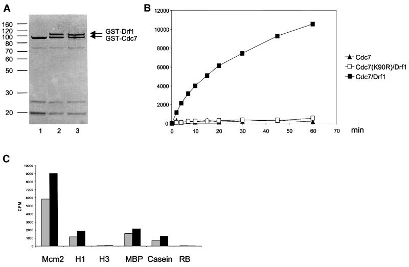
Studies in model organisms have contributed to elucidate multiple levels at which regulation of eukaryotic DNA replication occurs. Cdc7, an evolutionarily conserved serine-threonine kinase, plays a pivotal role in linking cell cycle regulation to genome duplication, being essential for the firing of DNA replication origins. Binding of the cell cycle-regulated subunit Dbf4 to Cdc7 is necessary for in vitro kinase activity. This binding is also thought to be the key regulatory event that controls Cdc7 activity in cells. Here, we describe a novel human protein, Drf1, related to both human and yeast Dbf4. Drf1 is a nuclear cell cycle-regulated protein, it binds to Cdc7 and activates the kinase. Therefore, human Cdc7, like cyclin-dependent kinases, can be activated by alternative regulatory subunits. Since the Drf1 gene is either absent or not yet identified in the genome of model organisms such as yeast and Drosophila, these findings introduce a new level of complexity in the regulation of DNA replication of the human genome.


Figures

None
Fig. 1. Drf1 sequence analysis. (A) Sequence alignment of human Drf1 (top) and Dbf4 (bottom) proteins. Identical amino acids are on a black background; conserved substitutions are in gray. The N, M and C motifs conserved in the Dbf4 protein family are indicated by the boxes. The Drf1 putative bipartite nuclear localization signal is underlined. (B) Phylogenetic tree of Dbf4-related proteins: Hs: Homo sapiens, Mm: Mus musculus, Dm: Drosophila melanogaster, Sc: Saccharomyces cerevisiae, An: Aspergillus nidulans, Sp: Schizosaccharomyces pombe.
None
Fig. 2. Purification and characterization of the Drf1–Cdc7 kinase complex. (A) Cdc7 (lane 1), Cdc7–Drf1 (lane 2) and Cdc7(K90R)/Drf1 (lane 3) recombinant proteins purified from insect cells as a fusion with GST were separated on a 10% polyacrylamide gel and stained with silver. (B) For each time point, 50 ng of recombinant Cdc7 (closed triangles), Cdc7–Drf1 complex (closed squares) or Cdc7(K90R)/Drf1 (open squares) were incubated in the presence of the Mcm2 N-terminal fragment (amino acids 1–285) and labeled ATP. Protein kinase activity of these proteins was measured using multiscreen plates as described in Materials and methods. (C) The Cdc7–Drf1 complex was incubated in the presence of 6 µM of the indicated proteins as substrate. After 30 (gray bars) or 60 (black bars) min, the reaction was stopped and proteins separated on a polyacrylamide gel. The amount of radioactivity in the different substrates was measured with a Storm 840 Phosphoimager. Mcm2 corresponds to the N-terminal 285 amino acid fragment of the Mcm2 protein, H1 and H3 to histone H1 and H3, respectively, MBP to myelin basic protein; and RB to the retinoblastoma protein.
None
Fig. 3. Characterization of Cdc7–Drf1 complexes in human cells. (A) Drf1 is phosphorylated when co-transfected with functional Cdc7. 293 cells were transiently transfected with HA-Drf1 in combination with either wild-type or kinase-deficient (K90R)Cdc7 constructs as indicated. Protein samples were either kept on ice (lanes 1 and 2) or incubated with or without calf intestinal phosphatase in the absence or presence of phosphatase inhibitors as indicated (lanes 3–5) before western blot analysis with anti-HA antibodies. (B and C) Formation of active Cdc7–Drf1 complexes in human cells. Protein extracts from 293 cells transfected with the indicated constructs were immunoprecipitated with either anti-HA (lanes 1–4) or anti-Flag (lanes 5–8) antibodies and subsequently incubated with radioactive ATP and a fragment of Mcm2 protein for 15 min before SDS–PAGE. In (B), Drf1 and Cdc7 were visualized with anti-HA or anti-Cdc7 antibodies, respectively. In (C), labeled proteins were visualized by autoradiography. Images correspond to different exposure times: a longer exposure was required to detect phosphorylated Drf1 and Dbf4 compared with phospho-MCM2. (D) Dbf4 does not immunoprecipitate with Drf1. Protein extracts from HeLa cells transfected with the indicated constructs were probed with anti-HA and anti-Cdc7 antibodies in western blot experiments in lanes 1 and 2. Drf1 and Dbf4, both tagged with the HA epitope, are indicated and migrate differentially in these gels. In lanes 3–5, extracts were immunoprecipitated with either anti-Drf1 mAb 5G4 or unrelated murine IgGs before western blot analysis. (E) Anti-Drf1 mAbs pull-down Cdc7 from extracts prepared from exponentially growing HeLa cells. In each lane, 4 mg of whole-cell extract were used in immunoprecipitation experiments with the anti-Drf1 mAbs 5G4 and 5H4 or control IgG. Western blot was performed with affinity-purified anti-Cdc7 polyclonal antibodies.
None
Fig. 4. Cell cycle analysis of Drf1 and Dbf4 expression. (A and B) Drf1 and Dbf4 mRNAs are induced upon re-entry into the cell cycle. NHDFs were arrested in G0 by serum starvation and then stimulated by addition of 10% serum. At the indicated times, RNA was prepared and Drf1 and Dbf4 mRNA levels analyzed by northern blotting. (B) The DNA content at the indicated times measured by FACS. (C, D and E) Fluctuation of S-phase cyclins and Cdc7 regulatory subunits during the cell cycle in normal human fibroblasts. NHDFs were blocked in S phase with thymidine and released into fresh medium. (C) Drf1, Dbf4 and β-actin mRNA levels were analyzed by northern blotting. The bar chart represents the quantification of Drf1 and Dbf4 levels at different times after normalization with respect to β-actin. Arbitrary units are given on the y-axis. Data were obtained by analyzing radioactivity present in each lane for each hybridization using the Molecular imager FX phosphoimager and Quantity One software (Bio-Rad). (D) Western blot analysis of Drf1, cyclin E, cyclin A and cdk2 levels. (E) The DNA content at the indicated times measured by FACS.
None
Fig. 5. Drf1 is a protein with a short half-life. HeLa cells overexpressing HA-Drf1 were either treated with cycloheximide or mock treated. At the indicated times, protein extracts were prepared and levels of the indicated protein analyzed by western blot.
None
Fig. 6. Drf1 is a nuclear protein. (A) Staining of HeLa cells expressing Ha-Drf1. Staining with anti-HA antibodies is shown in red, while DAPI staining is shown in blue. (B) Confocal microscopy analysis of Drf1 and Cdc7 localization in HeLa cells overexpressing HA-Drf1 and V5-Cdc7. Staining with anti-V5 antibodies is shown in red, while staining with anti-HA antibodies is shown in green. The last picture represents a merged image of the previous two. (C) Confocal microscopy analysis of Drf1 staining and replication foci. HA-Drf1 is shown in red and the BrdU staining in green. The last picture represents a merged image of the previous two.

References

    1. Bousset K. and Diffley,J.F. (1998) The Cdc7 protein kinase is required for origin firing during S phase. Genes Dev., 12, 480–490. - PMC - PubMed
    1. Brown G.W. and Kelly,T.J. (1998) Purification of Hsk1, a minichromosome maintenance protein kinase from fission yeast. J. Biol. Chem., 273, 22083–22090. - PubMed
    1. Diffley J.F. and Labib,K. (2002) The chromosome replication cycle. J. Cell Sci., 115, 869–872. - PubMed
    1. Dimitrova D.S. and Gilbert,D.M. (1999) The spatial position and replication timing of chromosomal domains are both established in early G1 phase. Mol. Cell, 4, 983–993. - PubMed
    1. Donaldson A.D., Fangman,W.L. and Brewer,B.J. (1998) Cdc7 is required throughout the yeast S phase to activate replication origins. Genes Dev., 12, 491–501. - PMC - PubMed

Publication types

MeSH terms