. 2002 Jun 17;21(12):3182-91.
doi: 10.1093/emboj/cdf309.

A novel uracil-DNA glycosylase with broad substrate specificity and an unusual active site

Affiliations

A novel uracil-DNA glycosylase with broad substrate specificity and an unusual active site

Alessandro A Sartori et al. EMBO J. .

Abstract

Uracil-DNA glycosylases (UDGs) catalyse the removal of uracil by flipping it out of the double helix into their binding pockets, where the glycosidic bond is hydrolysed by a water molecule activated by a polar amino acid. Interestingly, the four known UDG families differ in their active site make-up. The activating residues in UNG and SMUG enzymes are aspartates, thermostable UDGs resemble UNG-type enzymes, but carry glutamate rather than aspartate residues in their active sites, and the less active MUG/TDG enzymes contain an active site asparagine. We now describe the first member of a fifth UDG family, Pa-UDGb from the hyperthermophilic crenarchaeon Pyrobaculum aerophilum, the active site of which lacks the polar residue that was hitherto thought to be essential for catalysis. Moreover, Pa-UDGb is the first member of the UDG family that efficiently catalyses the removal of an aberrant purine, hypoxanthine, from DNA. We postulate that this enzyme has evolved to counteract the mutagenic threat of cytosine and adenine deamination, which becomes particularly acute in organisms living at elevated temperatures.


Figures

None
Fig. 1. Identification of Pa-UDGb and its orthologues. (A) Complete amino acid sequence alignment of P.aerophilum uracil-DNA glycosylase b (Pa-UDGb) with homologues from Sulfolobus solfataricus (EMBL: AE006867), Thermoplasma volcanium (EMBL: AP000994), Streptomyces coelicolor (Swiss-Prot: Q9S2L3) and Mycobacterium tuberculosis (Swiss-Prot: Q11059). Identical residues are shaded and the two putative active site motifs, corresponding to motifs A and B in (B), are underlined. The conserved phenylalanine that interacts in the binding pocket of the enzyme with the flipped-out base through π–π interactions is indicated by an asterisk. The sequence alignment shown was performed using the MultAlin software (Corpet, 1988) available at www.toulouse.infra.fr. (B) Partial amino acid sequence alignment of the active site motifs A and B of representatives of the five classes of uracil-DNA glycosylases: uracil-DNA glycosylase from E.coli (udg_ecoli, EMBL: J03725), human TDG (tdg_human, EMBL: U51166), SMUG1 from Homo sapiens (smug_human, EMBL: AF125182), P.aerophilum UDGa (Sartori et al., 2001) and UDGb (this work). Highly conserved residues are shown in black boxes, residues implicated in activating the catalytic water molecule are in open boxes and the hydrophobic residues preceding motif A are shown in grey boxes. Note that motif A of the putative active site of Pa-UDGb lacks a polar amino acid residue capable of activating a water molecule towards a nucleophilic attack on the C1′ of the sugar. The two mutated sites (A68D and H196N) are indicated by arrows.
None
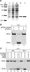
Fig. 2. (A) Expression and purification of the recombinant His-tagged Pa-UDGb (see Materials and methods). I, total extract of the E.coli strain BL21(DE3)pET28c(+)-paudgb; II, total extract of the same cells, following induction with IPTG; III, cleared lysate of the same cells; IV, proteins eluted from the Ni-NTA column with 250 mM imidazole; V, Pa-UDGb eluted from a Mono-S column; M, molecular size marker. The panel shows a 12% Coomassie blue-stained denaturing polyacrylamide gel. (B) Processing of G·U mispairs by fraction IV obtained from E.coli BL21 cells transfected with the pET28c(+)-paudgb plasmid (lane 2) or with the empty pET28c(+) vector (lane 3). This experiment shows that no E.coli uracil-processing activity is present in this fraction. The 60mer oligonucleotide substrate G·U was incubated for 1 h at 37°C with 6 µl of fraction IV as described in Materials and methods. (CPa-UDGb is a heat-stable, monofunctional uracil-DNA glycosylase. The enzyme alone removes uracil at both indicated temperatures, but does not cleave the sugar–phosphate backbone of the mispaired DNA substrate, as witnessed by the absence of the 23mer product band in the reaction where the G·U substrate was treated with Pa-UDGb alone (lane 1). Cleavage occured only upon the addition of hot alkali (lanes 2 and 6) or of human HAP1 (lane 3). The faint product band in lane 5 is due to heat-induced spontaneous β-elimination at the labile AP sites. Incubation at 70°C significantly increased the activity of Pa-UDGb (lane 6), whereas the E.coli UDG was completely inactivated at this temperature (lane 7, cf. lane 4).
None
Fig. 3. Comparison of processing efficiencies and binding of different substrates by Pa-UDGa and Pa-UDGb. (A) Processing of 20 pmol of the fluorescently labelled 60mer substrates G·U (squares), A·U (diamonds) and G·hmU (triangles) with 2 pmol of Pa-UDGa (solid lines) or Pa-UDGb (dashed lines). At the indicated time points, aliquots of the reaction mixture were removed and immediately quenched with 100 mM NaOH (10 min at 90°C) to inactivate the enzyme and to cleave the resulting AP sites. The substrate and product were separated on 20% denaturing polyacrylamide gels and the band intensity was quantified using a Storm 860 PhosphorImager with ImageQuant software. The values shown represent the average of at least three independent experiments. (B) Comparison of DNA-binding specificities of Pa-UDGa, Pa-UDGb and Pa-MIG. The enzymes were incubated with the fluorescently labelled 60mer substrates under conditions (15 min at 4°C) where the base removal does not take place (data not shown). A stable protein–DNA complex was formed only between Pa-UDGb and Pa-MIG and a duplex substrate containing an AP site opposite a guanine (lane G·AP). Data were obtained from a Storm 860 PhosphorImager scan of a 6% native polyacrylamide gel.
None
Fig. 4. Processing efficiency of various substrates by Pa-UDGb. (A) Processing of 20 pmol of the labelled substrates G·hmU (solid line, filled squares), G·εC (dashed line, filled diamonds), G·U (solid line, filled triangles), A·hmU (solid line, open triangles), A·U (solid line, filled circles), G·FU (dashed line, filled circles), T·Hx (solid line, open squares), ssU (solid line, open diamonds) and G·T (solid line, crosses) with 4 pmol of Pa-UDGb. (εC, ethenocytosine; FU, 5-fluorouracil; Hx, hypoxanthine). At the indicated time points, aliquots of the reaction mixture were removed and quenched immediately with 100 mM NaOH (10 min at 90°C) to inactivate the enzyme and to cleave the resulting AP sites. In the case of the base-labile ethenocytosine substrate, the AP sites were processed with 50 nM HAP1 (10 min at 37°C) in the presence of 2.5 mM MgCl2. The AP sites produced spontaneously under the reaction conditions in the case of the labile G·εC substrate were subtracted. The values shown represent the average of at least three independent experiments. (B) The labelled substrates (20 pmol) T·Hx (squares), C·Hx (triangles), T·G (circles) and ssU (diamonds), were incubated with 4 pmol of Pa-UDGb for 2, 4 and 24 h. The substrate and product were separated on 20% denaturing polyacrylamide gels and the band intensity was quantified using a Storm 860 PhosphorImager with ImageQuant software. The values shown represent the average of at least three independent experiments. (C) Processing of hypoxanthine-containing substrates by six different uracil-processing enzymes, Pa-UDGa (Sartori et al., 2001), Pa-UDGb, Pa-MIG (Yang et al., 2000), Ec-UDG, Hs-TDG (Neddermann and Jiricny, 1994) and Hs-MBD4 (Hendrich et al., 1999). For the G·U substrate, a 1:1 molar ratio of enzyme versus substrate was used, whereas for the T·Hx we used a 10-fold excess of enzyme over substrate. The incubations with the hyperthermophilic enzymes from P.aerophilum were carried out for 1 h at 70°C, and those with the mesophilic enzymes from E.coli and H.sapiens for 1 h at 37°C. The panel shows a 20% denaturing polyacrylamide gel scanned with a Storm 860 PhosphorImager.
None
Fig. 5. Processing of 20 pmol of the labelled G·hmU substrate with 4 pmol (solid lines, 1:5) of wild-type Pa-UDGb (squares), Pa-UDGb A68D (triangles) and Pa-UDGb H196N (circles). Processing of 10 pmol of G·hmU substrate with 10 pmol (dashed lines, 1:1) of Pa-UDGb A68D (triangles) and Pa-UDGb H196N (circles). At the indicated time points, aliquots of the reaction mixture were removed and immediately quenched with 100 mM NaOH (10 min at 90°C) to inactivate the enzyme and to cleave the resulting AP sites. The substrate and product were separated on 20% denaturing polyacrylamide gels and the band intensity was quantified using a Storm 860 PhosphorImager with ImageQuant software. The values shown represent the average of at least three independent experiments.
None
Fig. 6. Putative mechanisms of ‘associative’ and ‘dissociative’ cleavage of glycosidic bonds.

Similar articles

Cited by

References

    1. Aravind L. and Koonin,E.V. (2000) The α/β fold uracil DNA glycosylases: a common origin with diverse fates. Genome Biol., 1, RESEARCH0007. - PMC - PubMed
    1. Barrett T.E., Savva,R., Panayotou,G., Barlow,T., Brown,T., Jiricny,J. and Pearl,L.H. (1998) Crystal structure of a G:T/U mismatch-specific DNA glycosylase: mismatch recognition by complementary-strand interactions. Cell, 92, 117–129. - PubMed
    1. Brown T.C. and Brown-Luedi,M.L. (1989) G/U lesions are efficiently corrected to G/C in SV40 DNA. Mutat. Res., 227, 233–236. - PubMed
    1. Brown T.C. and Jiricny,J. (1987) A specific mismatch repair event protects mammalian cells from loss of 5-methylcytosine. Cell, 50, 945–950. - PubMed
    1. Corpet F. (1988) Multiple sequence alignment with hierarchical clustering. Nucleic Acids Res., 16, 10881–10890. - PMC - PubMed

Publication types

MeSH terms