. 2002 Jul;70(7):3468-78.
doi: 10.1128/IAI.70.7.3468-3478.2002.

Changes in temporal and spatial patterns of outer surface lipoprotein expression generate population heterogeneity and antigenic diversity in the Lyme disease spirochete, Borrelia burgdorferi

Affiliations

Changes in temporal and spatial patterns of outer surface lipoprotein expression generate population heterogeneity and antigenic diversity in the Lyme disease spirochete, Borrelia burgdorferi

P Scott Hefty et al. Infect Immun. 2002 Jul.

Abstract

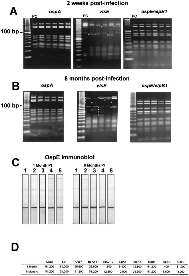
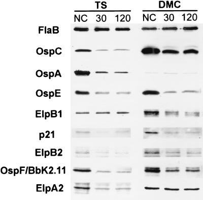
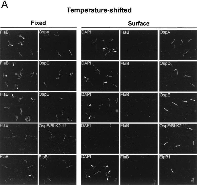
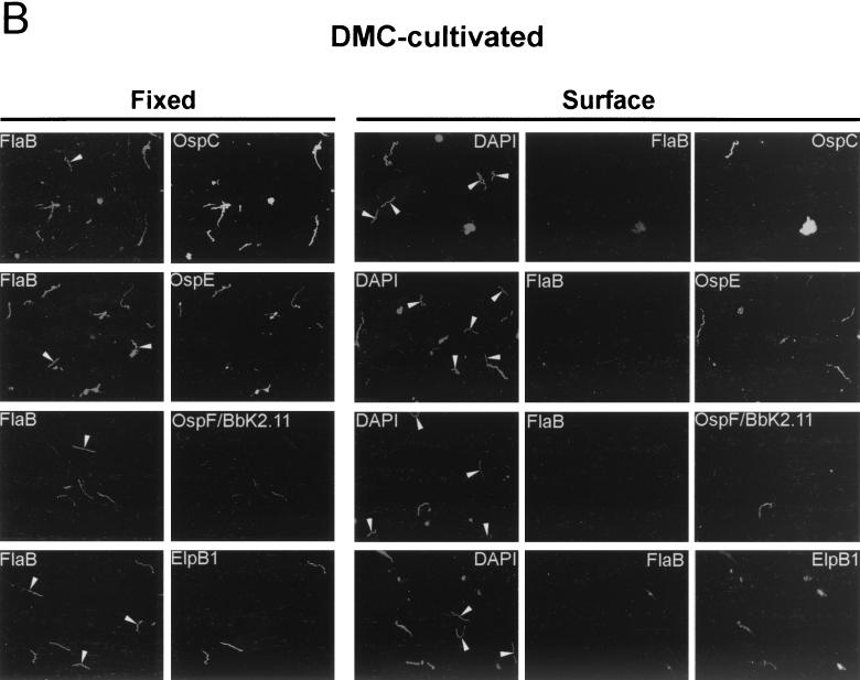
Borrelia burgdorferi differentially expresses many of the OspE/F/Elp paralogs during tick feeding. These findings, combined with the recent report that stable B. burgdorferi infection of mammals occurs only after 53 h of tick attachment, prompted us to further analyze the expression of the OspE/F/Elp paralogs during this critical period of transmission. Indirect immunofluorescence analysis revealed that OspE, p21, ElpB1, ElpB2, and OspF/BbK2.11 are expressed in the salivary glands of ticks allowed to feed on mice for 53 to 58 h. Interestingly, many of the spirochetes in the salivary glands that expressed abundant amounts of these antigens were negative for OspA and OspC. Although prior reports have indicated that OspE/F/Elp orthologs are surface exposed, none of the individual lipoproteins or combinations of the lipoproteins protected mice from challenge infections. To examine why these apparently surface-exposed lipoproteins were not protective, we analyzed their genetic stability during infection and their cellular locations after cultivation in vitro and within dialysis membrane chambers, mimicking a mammalian host-adapted state. Combined restriction fragment length polymorphism and nucleotide sequence analyses revealed that the genes encoding these lipoproteins are stable for at least 8 months postinfection. Interestingly, cellular localization experiments revealed that while all of these proteins can be surface localized, there were significant populations of spirochetes that expressed these lipoproteins only in the periplasm. Furthermore, host-specific signals were found to alter the expression patterns and final cellular location of these lipoproteins. The combined data revealed a remarkable heterogeneity in populations of B. burgdorferi during tick transmission and mammalian infection. The diversity is generated not only by temporal changes in antigen expression but also by modulation of the surface lipoproteins during infection. The ability to regulate the temporal and spatial expression patterns of lipoproteins throughout infection likely contributes to persistent infection of mammals by B. burgdorferi.


Figures

FIG. 1.
FIG. 1.
Genetic stability of the ospE/elpB1 loci and antibody reactivity to the OspE-related, OspF-related, and Elp proteins during infection. (A and B) RFLP analyses of the ospA, vlsE, and ospE/elpB1 loci of isogenic clones generated from mice hyperimmunized with OspE 2 weeks postinfection (A) and from unimmunized mice 8 months postinfection (B) using restriction enzyme Tru9I. The results for five representative clones from the 20 to 40 clones analyzed for each locus are shown. PC, RFLP analysis performed on amplicons generated from parental strain 297. For the vlsE locus, fragments that vary from the fragments in the parental pattern are indicated by asterisks. The left lane in each panel contained a 10-bp DNA ladder. (C) Representative immunoblot of recombinant OspE with serum from mice used to generate clonal isolates 1 and 8 months postinfection (PI). (D) Reciprocal antibody titers for the OspE/F/Elp paralogs and OspC in pooled sera from mice used to generate clonal isolates. Titers were determined 1 and 8 months postinfection.
FIG. 2.
FIG. 2.
Immunoblot analysis of whole-cell lysates from proteinase K-treated B. burgdorferi. Spirochetes cultivated following a temperature shift (TS) and in DMCs were incubated for 30 or 120 min with 200 μg of proteinase K per ml; control samples (NC) were incubated for 120 min without proteinase K. Proteinase K degradation was assessed by immunoblotting using monoclonal antibodies directed against FlaB (1H6-33) or OspA (14D2-27) or affinity-purified rat polyclonal anti-OspE, anti-p21, anti-OspF/BbK2.11, anti-ElpA2, anti-ElpB1, anti-ElpB2, or anti-OspC using the Enhanced Chemiluminescence Plus Western blotting system as described in Materials and Methods.
FIG. 3.
FIG. 3.
Expression and cellular localization of OspA, OspC, OspE, OspF/BbK2.11, and ElpB1 within individual organisms. Spirochetes cultivated following a temperature shift (A) or in DMCs (B) were subjected to IFA after acetone fixation (Fixed) or directly in solution prior to fixation (Surface). Samples were probed with rabbit polyclonal serum to FlaB and rat polyclonal serum directed against OspA, OspC, OspE, OspF/BbK2.11, or ElpB1. OspA results are shown only for temperature-shifted organisms because so few organisms were found to express this protein in DMC-cultivated organisms. For surface labeling experiments, DAPI, a permeable DNA-binding dye, was used to identify all spirochetes in a field. The arrows indicate spirochetes labeled with OspE, OspF/BbK2.11, and ElpB1 antibodies. The arrowheads indicate individual spirochetes that either did not express the indicated lipoprotein (Fixed panels) or did not display it on the surface (Surface panels). Organisms were visualized by fluorescence microscopy at a magnification of ×1,000.
FIG. 3.
FIG. 3.
Expression and cellular localization of OspA, OspC, OspE, OspF/BbK2.11, and ElpB1 within individual organisms. Spirochetes cultivated following a temperature shift (A) or in DMCs (B) were subjected to IFA after acetone fixation (Fixed) or directly in solution prior to fixation (Surface). Samples were probed with rabbit polyclonal serum to FlaB and rat polyclonal serum directed against OspA, OspC, OspE, OspF/BbK2.11, or ElpB1. OspA results are shown only for temperature-shifted organisms because so few organisms were found to express this protein in DMC-cultivated organisms. For surface labeling experiments, DAPI, a permeable DNA-binding dye, was used to identify all spirochetes in a field. The arrows indicate spirochetes labeled with OspE, OspF/BbK2.11, and ElpB1 antibodies. The arrowheads indicate individual spirochetes that either did not express the indicated lipoprotein (Fixed panels) or did not display it on the surface (Surface panels). Organisms were visualized by fluorescence microscopy at a magnification of ×1,000.

References

    1. Akins, D. R., K. W. Bourell, M. J. Caimano, M. V. Norgard, and J. D. Radolf. 1998. A new animal model for studying Lyme disease spirochetes in a mammalian host-adapted state. J. Clin. Investig. 101:2240-2250. - PMC - PubMed
    1. Akins, D. R., M. J. Caimano, X. Yang, F. Cerna, M. V. Norgard, and J. Radolf. 1999. Molecular and evolutionary analysis of Borrelia burgdorferi 297 circular plasmid-encoded lipoproteins with OspE- and OspF-like leader peptides. Infect. Immun. 67:1526-1532. - PMC - PubMed
    1. Akins, D. R., S. F. Porcella, T. G. Popova, D. Shevchenko, S. I. Baker, M. Li, M. V. Norgard, and J. D. Radolf. 1995. Evidence for in vivo but not in vitro expression of a Borrelia burgdorferi outer surface protein F (OspF) homolog. Mol. Microbiol. 18:507-520. - PubMed
    1. Benach, J. L., J. L. Coleman, R. A. Skinner, and E. M. Bosler. 1987. Adult Ixodes dammini on rabbits: a hypothesis for the development and transmission of Borrelia burgdorferi. J. Infect. Dis. 155:1300-1306. - PubMed
    1. Bockenstedt, L. K., E. Hodzic, S. Feng, K. W. Bourell, A. de Silva, R. R. Montgomery, E. Fikrig, J. D. Radolf, and S. W. Barthold. 1997. Borrelia burgdorferi strain-specific Osp C-mediated immunity in mice. Infect. Immun. 65:4661-4667. - PMC - PubMed

Publication types

LinkOut - more resources