. 2002 Jul;70(7):3510-20.
doi: 10.1128/IAI.70.7.3510-3520.2002.

Effect of low- and high-virulence Yersinia enterocolitica strains on the inflammatory response of human umbilical vein endothelial cells

Affiliations

Effect of low- and high-virulence Yersinia enterocolitica strains on the inflammatory response of human umbilical vein endothelial cells

Geertrui Denecker et al. Infect Immun. 2002 Jul.

Abstract

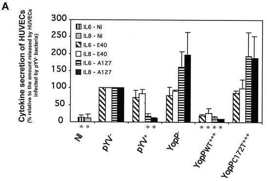
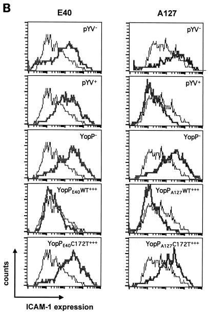
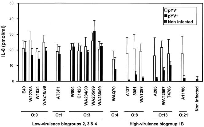
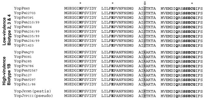
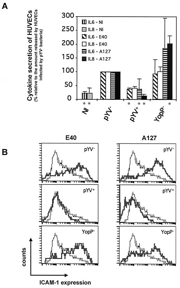
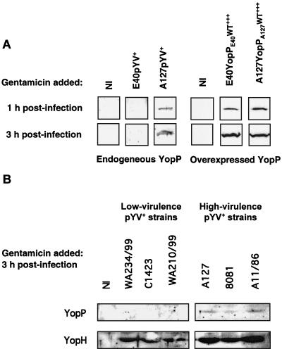
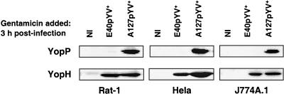
Pathogenic strains of Yersinia spp. inject a set of Yop effector proteins into eukaryotic cells by using a plasmid-encoded type III secretion system. In this study, we analyzed the inflammatory response of human umbilical vein endothelial cells (HUVECs) after infection with different Yersinia enterocolitica strains. We found that both expression of intercellular adhesion molecule 1 and release of the cytokines interleukin-6 (IL-6) and IL-8 by HUVECs are downregulated in a YopP-dependent way, demonstrating that YopP plays a major role in the inflammatory response of these cells. Infection of HUVECs with several low-virulence (biotype 2, 3, and 4) and high-virulence (biotype 1B) Y. enterocolitica strains showed that biotype 1B isolates are more efficient in inhibiting the inflammatory response than low-virulence Y. enterocolitica strains and that this effect depends on the time of contact. We extended the results of Ruckdeschel et al. and found that on the basis of the presence or absence of arginine-143 of YopP (K. Ruckdeschel, K. Richter, O. Mannel, and J. Heesemann, Infect. Immun. 69:7652-7662, 2001) all the Y. enterocolitica strains used fell into two groups, which correlate with the low- and high-virulence phenotypes. In addition, we found that high-virulence strains inject more YopP into the cytosol of eukaryotic target cells than do low-virulence strains.


Figures

FIG. 1.
FIG. 1.
Y. enterocolitica A127 is more efficient than Y. enterocolitica E40 in downregulating the inflammatory response of HUVECs. HUVECs were infected at an MOI of 50 with the following derivatives of Y. enterocolitica E40 and A127: wild type (pYV+), pYV plasmid cured (pYV), YopP deficient (YopP), YopP overexpressing the wild-type YopP (YopPWT+++), and YopP overexpressing the catalytically inactive mutant (YopPC172T+++) (Table 1; also see Materials and Methods). Noninfected (NI) HUVECs were used as a control. After 1 h of infection, gentamicin (50 μg/ml) was added to kill extracellular bacteria. (A) IL-6 secretion and IL-8 secretion analyzed by ELISA in cell culture supernatants collected 5 h after the beginning of the infection. An asterisks below a bar indicates that the cytokine secretion value for the strain was significantly different from the value for the corresponding pYV strain (Dunnett's post hoc analysis, P < 0.05) (see Materials and Methods). (B) ICAM-1 expression monitored by flow cytometry 24 h after the beginning of infection with mouse monoclonal antibodies against ICAM-1 conjugated to fluorescein. Thin lines, ICAM-1 expression of noninfected HUVECs; thick lines, ICAM-1 expression of infected HUVECs.
FIG. 1.
FIG. 1.
Y. enterocolitica A127 is more efficient than Y. enterocolitica E40 in downregulating the inflammatory response of HUVECs. HUVECs were infected at an MOI of 50 with the following derivatives of Y. enterocolitica E40 and A127: wild type (pYV+), pYV plasmid cured (pYV), YopP deficient (YopP), YopP overexpressing the wild-type YopP (YopPWT+++), and YopP overexpressing the catalytically inactive mutant (YopPC172T+++) (Table 1; also see Materials and Methods). Noninfected (NI) HUVECs were used as a control. After 1 h of infection, gentamicin (50 μg/ml) was added to kill extracellular bacteria. (A) IL-6 secretion and IL-8 secretion analyzed by ELISA in cell culture supernatants collected 5 h after the beginning of the infection. An asterisks below a bar indicates that the cytokine secretion value for the strain was significantly different from the value for the corresponding pYV strain (Dunnett's post hoc analysis, P < 0.05) (see Materials and Methods). (B) ICAM-1 expression monitored by flow cytometry 24 h after the beginning of infection with mouse monoclonal antibodies against ICAM-1 conjugated to fluorescein. Thin lines, ICAM-1 expression of noninfected HUVECs; thick lines, ICAM-1 expression of infected HUVECs.
FIG. 2.
FIG. 2.
Suppression of IL-8 release is dependent on the Y. enterocolitica serotype. HUVECs were infected with Y. enterocolitica strains and their corresponding pYV derivatives (Table 2) at an MOI of 50. Noninfected HUVECs were used as a control. After 1 h of infection, gentamicin (50 μg/ml) was added to kill extracellular bacteria. The amount of IL-8 released was assayed by using cell supernatants collected 24 h after the beginning of infection as described in the legend to Fig. 1. The ANOVA revealed a significant interaction between serotype and pYV (df = 1, MS = 896.8, F = 29.4, P < 0.001), supporting the conclusion that suppression of IL-8 release depends on the serotype.
FIG. 3.
FIG. 3.
Sequence comparison of YopP proteins from the different low- and high-virulence Y. enterocolitica strains: alignment of the conserved putative catalytic domains and the arginine/serine-143 of YopP from all the Y. enterocolitica strains listed in Table 1, as well as the sequences of YopJ from Y. pestis (accession no. NP047569) [yopjkym5 (pestis)] and YopJ from Y. pseudotuberculosis (accession no. AAA68488) [YopJypiii (pseudo)]. The arrow indicates arginine/serine-143, and the asterisks indicate the amino acid residues of the catalytic triad (histidine-109, glutamate-128, and cysteine-172).
FIG. 4.
FIG. 4.
Inhibition of the release of anti-inflammatory cytokines and the expression of ICAM-1 adhesion molecules by HUVECs infected by Y. enterocolitica for 3 h. HUVECs were infected at an MOI of 50 with the following derivatives of Y. enterocolitica E40 and A127: wild type (pYV+), pYV plasmid cured (pYV), and YopP deficient (YopP) (Table 1; also see Materials and Methods). Noninfected (NI) HUVECs were used as a control. After 3 h of infection, gentamicin (50 μg/ml) was added to kill extracellular bacteria. (A) IL-6 secretion and IL-8 secretion were analyzed by ELISA by using cell culture supernatants collected 5 h after the beginning of the infection. An asterisk below a bar indicates that the cytokine secretion value for the strain was significantly different from the value for the corresponding pYV strain (Dunnett's post hoc analysis, P < 0.05) (see Materials and Methods). (B) ICAM-1 expression monitored by flow cytometry 24 h after infection with mouse monoclonal antibodies against ICAM-1 conjugated to fluorescein. Thin lines, ICAM-1 expression of noninfected HUVECs; thick lines, ICAM-1 expression of infected HUVECs.
FIG. 5.
FIG. 5.
Differential injection of YopP proteins of low- and high-virulence Y. enterocolitica strains into the cytoplasm of HUVECs. HUVECs were infected with different Y. enterocolitica strains at an MOI of 50 (Tables 1 and 2; also see Materials and Methods). Noninfected (NI) HUVECs were used as a control. After 1 or 3 h of infection, gentamicin (50 μg/ml) was added to kill extracellular bacteria. Cytosolic cell lysates of HUVECs were prepared by digitonin lysis 5 h after the beginning of the infection and were analyzed by sodium dodecyl sulfate-polyacrylamide gel electrophoresis and Western blotting by using an anti-YopP antibody or an anti-YopH antibody, as indicated. For reasons of clarity the Western blots shown for detection of overexpressed YopP are short-term exposures, while the Western blots for detection of endogenous YopP are long-term exposures. (A) E40pYV+, A127pYV+, E40YopPE40WT+++, and A127YopPA127WT+++; (B) WA234/99pYV+, C1423pYV+, WA210/99pYV+, A127pYV+, 8081pYV+, and A11/86pYV+.
FIG. 6.
FIG. 6.
Injection of YopP and YopH into the cytoplasm of different cell types. Rat-1, HeLa, or J774A.1 cells were infected with E40pYV+ or A127pYV+ at an MOI of 50. Noninfected (NI) cells were used as a control. After 3 h of infection, gentamicin (50 μg/ml) was added to kill extracellular bacteria. Cytosolic cell lysates of were prepared by digitonin lysis 5 h after the beginning of the infection and were analyzed by sodium dodecyl sulfate-polyacrylamide gel electrophoresis and Western blotting by using an anti-YopP antibody or an anti-YopH antibody.

References

    1. Autenrieth, I. B., and R. Firsching. 1996. Penetration of M cells and destruction of Peyer's patches by Yersinia enterocolitica: an ultrastructural and histological study. J. Med. Microbiol. 44:285-294. - PubMed
    1. Bach, S., C. Buchrieser, M. Prentice, A. Guiyoule, T. Msadek, and E. Carniel. 1999. The high-pathogenicity island of Yersinia enterocolitica Ye8081 undergoes low-frequency deletion but not precise excision, suggesting recent stabilization in the genome. Infect. Immun. 67:5091-5099. - PMC - PubMed
    1. Beuscher, H. U., F. Rodel, A. Forsberg, and M. Rollinghoff. 1995. Bacterial evasion of host immune defense: Yersinia enterocolitica encodes a suppressor for tumor necrosis factor alpha expression. Infect. Immun. 63:1270-1277. - PMC - PubMed
    1. Bliska, J. B. 2000. Yop effectors of Yersinia spp. and actin rearrangements. Trends Microbiol. 8:205-208. - PubMed
    1. Boland, A., and G. R. Cornelis. 1998. Role of YopP in suppression of tumor necrosis factor alpha release by macrophages during Yersinia infection. Infect. Immun. 66:1878-1884. - PMC - PubMed

Publication types

MeSH terms

LinkOut - more resources