. 2002 Jul;70(7):3793-803.
doi: 10.1128/IAI.70.7.3793-3803.2002.

Chlamydia trachomatis induces remodeling of the actin cytoskeleton during attachment and entry into HeLa cells

Affiliations

Chlamydia trachomatis induces remodeling of the actin cytoskeleton during attachment and entry into HeLa cells

Reynaldo A Carabeo et al. Infect Immun. 2002 Jul.

Abstract

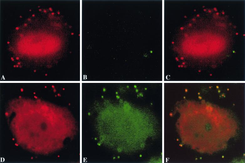
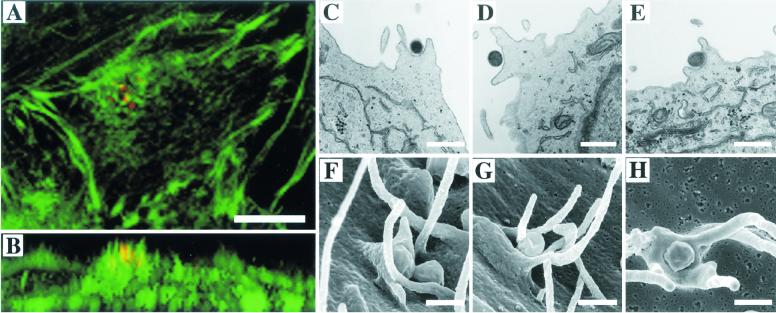
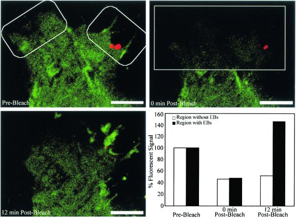
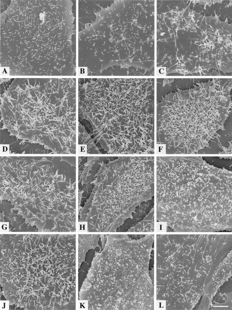
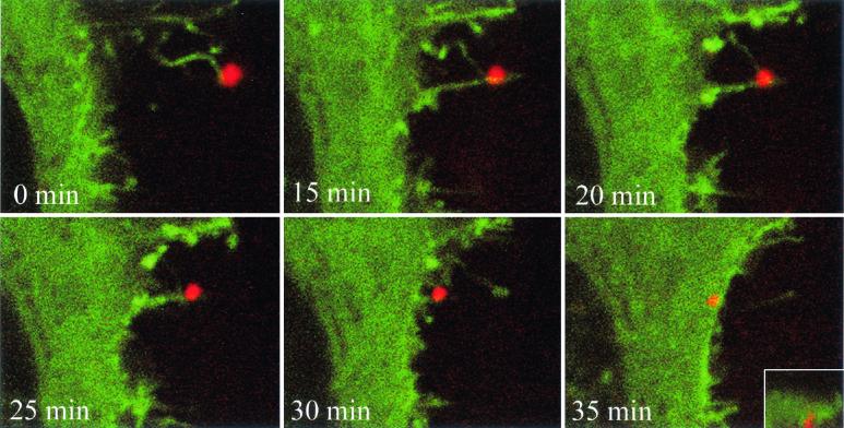
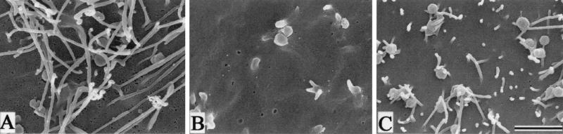
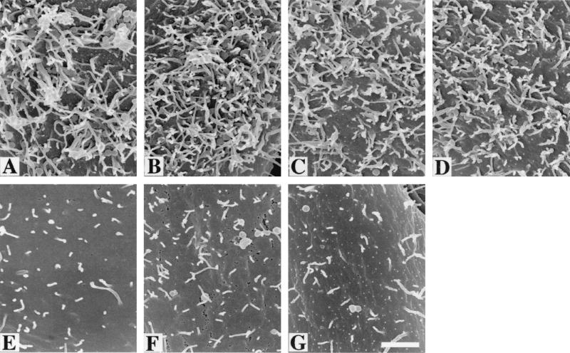
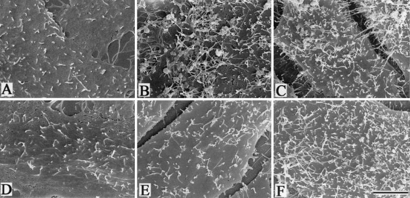
To elucidate the host cell machinery utilized by Chlamydia trachomatis to invade epithelial cells, we examined the role of the actin cytoskeleton in the internalization of chlamydial elementary bodies (EBs). Treatment of HeLa cells with cytochalasin D markedly inhibited the internalization of C. trachomatis serovar L2 and D EBs. Association of EBs with HeLa cells induced localized actin polymerization at the site of attachment, as visualized by either phalloidin staining of fixed cells or the active recruitment of GFP-actin in viable infected cells. The recruitment of actin to the specific site of attachment was accompanied by dramatic changes in the morphology of cell surface microvilli. Ultrastructural studies revealed a transient microvillar hypertrophy that was dependent upon C. trachomatis attachment, mediated by structural components on the EBs, and cytochalasin D sensitive. In addition, a mutant CHO cell line that does not support entry of C. trachomatis serovar L2 did not display such microvillar hypertrophy following exposure to L2 EBs, which is in contrast to infection with serovar D, to which it is susceptible. We propose that C. trachomatis entry is facilitated by an active actin remodeling process that is induced by the attachment of this pathogen, resulting in distinct microvillar reorganization throughout the cell surface and the formation of a pedestal-like structure at the immediate site of attachment and entry.


Figures

FIG. 1.
FIG. 1.
EBs of serovars L2 and D were allowed to attach to HeLa cells that were untreated or pretreated with 2.5 μg of cytochalasin D/ml at 4°C and shifted to 37°C for 30 min to allow chlamydial entry. Cytochalasin D, when required, was present at all times during the 37°C incubation. Internalized and uninternalized EBs were detected as described in Materials and Methods. Total EBs (red), uninternalized EBs (green), merged (yellow). (A to C) Serovar L2, untreated. (D to F) Serovar L2, cytochalasin D treated.
FIG. 2.
FIG. 2.
Actin aggregation and associated cell surface changes at the sites of EB attachment and entry. HeLa cells infected with serovar L2 EBs (red) were either fixed with 4% paraformaldehyde at 30 min postinfection, permeabilized with 0.1% Triton X-100, and incubated with fluorescently tagged phalloidin to visualize F-actin by confocal fluorescence microscopy or processed for transmission electron microscopy. (A) x-y plane. (B) x-z plane. (C and D) TEM. (F and G) SEM images of pedestal-like structures in serovar L2-infected HeLa cells. (E and H) TEM (E) and SEM (H) images of serovar D-infected HeLa cells. Note the F-actin (green)-rich cell surface projection at the site of EB (red) attachment in panel B. Scale bars: panels A and B, 5 μm; panels C, D, and E, 0.8 μm; panels F, G, and H, 0.5 μm.
FIG. 3.
FIG. 3.
FRAP. HeLa cells expressing GFP-actin were incubated with fluorescently tagged serovar L2 EBs at 4°C to allow attachment. GFP-actin fluorescence in a small area of a cell containing EBs was bleached. The cells were transferred to a 37°C incubator, and images were taken at 60 s intervals using the confocal microscope to monitor fluorescence recovery. Images shown represent time points prior to bleaching, 0 min postbleaching, and 15 min postbleaching. Fluorescence recovery in areas designated by boxes within the bleached region was quantitated. Scale bar, 1 μm.
FIG. 4.
FIG. 4.
SEM of HeLa cells infected with serovar L2 or D EBs. HeLa cells were infected at an MOI of 100 at 4°C for 1 h and shifted to 37°C. Fixation to inhibit infection was performed at the indicated time points, and samples were processed for SEM. (A) Uninfected, 30 min. (B) Serovar L2, 0 min. (C) Serovar L2, 10 min. (D) Serovar L2, 20 min. (E) Serovar L2, 30 min. (F) Serovar L2, 40 min. (G) Serovar L2, 50 min. (H) Serovar L2, 60 min. (I) Serovar D, 0 min. (J) Serovar D, 30 min. (K) Zymosan, 0 min. (L) Zymosan, 30 min. Note the phases of morphological changes in and distribution of microvilli. Scale bar, 0.8 μm.
FIG. 5.
FIG. 5.
Interaction of a HeLa cell with a C. trachomatis serovar L2 EB. Time-lapse confocal images of HeLa cells transfected with a GFP-actin expression vector reveal cell surface projections interacting with a fluorescently tagged EB and leading to the retraction of the microvillus-like structure and subsequent internalization of the EB. The site of entry is shown as an x-z image in the inset panel.
FIG. 6.
FIG. 6.
Inhibitory effects of cytochalasin D treatment on microvillar hypertrophy. HeLa cells were either treated with cytochalasin D or left untreated at 4°C during infection with serovar L2 EBs. When required, cytochalasin D was present during the 37°C incubation for 30 min. Samples were processed for SEM. (A) Untreated. (B) Cytochalasin D treated. (C) Cytochalasin D removal and recovery (10 min). Scale bar, 1.5 μm.
FIG. 7.
FIG. 7.
Sensitivity of serovar L2 EBs to different treatments. HeLa cells were infected with serovar L2 EBs that were untreated (A), DTT treated (B), IAA treated (C), DTT-IAA treated (D), mock infected (E), heat killed (F), or glutaraldehyde fixed (G). Scanning electron micrographs reveal the effects of different EB treatments on the induction or lack thereof of microvillus hypertrophy. Scale bar, 1 μm.
FIG. 8.
FIG. 8.
Attachment to the secondary receptor is required for induction of microvillar hypertrophy. Wild-type CHO-K1 and D4.1-3 cells were examined for their ability to support microvillar hypertrophy during infection with serovar L2 EBs. (A-C) CHO-K1. (D-F) D4.1-3. (A and D) Uninfected. (B and E) Serovar L2 infected. (C and F) Serovar D infected. Scale bar, 5 μm.
FIG. 9.
FIG. 9.
EBs enter via a PI-3 kinase-independent mechanism. Wortmannin was tested for its ability to inhibit entry of untreated or opsonized C. trachomatis L2 EBs via the normal or Fc receptor-mediated endocytic pathway. Internalization efficiencies in both experimental groups were determined as described in Materials and Methods. Data are from a minimum of 60 cells and expressed as means ± SD.

References

    1. Adam, T., M. Arpin, M. C. Prevost, P. Gounon, and P. J. Sansonetti. 1995. Cytoskeletal rearrangements and the functional role of T-plastin during entry of Shigella flexneri into HeLa cells. J. Cell Biol. 129:367-381. - PMC - PubMed
    1. Adam, T., M. Giry, P. Boquet, and P. Sansonetti. 1996. Rho-dependent membrane folding causes Shigella entry into epithelial cells. EMBO J. 15:3315-3321. - PMC - PubMed
    1. Allison, A. C., P. Davies, and S. De Petris. 1971. Role of contractile microfilaments in macrophage movement and endocytosis. Nat. New Biol. 232:153-155. - PubMed
    1. Batt, R. M., C. A. Hart, L. McLean, and J. R. Saunders. 1987. Organ culture of rabbit ileum as a model for the investigation of the mechanism of intestinal damage by enteropathogenic Escherichia coli. Gut 28:1283-1290. - PMC - PubMed
    1. Bavoil, P. M., and R. C. Hsia. 1998. Type III secretion in Chlamydia: a case of deja vu? Mol. Microbiol. 28:860-862. - PubMed

MeSH terms

LinkOut - more resources