. 2002 Jul;70(7):3804-15.
doi: 10.1128/IAI.70.7.3804-3815.2002.

Candida albicans expresses a focal adhesion kinase-like protein that undergoes increased tyrosine phosphorylation upon yeast cell adhesion to vitronectin and the EA.hy 926 human endothelial cell line

Affiliations

Candida albicans expresses a focal adhesion kinase-like protein that undergoes increased tyrosine phosphorylation upon yeast cell adhesion to vitronectin and the EA.hy 926 human endothelial cell line

Giorgio Santoni et al. Infect Immun. 2002 Jul.

Abstract

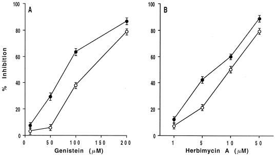
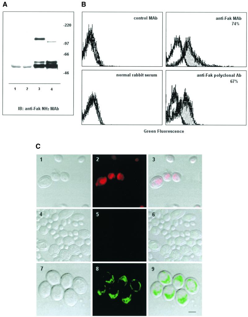
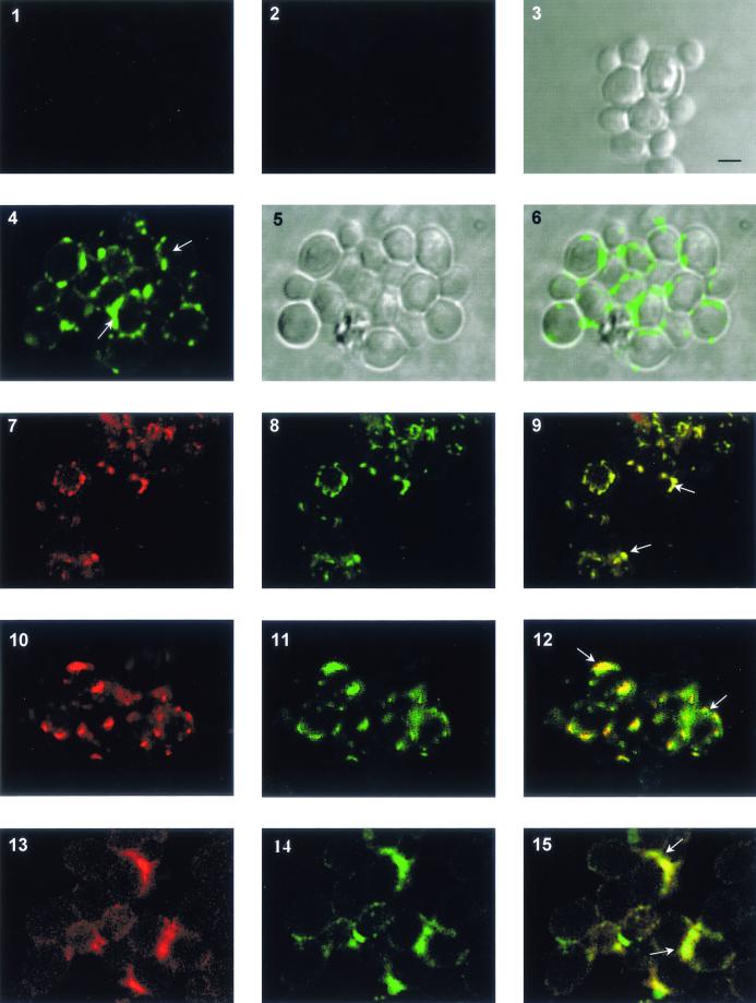
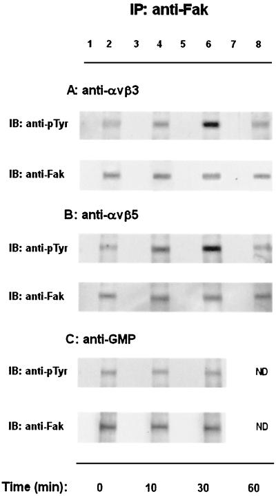
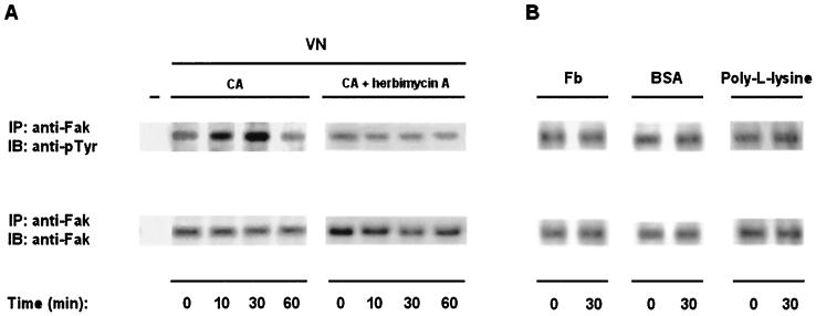
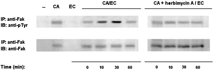
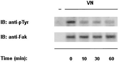
The signaling pathways triggered by adherence of Candida albicans to the host cells or extracellular matrix are poorly understood. We provide here evidence in C. albicans yeasts of a p105 focal adhesion kinase (Fak)-like protein (that we termed CaFak), antigenically related to the vertebrate p125Fak, and its involvement in integrin-like-mediated fungus adhesion to vitronectin (VN) and EA.hy 926 human endothelial cell line. Biochemical analysis with different anti-chicken Fak antibodies identified CaFak as a 105-kDa protein and immunofluorescence and cytofluorimetric analysis on permeabilized cells specifically stain C. albicans yeasts; moreover, confocal microscopy evidences CaFak as a cytosolic protein that colocalizes on the membrane with the integrin-like VN receptors upon yeast adhesion to VN. The protein tyrosine kinase (PTK) inhibitors genistein and herbimycin A strongly inhibited C. albicans yeast adhesion to VN and EA.hy 926 endothelial cells. Moreover, engagement of alpha v beta 3 and alpha v beta 5 integrin-like on C. albicans either by specific monoclonal antibodies or upon adhesion to VN or EA.hy 926 endothelial cells stimulates CaFak tyrosine phosphorylation that is blocked by PTK inhibitor. A role for CaFak in C. albicans yeast adhesion was also supported by the failure of VN to stimulate its tyrosine phosphorylation in a C. albicans mutant showing normal levels of CaFak and VNR-like integrins but displaying reduced adhesiveness to VN and EA.hy 926 endothelial cells. Our results suggest that C. albicans Fak-like protein is involved in the control of yeast cell adhesion to VN and endothelial cells.


Figures

FIG. 1.
FIG. 1.
Effect of the PTK inhibitors genistein and herbimycin A on C. albicans yeast cell adhesion to VN and EA.hy 926 human endothelial cell line. Adhesion of [3H]glucose-labeled C. albicans to VN (1 μg/ml) (•) and EA.hy 926 cells (○) evaluated in the absence (control medium) or presence of different doses of genistein (A), herbimycin A (B), or vehicle (DMSO) (data not shown) at 30 min after incubation at 37°C. Results are the mean percent specific inhibition ± the standard deviation (three separate experiments). The mean percent cell yeast adhesions to VN and EA.hy 926 cells were 48 ± 1 and 63 ± 2, respectively. Since no differences were observed between adhesion in the presence of control medium and vehicle, only data in the presence of medium are shown for sake of simplicity.
FIG.2.
FIG.2.
C. albicans yeast cells express a protein antigenically related to p125Fak. (A) C. albicans yeast (lane 4) and human endothelial EA.hy 926 (lane 3) cell lysates immunoprecipitated with a rabbit anti-Fak COOH polyclonal Ab and immunoblotted with a mouse anti-Fak NH2 MAb. Lanes 2 and 1 represent normal rabbit serum immunoprecipitates from C. albicans and EA.hy 926 cells used as negative control, respectively. Sizes are indicated in kilodaltons. The results shown are representative of one of three separate experiments. (B) Immunofluorescence and FACS analysis on permeabilized C. albicans yeast cells with mouse anti-Fak NH2 MAb or rabbit anti-Fak COOH polyclonal Ab as primary Ab; an irrelevant isotype-matched anti-rat CD5 MAb or normal rabbit serum was used as control Ab. FITC-conjugated GAM or GAR were used as the second-step Ab. The dotted area represents positive stain. The results are representative of one of three separate experiments. (C) Confocal laser-scanning microscopy analysis on permeabilized C. albicans yeast cells with anti-Fak NH2 MAb as primary Ab and FITC-GAM as a second-step Ab. Panels: 1, 4, and 7, C. albicans yeast cells on a bright field; panel 2, C. albicans yeast cells pretreated with propidium iodide and acquired as confocal image; panel 3, merge of bright field and confocal image; panel 5, C. albicans yeast cells incubated with an isotype-matched irrelevant (anti-rat CD5) MAb and FITC-GAM; panel 6, merge of images in panels 4 and 5; panel 8, C. albicans yeast cells incubated with a mouse anti-Fak NH2 MAb and FITC-GAM; panel 9, merge of images in panels 8 and 9. The results are representative of one of three separate experiments. Bar, 1 μm.
FIG. 3.
FIG. 3.
CaFak and integrin-like receptors colocalize into the focal adhesions in C. albicans yeast cells adherent to VN. Confocal laser-scanning microscopy analysis on permeabilized C. albicans yeast cells with anti-Fak COOH, anti-αv, anti-β3, or anti-β5 integrin rabbit antiserum or anti-Fak NH2 mouse MAb as the primary Ab and FITC-GAR and Texas Red-GAM as second-step Abs. Panels: 3 and 5, C. albicans yeast cells on a bright field; 1 and 2, C. albicans yeast cells incubated with Texas Red-GAM or FITC-GAR, respectively; 4, C. albicans yeast cells stained with anti-Fak COOH and FITC-GAR; 6, merge of images in panels 4 and 5; 7, 10, and 13, C. albicans yeast cells stained with anti-Fak NH2 MAb and Texas Red-GAM; C. albicans yeast cells stained with anti-αv (panel 8), anti-β3 (panel 11), or anti-β5 integrin (panel 14) Ab and FITC-GAR, respectively; 9, 12, and 15, merge of images in panels 7 and 8, 10 and 11, and 13 and 14, respectively. The results are representative of one of three separate experiments. White arrows indicate CaFak localization at focal adhesion sites. Bar, 1 μm.
FIG. 4.
FIG. 4.
Engagement of αvβ3 and αvβ5 integrin-like VNRs on C. albicans yeast cells stimulates CaFak tyrosine phosphorylation. C. albicans yeast cells were left untreated (time zero) or stimulated for the indicated times at 37°C with mouse anti-αvβ3 (LM609) (A), anti-αvβ5 (P1F6) (B), or anti-GMP (GF2) control MAb (C). Cell lysates were immunoprecipitated with an anti-Fak COOH rabbit polyclonal Ab. The resulting protein complexes were resolved by SDS-7% polyacrylamide gel electrophoresis, transferred to polyvinylidene difluoride membrane, and immunoblotted with anti-pTyr (4G10) (top panel) or anti-Fak NH2 MAb (bottom panel). Lanes 1, 3, 5, and 7 represent normal rabbit serum immunoprecipitates used as negative control. The results shown are representative of one of three separate experiments. As evaluated by densitometric analysis, upon αvβ3 stimulation, a 0.7-fold increase of CaFak tyrosine phosphorylation was observed at 10 min, a 5-fold increase was observed at 30 min, a 0.5-fold increase was observed at 60 min and upon αvβ5 stimulation, a 1.2-fold increase of CaFak tyrosine phosphorylation was observed at 10 min, a 5-fold increase was observed at 30 min, and a 0.3-fold increase was observed at 60 min.
FIG. 5.
FIG. 5.
Tyrosine kinase activation is required for stimulation of CaFak tyrosine phosphorylation upon adhesion of C. albicans yeast cells to VN. (A) C. albicans yeast cells preincubated or not with herbimycin A (10 μg/ml) for 16 h at RT in YPD medium were left untreated or were allowed to adhere to VN for the indicated times at 37°C. Cell lysates were analyzed as indicated in Fig. 4. Lane − represents RAM immunoprecipitates used as a negative control. The results shown are representative of one of three separate experiments. As evaluated by densitometric analysis, VN stimulates a twofold increase of CaFak tyrosine phosphorylation at 10 min and a fourfold increase at 30 min. (B) C. albicans yeast cells were left untreated (time zero) or allowed to adhere to Fb, BSA, or poly-l-lysine (10 μg/ml) for the indicated times at 37°C. Cell lysates were analyzed as described above. The results shown are representative of one of three separate experiments.
FIG. 6.
FIG. 6.
Tyrosine kinase activation is required for stimulation of CaFak tyrosine phosphorylation upon adhesion of C. albicans yeast cells to the EA.hy 926 human endothelial cell line. C. albicans yeast cells pretreated or not pretreated with herbimycin A (10 μg/ml) for 16 at RT in YPD medium were allowed to bind to paraformaldehyde-prefixed EA.hy 926 human endothelial cell line (CA/EC) for the indicated times at 37°C. Cell lysates were analyzed as indicated in Fig. 4. Lane − represents RAM immunoprecipitates used as a negative control. Lanes CA and EC represent anti-Fak immunoprecipitates from C. albicans and prefixed EA.hy 926 cell lysates, respectively. The results shown are representative of one of three separate experiments. As evaluated by densitometric analysis, binding of C. albicans to EA.hy 926 cells stimulates a 2.5-fold increase of CaFak tyrosine phosphorylation at 10 min and a 4-fold increase at 30 min.
FIG. 7.
FIG. 7.
Reduced adhesiveness of yeast cells from C. albicans 3153 mutant strain to VN and EA.hy 926 human endothelial cell line. (A) C. albicans clinical isolate (▪) or 3153 mutant (▨) yeast cells were stained with anti-Fak COOH polyclonal Ab or anti-Fak NH2 MAb after permeabilization, and unpermeabilized cells were stained with anti-αvβ3 (LM609) or anti-αvβ5 (P1F6) MAb and evaluated by FACS analysis. Irrelevant isotype-matched anti-rat CD5 MAb or normal rabbit serum was used as control Ab. FITC-GAM or FITC-GAR was used as secondary Ab. (B) Adhesion of [3H]glucose-labeled C. albicans clinical isolate (○) or 3153 mutant (•) yeast cells to VN (1 μg/ml) or human EA.hy 926 endothelial cell line was tested at different times (15, 30, and 60 min and 15, 30, 60, and 90 min, respectively) at 37°C. The results are the mean percent cell adhesion ± the standard deviation of three separate experiments. ✽, P < 0.01.
FIG. 8.
FIG. 8.
Adhesion to VN fails to stimulate CaFak tyrosine phosphorylation in yeast cells from C. albicans 3153 mutant strain. Yeast cells from C. albicans 3153 mutant strain were allowed to adhere to VN (10 μg/ml) for the indicated times at 37°C. Cell lysates were analyzed as indicated in Fig. 4. Lane − represents RAM immunoprecipitates used as negative control.The results shown are representative of one of three separate experiments.

Similar articles

Cited by

References

    1. Akiyama, T., and H. Ogawara. 1998. Use and specificity of genistein as inhibitor of protein-tyrosine kinases. Methods Enzymol. 201:553-561. - PubMed
    1. Bailey, A., E. Wadsworth, and R. Calderone. 1995. Adherence of Candida albicans to human buccal epithelial cells: host-induced protein synthesis and signaling events. Infect. Immun. 63:569-572. - PMC - PubMed
    1. Blanchard, M. M., and V. Nowotny. 1994. High throughput rapid yeast DNA extraction: application to yeast artificial chromosomes as polymerase chain reaction templates. Genet. Anal. Tech. Appl. 11:7-11. - PubMed
    1. Bruckmann, A., W. Kunkel, A. Hartl, R. Wetzker, and R. Eck. 2000. A phosphatidylinositol 3-kinase of Candida albicans influences adhesion, filamentous growth, and virulence. Microbiology 146:2755-2764. - PubMed
    1. Burridge, C. A., C. Turner, and L. Romer. 1992. Tyrosine phosphorylation of paxillin and pp125FAK accompanies cell adhesion to extracellular matrix: a role in cytoskeletal assembly. J. Cell Biol. 119:893-904. - PMC - PubMed

Publication types

MeSH terms

LinkOut - more resources