

Review
doi: 10.1136/heart.88.1.104.
Development and structure of the atrial septum
Affiliations
- PMID: 12067964
- PMCID: PMC1767197
- DOI: 10.1136/heart.88.1.104
Item in Clipboard
Review
Development and structure of the atrial septum
Heart.
2002 Jul.
No abstract available
Figures
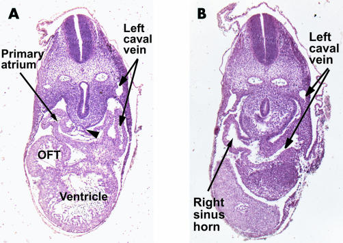
These sections are taken from a human embryo at Carnegie stage 12, sectioned in the short axis of the body. Section A shows how the walls of the primary atrium are continuous with the body of the embryo through the so-called “dorsal mesocardium” (arrowhead). Section B shows the continuity between the primary atrial component and the sinus horns, which are asymmetrical as they join the heart, the left horn being continuous with the left cardinal vein, which becomes the left superior caval vein.
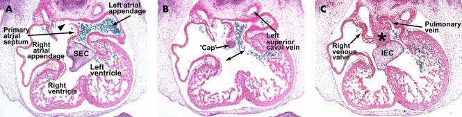
These three sections are from a human embryo at Carnegie stage 16, and are cut in the frontal plane of the heart. Section A is through the superior atrioventricular endocardial cushion (SEC). Note that the upper end of the primary septum has broken down to form the secondary foramen (arrowhead). The endocardial cushions have yet to fuse at this stage, and section B is taken between the cushions. Note the mesenchymal cap on the leading edge of the primary septum. Section C is through the inferior cushion (IEC) and shows how tissue from the body of the embryo enters the heart through the vestibular spine (asterisk). Note the solitary pulmonary vein joining the left side of the primary atrium.
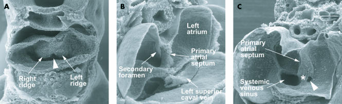
These pictures are scanning electron micrographs of the developing mouse heart. Picture A, from a mouse at embryonic day 9.5, with 28 somites, shows the stage before canalisation of the pulmonary vein. When formed, the vein will enter heart through the so-called “pulmonary pit”, arrowed, which at this stage is flanked by prominent right and left pulmonary ridges. Picture B, at embryonic day 10.5, when the embryo has 42 somites, shows the primary septum growing down towards the atrioventricular cushions. Its upper edge has broken down to form the secondary foramen. Note that the left superior caval vein, now formed from the left cardinal vein, is a discrete structure within the left atrioventricular groove. Picture C, again from an embryo of 10.5 days, but now with 45 somites, shows the pulmonary vein opening as a solitary channel inferiorly to the left atrium, the systemic venous sinus, enclosed by the venous valves, now having become incorporated into the right atrium. The right pulmonary ridge has now expanded to become the vestibular spine (asterisk).
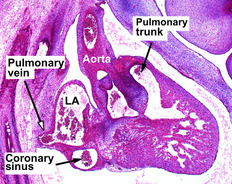
This section, taken in the sagittal plane from a human embryo at the 20th Carnegie stage, shows the solitary pulmonary vein entering inferiorly to the left atrium. Only with subsequent growth do four pulmonary veins enter that atrial roof, but this process is necessary to produce the so-called “septum secundum”, in reality the infolded atrial roof (see fig 6).
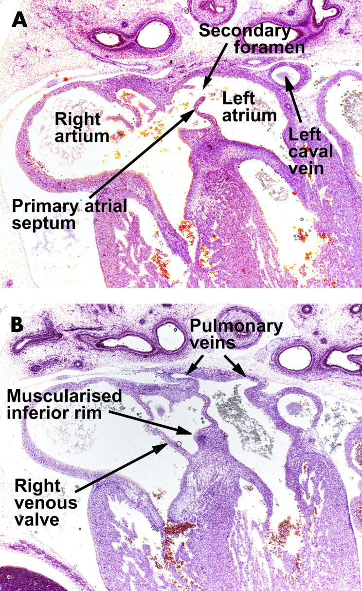
These sections are from a human embryo at Carnegie stage 20/21, showing the beginnings of the infolding of the atrial wall which will produce the rims of the oval fossa. Section A is taken cranial to section B. Note the ongoing muscularisation of the antero-inferior rim of the oval fossa (see also fig 10).
This section, from a human embryo at 11 weeks of development, shows the continuing infolding of the atrial roof as the pulmonary veins become incorporated into the left atrium.
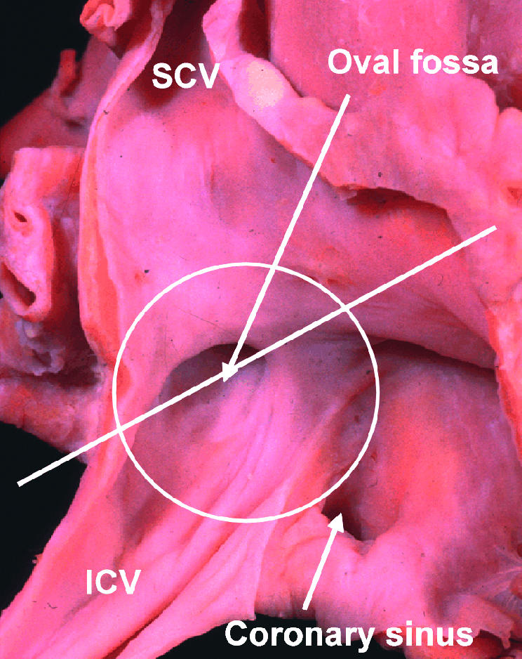
This picture of the human right atrium, taken from posteriorly and the right, shows how, at first sight, an extensive septal area, within the circle, separates the right from the left atrium. The true situation is shown in fig 8, which is a cross section along the line shown in the figure. SCV, superior caval vein; ICV, inferior caval vein.
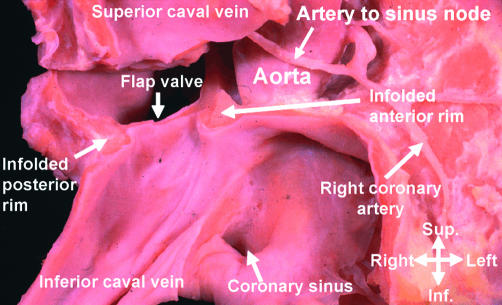
This section, along the line shown in fig 7, reveals how the rims of the oval fossa anteriorly and posteriorly are folds of the atrial wall. Note the relation of the anterior atrial wall to the aortic root.
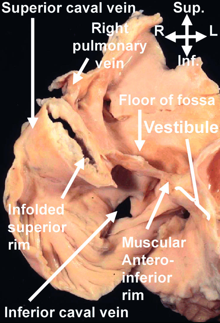
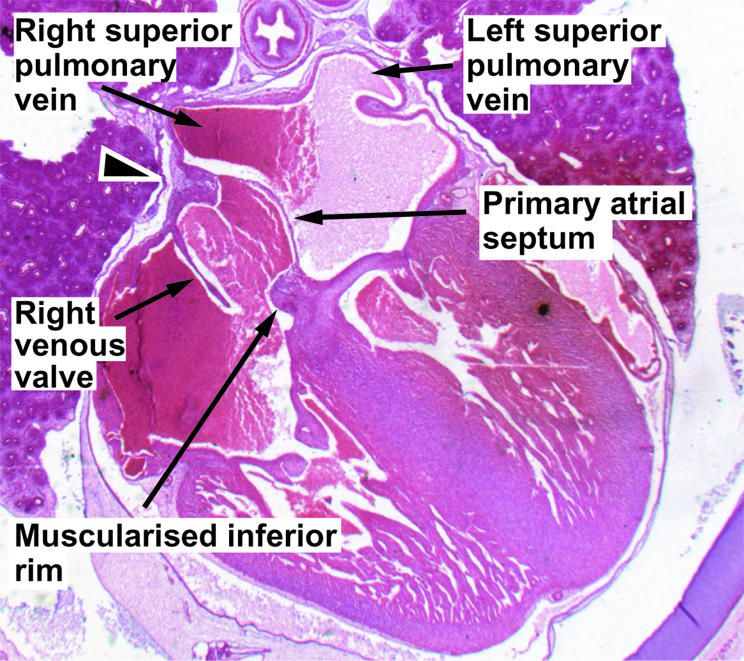
This section, taken in “four chamber” plane through an adult human heart, shows how the so-called “septum secundum” is a deep infolding between the connections of the pulmonary veins to the left atrium, and the superior caval vein to the right atrium. The “compass” shows the orientation. Sup, superior; Inf, inferior; R, right; L, left.
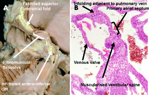
The left panel shows another “four chamber” section through an adult human heart. It shows not only the accumulation of fat within the superior interatrial fold, but also the fat which is infiltrating through the inferior atrioventricular groove into the inferior rim of the oval fossa. The right panel shows a section from a human embryo of 7–8 weeks' development, when two pulmonary veins have been incorporated into the left atrium. It shows the muscularisation of the vestibular spine, which forms the inferior rim of the oval fossa.
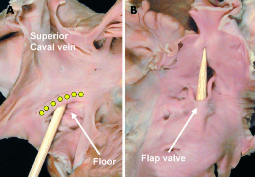
The pictures show a probe-patent oval foramen in a human heart, viewed from the right (left panel) and left (right panel) atrial aspects. The probe is placed through the foramen, separating the flap valve in the left atrium from the rims of the oval fossa (yellow dots).
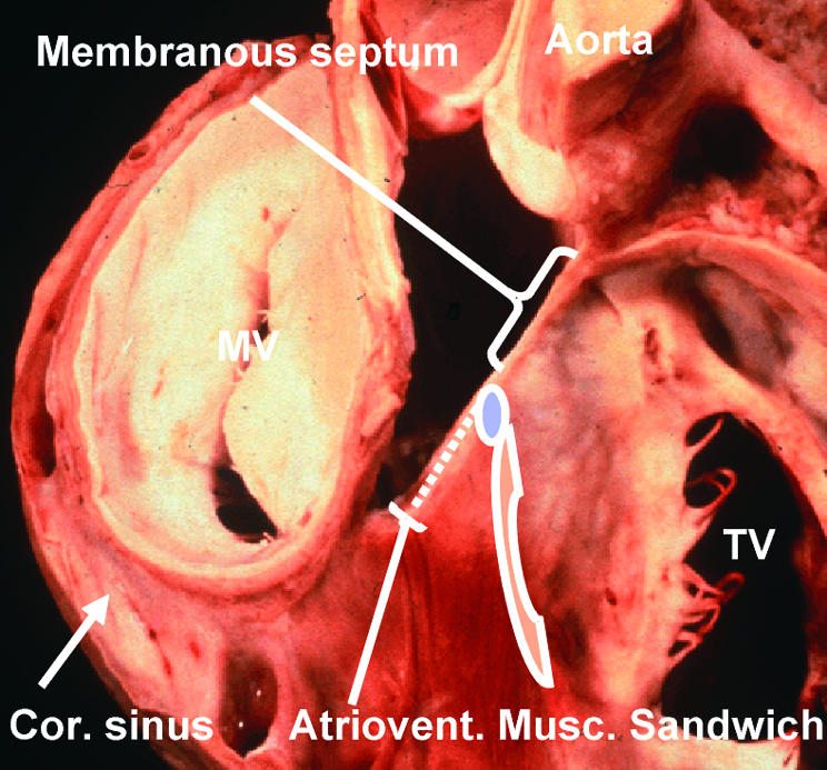
This dissection of a human heart shows the relations between the right and left atrioventricular junctions and the subaortic outflow tract. It has been made by removing the atrial walls, along with the non-coronary sinus of the aortic root. It shows the location of the membranous septum, which is penetrated by the atrioventricular conduction axis as it passes from the atrioventricular node (blue oval) to reach the crest of the muscular ventricular septum. The dots show the tissues of the inferior atrioventricular groove which interpose between the atrial and ventricular layers of the muscular atrioventricular sandwich. The red cross hatched area is the vestibule of the tricuspid valve (TV). MV, mitral valve; Cor sinus, coronary sinus.
References
-
- Röse C. Zur Entwicklungsgeschichte des Saugerthierherzens. Morphol Jahr 1899;15:436–56.
-
- Christie GA. The development of the limbus fossae ovalis in the human heart: a new septum. J Anat 1963;97:45–54. ▸ These works show that it has long been recognised that the so-called “septum secundum” is, in reality, no more than an infolding between the connections of the pulmonary veins to the left atrium, and the caval veins to the right atrium. - PMC - PubMed
-
- Basso C, Barbazza R, Thiene G. Images in cardiovascular medicine. Lipomatous hypertrophy of the atrial septum. Circulation 1998;97:1423. ▸ Although the authors claim that their image demonstrates the collection of fat within the atrial septum, in fact it is clear that the fat has accumulated within the superior interatrial groove. - PubMed
-
- Li J, Ho SY, Becker AE, et al. Multiple cardiac lipomas and sudden death – a case report and literature review. Cardiovasc Pathol 1998;7:349–52. ▸ The authors review an amazing heart which shows multiple lipomas, and show how the fat accumulates within the grooves of the heart, albeit that there is also infiltration of the myocardium. - PubMed
-
- His W. Die Area interposita, die Eustachi'sche Klappe und die Spina vestibuli. In: Anatomie Menschlicher Embryonen. Leipzig: von FCW Vogel, 1880:49–152. ▸ The initial description of the “spina vestibuli”. His's reconstructions are remarkably accurate, although he does not specify the nature of the vestibule in which the spine is formed.
Publication types
MeSH terms
LinkOut - more resources
Full Text Sources