. 2002 Jun 15;541(Pt 3):701-16.
doi: 10.1113/jphysiol.2001.016139.

Cocaine binds to a common site on open and inactivated human heart (Na(v)1.5) sodium channels

Affiliations

Cocaine binds to a common site on open and inactivated human heart (Na(v)1.5) sodium channels

M E O'Leary et al. J Physiol. .

Abstract

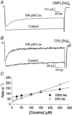
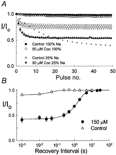
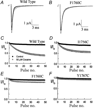
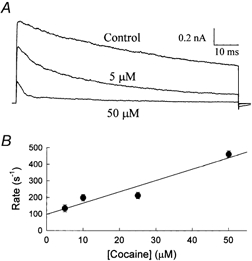
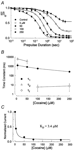
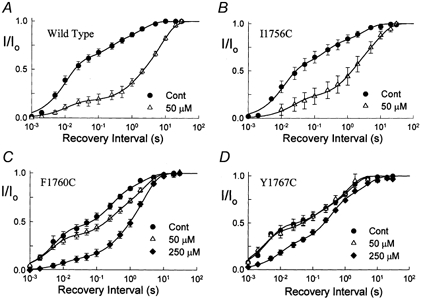
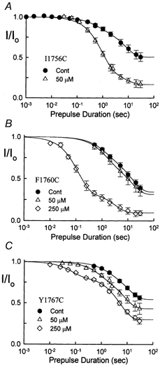
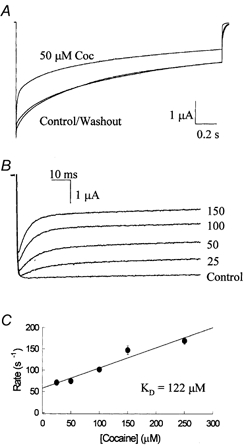
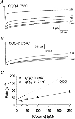
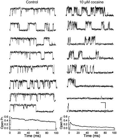
The inhibition by cocaine of the human heart Na+ channel (Na(v)1.5) heterologously expressed in Xenopus oocytes was investigated. Cocaine produced little tonic block of the resting channels but induced a characteristic, use-dependent inhibition during rapid, repetitive stimulation, suggesting that the drug preferentially binds to the open or inactivated states of the channel. To investigate further the state dependence, depolarizing pulses were used to inactivate the channels and promote cocaine binding. Cocaine produced a slow, concentration-dependent inhibition of inactivated channels, which had an apparent K(D) of 3.4 microM. Mutations of the interdomain III-IV linker that remove fast inactivation selectively abolished this high-affinity component of cocaine inhibition, which appeared to be linked to the fast inactivation of the channels. A rapid component of cocaine inhibition persisted in the inactivation-deficient mutant that was enhanced by depolarization and was sensitive to changes in the concentration of external Na+, properties that are consistent with a pore-blocking mechanism. Cocaine induced a use-dependent inhibition of the non-inactivating mutant and delayed the repriming at hyperpolarized voltages, indicating that the drug slowly dissociated when the channels were closed. Mutation of a conserved aromatic residue (Y1767) of the D4S6 segment weakened both the inactivation-dependent and the pore-blocking components of the cocaine inhibition. The data indicate that cocaine binds to a common site located within the internal vestibule and inhibits cardiac Na+ channels by blocking the pore and by stabilizing the channels in an inactivated state.


Figures

Figure 6
Figure 6. Cocaine block is sensitive to changes in the concentration of external Na+
A, whole-cell currents of the QQQ mutant measured in normal Ringer solution (116 mm Na+) before and after the application 100 μM cocaine. Currents were activated by depolarizing to −25 mV from a holding potential of −100 mV. B, same pulsing protocol was applied in reduced external Na+ Ringer solution (29 mm Na+). C, the blocking rates were determined from the rapid decay of the currents as described previously. The association and dissociation rate constants were 3.1 × 10⁵m−1 s−1 and 66.3 s−1 for 100 % Na+ Ringer solution and 6.3 × 10⁵m−1 s−1 and 27.3 s−1 for the 25 % Na+ conditions. The KD of cocaine inhibition at −25 mV is 214 μM in the high external Na+ and 43 μM in low external Na+ Ringer solution.
Figure 7
Figure 7. Use-dependent inhibition of the QQQ mutant Na+ channels
A, a series of 50 depolarizing pulses to −10 mV for 20 ms were applied at a frequency of 5 Hz. Currents elicited by individual pulses were normalized and plotted versus pulse number. The use-dependent inhibition was measured in either control 100 % Na+ Ringer solution (116 mm) or 25 % external Na+ Ringer solution (29 mm). The dotted line is the cocaine inhibition of the wild-type channel in 100 % Na+ solution replotted from Fig. 1C. B, repriming time course of cocaine-blocked channels. Cells were depolarized for 100 ms to −10 mV in the absence and presence of 150 μM cocaine before returning to −100 mV for a variable duration (1 ms-30 s). A standard −10 mV test pulse was used to assay availability after the completion of the recovery interval. In the presence of cocaine the recovery time course is well fitted by a single exponential with a time constant of 1.8 ± 0.1 s and a steady-state amplitude of 0.43 ± 0.05 (n = 8). Also plotted is the time course of recovery from slow inactivation which has a time constant of 49.3 ± 5.0 ms and a steady-state current amplitude of 0.91 ± 0.01 (n = 9).
Figure 1
Figure 1. Use-dependent inhibition of Nav1.5 Na+ channels expressed in Xenopus oocytes
The use-dependent inhibition was induced by a series of 50 depolarizing pulses to −10 mV for 20 ms applied at a frequency of 5 Hz. A and B, currents of the wild-type (A) and F1760C mutant (B) after bath application of 50 μM cocaine. Current traces correspond to the pulse numbers 1, 5, 10, and 50 of the stimulation train. C-F, the peak current elicited by each pulse within the train was normalized to the current of the first pulse and plotted versus the pulse number. The normalized currents were measured before (filled symbols) and after (open symbols) application of 50 μM cocaine for wild-type (n = 7), I1756C (n = 4), F1760C (n = 14), and Y1767C (n = 7).
Figure 9
Figure 9. Cocaine block of Na+ current in excised patches
Outside-out patches were excised from oocytes expressing the QQQ mutant. The Na+ in the internal pipette solution was increased to 140 mm and the external Na+ was replaced with choline to facilitate the measurement of outward current. A, macroscopic currents from a patch elicited by a depolarizing pulse to +40 mV for 100 ms before and after bath application of 5 and 50 μM cocaine. (VH =−100 mV). B, the decay of the current was fitted with the sum of two exponentials and the apparent blocking rate (1/τf) plotted versus cocaine concentration. The linear relationship is consistent with a bimolecular interaction with association and dissociation rate constants of 6.8 × 10⁶m−1 s−1 and 97.8 s−1, respectively, yielding a KD of 14.3 μM. Data are the means ±s.e.m. of six to nine determinations.
Figure 2
Figure 2. Time course of cocaine binding to inactivated channels
A, the time course of cocaine inhibition was measured using a triple pulse protocol consisting of a conditioning pulse to −10 mV of variable duration (1 ms-60 s), a short hyperpolarization to −100 mV for 150 ms and a test pulse to −10 mV. the peak currents elicited by test pulses were normalized to controls (I/Io) and plotted versus the prepulse duration. The decay of the current is best described by the sum of two exponentials : I/Io =A1exp(-t1) +A2exp(-tS) +A, where A1, A2 and A are the relative amplitudes of the fast, slow and steady state components, respectively. The fast (τ1) and slow (τ2) time constants of 158 ± 76 and 6461 ± 932 ms for control (n = 11), 1254 ± 737 and 6284 ± 1559 ms for 5 μM (n = 5), 855 ± 139 and 3494 ± 1592 ms for 50 μM (n = 6), 6 ± 4 and 581 ± 21 ms for 100 μM (n = 11), 8 ± 6 ms and 267 ± 25 ms (n = 5) for 250 μM cocaine. B, time constants of the fast, intermediate, and slow components of current decay plotted versus the cocaine concentration (see text). C, plot of the normalized steady-state current amplitude (A) versus the cocaine concentration. The smooth curve is a fit to a single site model with a KD of 3.4 ± 0.4 μM.
Figure 4
Figure 4. Time course of recovery from cocaine inhibition
Channels were depolarized for 10 s to −10 mV before returning to −100 mV for intervals ranging from 1 ms to 30 s. A −10 mV test pulse was then used to assay the fraction of recovered current, which was normalized to control test currents and plotted versus the recovery interval. The recovery time course was measured before (•) and after bath application of 50 μM (▵) (A-D) or 250 μM (♦) (C and D)cocaine. The smooth curves are fits to the sum of three exponentials with the parameters listed in Table 1.
Figure 3
Figure 3. Effects of D4S6 mutations on cocaine inhibition
The onset of cocaine inhibition was measured using the same pulsing protocol as in Fig. 2. In the absence of cocaine, the development of slow inactivation was biexponential with fast and slow time constants of 0.6 ± 0.1 and 7.6 ± 0.9 s for I1756C (panel A; A1 = 0.13, n = 7), 1.2 ± 0.5 and 13.8 ± 3.8 s for F1760C (panel B; A1 = 0.22, n = 11), 1.4 ± 0.9 and 10.6 ± 2.1 s for Y1767C (panel C; A1 = 0.10, n = 17). After application of 50 μM cocaine the time constants were 0.9 ± 0.2 and 7.3 ± 5.2 s for I1756C (A1 = 0.65, n = 4), 1.3 ± 0.4 and 13.3 ± 4.4 s for F1760C (A1 = 0.29, n = 10) and 0.7 ± 0.3 and 7.5 ± 1.8 s Y1767C (A1 = 0.2, n = 7). The fraction of non-inhibited current (A) before and after addition of 50 μM cocaine was 0.51 ± 0.01 and 0.171 ± 0.02 for I1756C, 0.34 ± 0.04 and 0.31 ± 0.04 for F1760C, and 0.54 ± 0.01 and 0.43 ± 0.02 for Y1767C. Also plotted is the inhibition of the F1760C and Y1767C mutants by 250 μM cocaine with fast and slow time constants of 0.12 ± 0.01 and 3.2 ± 0.9 s for F1760C (A1 = 0.65, n = 5) and 0.07 ± 0.02 s and 5.3 ± 0.4 s for Y1767C (A1 = 0.18, n = 4).
Figure 5
Figure 5. The time course of cocaine block of the QQQ mutant channels
The inactivation-deficient mutant (IFM→QQQ) was constructed by replacing a series of hydrophobic residues of the interdomain III-IV linker with glutamines (see text). A, currents were elicited by depolarizing for 2 s to −10 mV from a holding potential of −100 mV. In the absence of cocaine, the decay of the current is biexponential with time constants of 85.6 ± 6.2 and 1063.4 ± 98.4 ms (n = 6). After application of 50 μM cocaine the decay of the current was fitted with the sum of three exponentials with time constants of 15.2 ± 0.6, 181.5 ± 7.0 and 1519.3 ± 85.7 ms (n = 6). The cocaine inhibition was completely reversible upon washout with time constants of 75.4 ± 6.8 and 1154.6 ± 180.5 ms (n = 6). B, the time course of cocaine inhibition during 200 ms depolarizations was measured over a range of concentrations and the decay time constants determined from biexponential curve fits. C, the apparent blocking rate (1/τf) plotted versus the cocaine concentration is linear with slope (kon) and y-intercept (koff) of 4.7 × 10⁵m−1 s−1 and 57.9 s−1, respectively. The inhibition constant (KD =koff/kon) at −10 mV is 122 μM.
Figure 8
Figure 8. Effects of D4S6 mutations on cocaine block
The I1756C and Y1767C mutations were transferred to the QQQ mutant background and the time course of cocaine block was determined by applying 900 ms depolarizing pulses to −10 mV. In the absence of cocaine, the decay of the current is biexponential reflecting the slow inactivation of the channels (see text). A, cocaine (25-250 μM) induces the characteristic accelerated decay in the QQQ-I1756C mutant current. B, same protocol with the QQQ-Y1767C double mutant indicates that this channel is relatively insensitive to cocaine. C, the decay of the QQQ-I1756C current was fitted with the sum of two exponentials and the apparent blocking rates (1/τf) plotted versus the cocaine concentration. The QQQ-I1756C mutant has a kon and koff of 2.6 × 10⁵m−1 s−1 and 26.3 s−1, respectively, yielding a KD of 101 μM. The data suggest a KD of > 600 μM for the QQQ-Y1767C mutant.
Figure 10
Figure 10. Cocaine reduces the single-channel open times
Recordings of a single QQQ mutant channel before and after application of 10 μM cocaine. Recording configuration is identical to that described in Fig. 9. Currents were elicited by a depolarizing voltage pulse to +40 mV from a holding potential of −100 mV. Upward deflections represent channel openings. Cocaine (10 μM) reduced the mean open times but did not alter the single channel current amplitude (see text). Bottom, ensemble average open probability constructed from 300 depolarizations for the control and from 334 depolarizations in the presence of cocaine. Calibration bars are 10 ms and 2 pA.

Similar articles

Cited by

References

    1. Alpert LA, Fozzard HA, Hanck DA, Makielski JC. Is there a second external lidocaine binding site on mammalian cardiac cells? American Journal of Physiology. 1989;257:H79–84. - PubMed
    1. Armstrong CM, Bezanilla F, Rojas E. Destruction of sodium conductance inactivation in squid axons perfused with pronase. Journal of General Physiology. 1973;62:375–391. - PMC - PubMed
    1. Bean BP, Cohen CJ, Tsien RW. Lidocaine block of cardiac sodium channels. Journal of General Physiology. 1983;81:613–642. - PMC - PubMed
    1. Bennett PB, Valenzuela C, Chen LQ, Kallen RG. On the molecular nature of the lidocaine receptor of cardiac Na+ channels. Modification of block by alterations in the alpha-subunit III-IV interdomain. Circulation Research. 1995;77:584–592. - PubMed
    1. Billman GE. Cocaine: a review of its toxic actions on cardiac function. Critical Review of Toxicology. 1995;25:113–132. - PubMed

Publication types

MeSH terms

LinkOut - more resources