

Ionic currents in isolated and in situ squid Schwann cells
- PMID: 12068039
- PMCID: PMC2290350
- DOI: 10.1113/jphysiol.2002.019638
Ionic currents in isolated and in situ squid Schwann cells
Abstract
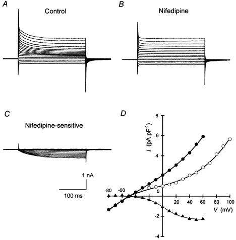
Ionic currents from Schwann cells isolated enzymatically from the giant axons of the squids Loligo forbesi, Loligo vulgaris and Loligo bleekeri were compared with those obtained in situ. Macroscopic and single channel ionic currents were recorded using whole-cell voltage and patch clamp. In the whole-cell configuration, depolarisation from negative holding potentials evoked two voltage-dependent currents, an inward current and a delayed outward current. The outward current resembled an outwardly rectifying K+ current and was activated at -40 mV after a latent period of 5-20 ms following a step depolarisation. The current was reduced by externally applied nifedipine, Co2+ or quinine, was not blocked by addition of apamin or charibdotoxin and was insensitive to externally applied L-glutamate or acetylcholine. The voltage-gated inward current was activated at -40 mV and was identified as an L-type calcium current sensitive to externally applied nifedipine. Schwann cells were impaled in situ in split-open axons and voltage clamped using discontinuous single electrode voltage clamp. Voltage dependent outward currents were recorded that were kinetically identical to those seen in isolated cells and that had similar current-voltage relations. Single channel currents were recorded from excised inside-out patches. A single channel type was observed with a reversal potential close to the equilibrium potential for K+ (E(K)) and was therefore identified as a K+ channel. The channel conductance was 43.6 pS when both internal and external solutions contained 150 mM K+. Activity was weakly dependent on membrane voltage but sensitive to the internal Ca2+ concentration. Activity was insensitive to externally or internally applied L-glutamate or acetylcholine. The results suggest that calcium channels and calcium-activated K+ channels play an important role in the generation of the squid Schwann cell membrane potential, which may be controlled by the resting intracellular Ca2+ level.
Figures


Similar articles
-
Voltage-gated potassium currents in myelinating Schwann cells in the mouse.J Physiol. 1990 Dec;431:123-39. doi: 10.1113/jphysiol.1990.sp018323. J Physiol. 1990. PMID: 2100304 Free PMC article.
-
A patch-clamp study of potassium channels and whole-cell currents in acinar cells of the mouse lacrimal gland.J Physiol. 1984 May;350:179-95. doi: 10.1113/jphysiol.1984.sp015195. J Physiol. 1984. PMID: 6086894 Free PMC article.
-
Calcium-activated potassium channels in native endothelial cells from rabbit aorta: conductance, Ca2+ sensitivity and block.J Physiol. 1992 Sep;455:601-21. doi: 10.1113/jphysiol.1992.sp019318. J Physiol. 1992. PMID: 1484364 Free PMC article.
-
Potassium current in the squid giant axon.Int Rev Neurobiol. 1985;27:363-84. doi: 10.1016/s0074-7742(08)60562-0. Int Rev Neurobiol. 1985. PMID: 2417975 Review.
-
Model experiments on squid axons and NG108-15 mouse neuroblastoma x rat glioma hybrid cells.J Physiol Paris. 1995;89(4-6):181-93. doi: 10.1016/0928-4257(96)83635-7. J Physiol Paris. 1995. PMID: 8861817 Review.
Cited by
-
Neurons and Glia Cells in Marine Invertebrates: An Update.Front Neurosci. 2020 Feb 18;14:121. doi: 10.3389/fnins.2020.00121. eCollection 2020. Front Neurosci. 2020. PMID: 32132895 Free PMC article. Review.
References
-
- Abbott NJ, Brown ER, Kukita F, Tsutsui I, Inoue I. Axon-Schwann cell signalling in the squid. Japanese Journal of Physiology. 1995a;45(suppl. 2):I–4.
-
- Abbott NJ, Brown ER, Pichon Y, Kukita F. Electrophysiology of squid Schwann cells. In: Abbott NJ, Williamson R, Maddock L, editors. Cephalopod Neurobiology. UK: Oxford University Press; 1995b. pp. 197–212.
-
- Abbott NJ, Pichon Y, Brown ER. Long-term microelectrode recording from Schwann cells surrounding the isolated giant axon of the squid; steady and oscillating potentials. Journal of Physiology. 1990;430.P:128P.
Publication types
MeSH terms
Substances
LinkOut - more resources
Full Text Sources
Research Materials
Miscellaneous