. 2002 Jun 15;541(Pt 3):797-810.
doi: 10.1113/jphysiol.2002.018796.

Voltage-dependent inward currents of interstitial cells of Cajal from murine colon and small intestine

Affiliations

Voltage-dependent inward currents of interstitial cells of Cajal from murine colon and small intestine

Young Chul Kim et al. J Physiol. .

Abstract

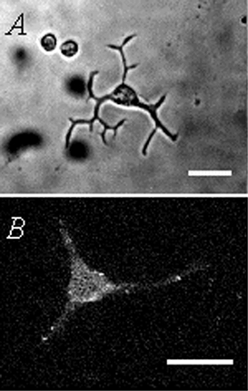
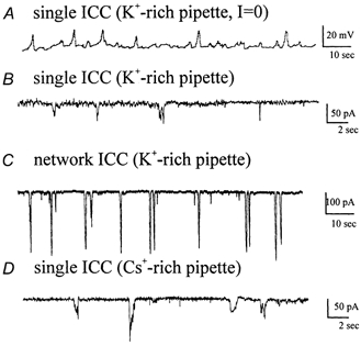
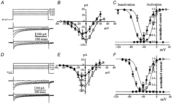
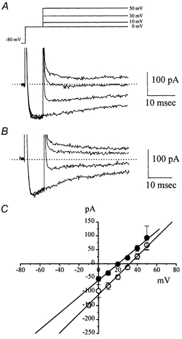
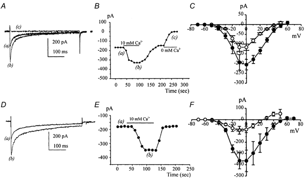
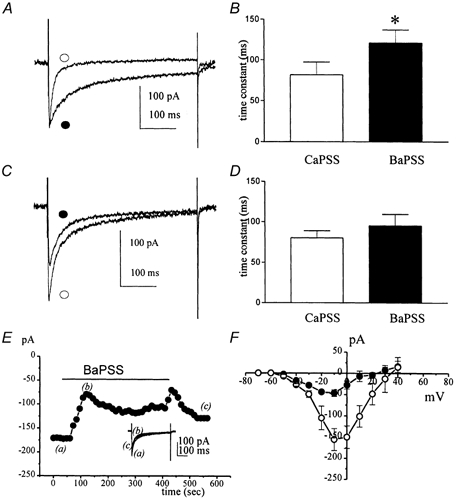
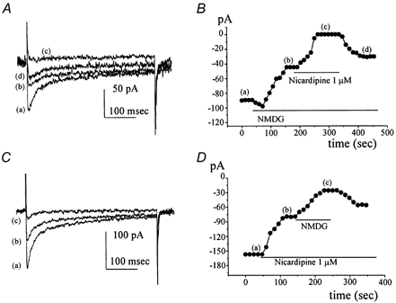
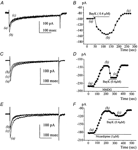
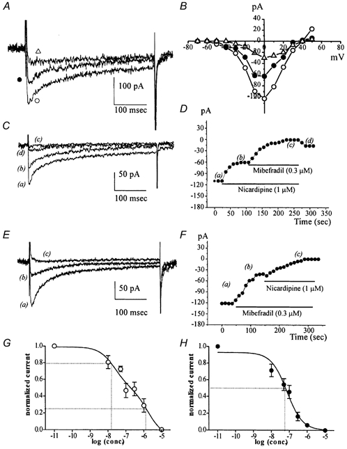
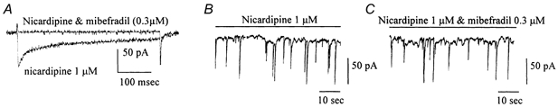
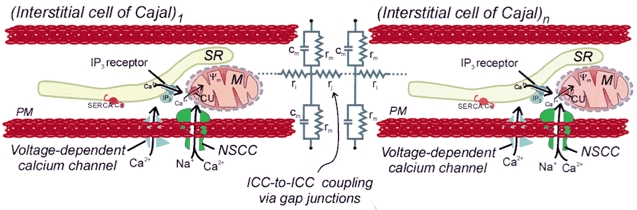
Electrical slow waves in gastrointestinal (GI) muscles are generated by pacemaker cells, known as interstitial cells of Cajal (ICC). The pacemaker conductance is regulated by periodic release of Ca2+ from inositol 1,4,5-trisphosphate (IP(3)) receptor-operated stores, but little is known about how slow waves are actively propagated. We investigated voltage-dependent Ca2+ currents in cultured ICC from the murine colon and small intestine. ICC, identified by kit immunohistochemistry, were spontaneously active under current clamp and generated transient inward (pacemaker) currents under voltage clamp. Depolarization activated inward currents due to entry of Ca2+. Nicardipine (1 microM) blocked only half of the voltage-dependent inward current. After nicardipine, there was a shift in the potential at which peak current was obtained (-15 mV), and negative shifts in the voltage dependence of activation and inactivation of the remaining voltage-dependent inward current. The current that was resistant to dihydropyridine (I(VDDR)) displayed kinetics, ion selectivity and pharmacology that differed from dihydropyridine-sensitive Ca2+ currents. I(VDDR) was increased by elevating extracellular Ca2+ from 2 to 10 mM, and this caused a +30 mV shift in reversal potential. I(VDDR) was blocked by Ni2+ (100 microM) or mebefradil (1 microM) but was not affected by blockers of N-, P- or Q-type Ca2+ channels. Equimolar replacement of Ca2+ with Ba2+ reduced I(VDDR) without effects on inactivation kinetics. BayK8644 had significantly less effect on I(VDDR) than on I(VDIC). In summary, two components of inward Ca2+ current were resolved in ICC of murine small intestine and colon. Since slow waves persist in the presence of dihydropyridines, the dyhydropyridine-resistant component of inward current may contribute to slow wave propagation.


Figures

Figure 1
Figure 1. Phase contrast and fluorescence micrographs of cultured ICC from the murine colon
A, a phase contrast image of a single multi-process ICC grown for 1 day in culture from a dispersion of cells from the tunica muscularis of the proximal colon. Note the fusiform cell body, prominent nucleus, and multiple thin processes extending from the nuclear region. Cells with this morphology expressed Kit-like immunoreactivity, as shown in the fluorescence micrograph, B. Scale bars in A and B, 25 μm.
Figure 2
Figure 2. Spontaneous inward currents from cultured single and networked ICC in murine colon
A, spontaneous membrane potential oscillations were recorded from a single ICC under current clamp (I = 0) in the presence of nicardipine (1 μM). The pipette solution contained 140 mm K+. B, spontaneous inward currents were recorded from a single ICC under voltage clamp in the presence of nicardipine (1 μM). The cell was held at −80 mV, and the pipette solution contained 140 mm K+. C, spontaneous inward currents were more robust and regular when recorded from ICC in networks. Cell was held at −80 mV with a K+-rich pipette solution in the pipette. D, Cs+-rich pipette solution used to enhance resolution of voltage-dependent Ca2+ currents did not inhibit spontaneous inward currents.
Figure 3
Figure 3. Voltage-dependent inward currents (IVDIC and IVDDR) in ICC under whole-cell configuration
IVDIC was generated by depolarizing pulses from −70 to +50 mV. Data were obtained using the perforated patch technique (A-C from colonic ICC and D-F from small intestinal ICC). A and D, currents were elicited by voltage steps from a holding potential of −80 mV (currents displayed resulted from test potentials from 0 to +40 mV, voltage protocol shown as inset in A). Currents were generated before (top current traces) and after nicardipine (1 μM; bottom traces in A and D). B and E, summaries of current-voltage relationships of IVDIC (○) and current remaining after nicardipine (IVDDR; •). C and F, voltage dependences of activation and inactivation of IVDIC (○) and IVDDR (•) are shown as plots of normalized peak currents. For steady-state activation, maximum conductance was determined from the peak inward current and corrected for the change in driving force at each test potential. Half-activation and inactivation potentials were determined from Boltzmann functions fitted to the data. Data are plotted as means ±s.e.m.
Figure 4
Figure 4. Reversal potential of IVDIC and IVDDR using instantaneous tail current analysis
A, cells were held at −80 mV, stepped to 0 mV for 7 ms to activate IVDIC, and then stepped to various potentials (-10 to +50 mV; data displayed were obtained with test potentials of 0 to +50 mV using voltage protocol shown in the inset). B, the same protocols were applied after nicardipine (1 μM) to determine the reversal of IVDDR tail currents. Representative currents in panels A and B are from different cells. C, a summary plot of the amplitude of tail currents as a function of test potential. ○, reversal of IVDIC (n = 6); •, the reversal of the current remaining after nicardipine (IVDDR; n = 17). Note that IVDDR reverses at more negative potentials.
Figure 5
Figure 5. The effects of external Ca2+on IVDIC and IVDDRin colonic ICC
A, representative traces shown in 2 (a), 10 (b) and 0 (c) mm Ca2+. B, changes in IVDIC caused by switching external solution from 2 to 10 to 0 mm Ca2+. Data were obtained from steps from −80 to 0 mV every 12 s. C, current-voltage relationship for IVDIC recorded from seven cells in 2 (○) and 10 (•) mm Ca2+. D, IVDDR in 2 (a) and 10 (b) mm Ca2+ in the presence of nicardipine (1 μM). E, changes in peak caused by switching external solution from 2 to 10 mm Ca2+. Data were obtained from steps from −80 to 0 mV every 10 s. F, current-voltage relationship for IVDDR recorded from five cells in 2 (○) and 10 (•) mm Ca2+.
Figure 6
Figure 6. The effect of external Ba2+on IVDIC and IVDDR in murine colon
A, representative traces showing IVDIC from a colonic ICC before (○) and after (•) equimolar Ba2+ replacement for Ca2+. Voltage steps from −80 to 0 mV were applied repetitively. B, the summary of inactivation time constants of IVDIC at 0 mV before (○) and after (•) equimolar Ba2+ replacement for Ca2+. C, representative traces of IVDDR from a colonic ICC before (○) and after (•) equimolar Ba2+ replacement for Ca2+. Voltage steps from −80 to 0 mV were applied in the presence of nicardipine (1 μM). D, the summary of inactivation time constants of IVDDR at 0 mV before (○) and after (•) equimolar Ba2+ replacement for Ca2+. Experiments were performed in the presence of nicardipine (1 μM) to isolate IVDDR. E, change in peak current due to equimolar Ba2+ replacement for Ca2+ on IVDDR as a function of time. Data were obtained for steps from −80 to 0 mV every 12 s. F, current-voltage relationship for peak IVDDR in Ca2+-PSS (○) and Ba2+-PSS (•).
Figure 7
Figure 7. The effect of external Na2+ replacement on IVDIC and IVDDR in colonic ICC
A, IVDIC elicited from a colonic ICC by voltage steps from −80 to 0 mV (a), after replacement of external Na+ with NMDG (b), after addition of nicardipine (1 μM) to isolate IVDDR (c) and after washout of nicardipine (d). Traces a-d were taken at the time points noted in B. B, peak currents elicited by stepping from −80 to 0 mV every 12 s during equimolar replacement of Na+ with NMDG and after addition of nicardipine. C, currents elicited from a colonic ICC by voltage steps from −80 to 0 mV (a), after addition of nicardipine (1 μM) to isolate IVDDR (b) and after replacement of external Na+ with NMDG (c). Traces a-c were taken at the time points noted in D. D, peak currents elicited by steps from −80 to 0 mV every 12 s during exposure to nicardipine and after equimolar replacement of Na+ with NMDG. NMDG markedly reduced IVDDR in a reversible manner.
Figure 8
Figure 8. The effect of BayK8644 on IVDIC and IVDDR in colonic ICC
A, IVDIC elicited from a colonic ICC by voltage steps from −80 to 0 mV at various times after addition of BayK8644 (0.4 μM). Traces a-c were taken at the time points noted in B. B, change in peak current after addition of BayK8644 (0.4 μM) as a function of time. Data obtained from steps from −80 to 0 mV every 12 s. C, IVDIC elicited from a colonic ICC by voltage steps from −80 to 0 mV after replacement of external Na+ with NMDG and after subsequent addition of BayK8644 (0.4 μM). Traces a-c were taken at the time points noted in D. D, peak currents elicited by steps from −80 to 0 mV every 12 s after replacement of Na+ with NMDG and after addition of BayK8644. E, effects of BayK8644 (0.4 μM) on IVDDR elicited from a colonic ICC (i.e. in the presence of nicardipine) by voltage steps from −80 to 0 mV. Traces a-c were taken at the time points noted by the same letters in F. F, peak currents elicited by steps from −80 to 0 mV every 12 s during exposure to BayK8644 in the presence of nicardipine. The effects of BayK8644 on IVDDR were significantly less than the effects of this compound on IVDIC (see text for details).
Figure 9
Figure 9. Inhibition of IVDDR in small intestinal ICC by nickel and mibefradil
A, representative currents elicited by test potentials from −80 to of 0 mV in control (IVDIC; ○), and after subsequent addition of nicardipine (1 μM) and nickel (100 μM). Traces displayed were obtained by steps to 0 mV. B, current-voltage relationship showing IVDIC (○), IVDDR (current after nicardipine, 1 μM; •), and the effects of nickel on IVDDR (100 μM; ▵). C, typical currents elicited from a small intestinal ICC by voltage steps from −80 to 0 mV after application of nicardipine (1 μM) and mibefradil (1 μM). Traces displayed (a-d) were taken at the time points noted in D. D, peak current elicited by voltage step from −80 to 0 mV applied every 12 s after addition of nicardipine (1 μM) and mibefradil (0.3 μM). Nicardipine blocked approximately half of IVDIC and mibefradil blocked the remaining current (IVDDR). The effects of mibefradil were difficult to wash out. E, typical currents elicited from a colonic ICC by voltage steps from −80 to 0 mV after application of mibefradil (0.3 μM) and nicardipine (1 μM) Traces displayed (a-d) were taken at the time points noted in D. F, peak current elicited by voltage step from −80 to 0 mV applied every 12 s after addition of mibefradil (0.3 μM) and nicardipine (1 μM). At this concentration, mibefradil blocked approximately half of IVDlC and nicardipine blocked the remaining current. G and H, the concentration-response relationship for the effects of mibefradil on IVIDC and IVDDR (i.e. mibefradil concentrations added in the presence of nicardipine, 1 μM), respectively. Data in G were fitted with a two-site competition equation, and dotted lines show IC50 values for the fit. In H the block by mibefradil was fitted with a one-site competition equation, and the IC50 is denoted by the dotted lines.
Figure 10
Figure 10. The effects of nicardipine and mibefradil on spontaneous inward currents in small intestinal ICC
A, representative traces elicited by test potentials from −80 to 0 mV in the presence of nicardipine (1 μM) and nicardipine plus mibefradil (0.3 μM). B and C, neither nicardipine nor nicardipine and mibefradil together blocked spontaneous inward currents in isolated ICC. The holding potential was −80 mV.
Figure 11
Figure 11. Role of voltage-dependent Ca2+ conductances in propagation of slow waves
Pacemaker activity is initiated in ICC by Ca2+ release from IP3 receptor-operated stores (Suzuki et al. 2000; Ward et al. 2000) in the sarcoplasmic reticulum (SR). The site where this first occurs in an ICC network is the primary pacemaker (ICC1). Leak of Ca2+ through a dihydropyridine-resistant conductance may increase the rate of firing of the pacemaker mechanism in the primary pacemaker since reduced extracellular Ca2+ slows the pacemaker frequency (cf. Ward & Sanders, 1992). Ca2+ released from IP3 receptors, which are in close physical proximity to mitochondria, stimulates Ca2+ uptake into mitochondria (M) via a Ca2+-dependent transporter (CU). Mitochondrial Ca2+ uptake transiently reduces Ca2+ activity in the space close to the plasma membrane (PM; and see Ward et al. 2000). Reducing Ca2+ in this space activates a Ca2+-inhibited, non-selective cation conductance (INSCC). Activation of these channels generates the pacemaker current (see Koh et al. 2002). Inward current depolarizes adjacent ICC within the network (ICCn) that are coupled to the primary pacemaker via gap junctions. The extent of depolarization depends upon the cable properties of the network, which depend upon parameters such as internal resistance (ri), junctional resistance due to gap junctions (rj), and membrane resistance (rm) and capacitance (cm). Depolarization of neighbouring ICC activates voltage-dependent Ca2+ channels. The present study demonstrates that both dihydropyridine-sensitive and dihydropyridine-resistant conductances are expressed by ICC (IVDIC) and might contribute to Ca2+ entry, but slow waves are propagated in the presence of dihydropyridines, suggesting that the dihydropyridine-resistant conductance (IVDDR) is sufficient. Ca2+ entry into the space near IP3 receptors promotes (i.e. phase advances) release of Ca2+ and regenerates the pacemaker mechanism. This process also depends upon re-uptake of Ca2+ into stores by the SR Ca2+-ATPase (SERCA) to reset the mechanism.

Similar articles

Cited by

References

    1. Arnoult C, Villaz M, Florman HM. Pharmacological properties of the T-type Ca2+ current of mouse spermatogenic cells. Molecular Pharmacology. 1998;53:1104–1111. - PubMed
    1. Bauer AJ, Publicover NG, Sanders KM. Origin and spread of slow waves in canine gastric antral circular muscle. American Journal of Physiology. 1985;249:G800–806. - PubMed
    1. Christensen J, Hauser RL. Circumferential coupling of electric slow waves in circular muscle of cat colon. American Journal of Physiology. 1971;221:1033–1037. - PubMed
    1. Dickens EJ, Hirst GD, Tomita T. Identification of rhythmically active cells in guinea-pig stomach. Journal of Physiology. 1999;514:515–531. - PMC - PubMed
    1. Ertel SI, Ertel EA. Low-voltage-activated T-type Ca2+ channels. Trends in Pharmological Sciences. 1997;18:37–42. - PubMed

Publication types

MeSH terms

Substances

LinkOut - more resources