. 2002 Jun 15;541(Pt 3):863-75.
doi: 10.1113/jphysiol.2001.013415.

Different regional effects of voluntary exercise on the mechanical and electrical properties of rat ventricular myocytes

Affiliations

Different regional effects of voluntary exercise on the mechanical and electrical properties of rat ventricular myocytes

A J Natali et al. J Physiol. .

Abstract

Short-term (6 weeks) voluntary wheel running exercise in young female rats that were in an active growth phase resulted in whole-heart hypertrophy and myocyte concentric hypertrophy, when compared to sedentary controls. The cross-sectional area of ventricular myocytes from trained rats was significantly greater than for those isolated from sedentary rats, with the greatest change in morphology seen in sub-endocardial cells. There was no statistically significant effect of training on cell shortening in the absence of external mechanical loading, in [Ca2+](i) transients, or in myofilament Ca2+ sensitivity (assessed during re-lengthening following tetanic stimulation). Under the external mechanical load of carbon fibres, absolute force developed in myocytes from trained rats was significantly greater than in those from sedentary rats. This suggests that increased myocyte cross-sectional area is a major contractile adaptation to exercise in this model. Training did not alter the passive mechanical properties of myocytes or the relative distribution of titin isomers, which was exclusively of the short, N2B form. However, training did increase the steepness of the active tension-sarcomere length relationship, suggesting an exercise-induced modulation of the Frank-Starling mechanism. This effect would be expected to enhance cardiac contractility. Training lengthened the action potential duration of sub-epicardial myocytes, reducing the transmural gradient in action potential duration. This observation may be important in understanding the cellular causes of T-wave abnormalities found in the electrocardiograms of some athletes. Our study shows that voluntary exercise modulates the morphological, mechanical and electrical properties of cardiac myocytes, and that this modulation is dependent upon the regional origin of the myocytes.


Figures
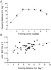
Figure 1
Figure 1. Daily voluntary running distance and cardiac hypertrophy in female rats
A, daily running distance for each week of the study (N = 37 animals). B, relationship between daily running distance and left ventricular weight (LVW) to body weight (BW) ratio at cell isolation(LVW:BW; linear regression, R = 0.50, P < 0.05).
Figure 2
Figure 2. Effect of training on the amplitude of [Ca2+]i transients and unloaded cell shortening
A, fura-2 fluorescence ([Ca2+]i; upper trace) and cell shortening (lower trace) in a representative myocyte. B, mean data for amplitude of the fura-2 transient (upper traces) and cell shortening expressed as a percentage of resting cell length (lower traces). Training condition (T = trained, S = sedentary) and region of origin (EPI = sub-epicardium, ENDO = sub-endocardium) of the cells did not significantly affect these parameters (P > 0.05, power 30 % for shortening and 20 % for fura-2 transient, n = number of cells, N = 10 sedentary and 10 trained animals).
Figure 3
Figure 3. Effect of region of origin and training on re-lengthening and fluorescence
A, change in fura-2 fluorescence and B, cell length in response to stimulation at 10 Hz for 10 s in the presence of 1 μM thapsigargin. C, relationship between fluorescence and cell length during re-lengthening (indicated by arrow) for traces shown in A and B. D, mean slope of re-lengthening-fluorescence (R-F) relationships. Training and region of origin did not significantly affect these parameters (P > 0.05, power 24 %, n = number of cells, N = 4 sedentary and 4 trained animals).
Figure 4
Figure 4. Mechanically unloaded cell shortening and force development of representative myocytes
A, unloaded cell shortening of an ENDO myocyte from a sedentary (S) and trained (T) rat with the same resting cell length and showing a similar degree of shortening. B, force developed by the same myocytes when mechanically loaded with carbon fibres.
Figure 5
Figure 5. Mechanically unloaded cell shortening and force development in myocytes
A, mean data for unloaded cell shortening and B, absolute force developed for the four types of cell. Cell shortening was similar in the four groups (P > 0.05, power 17 %), but trained cells developed greater force. C, mean force normalised to cell cross-sectional area. Most, but not all of the greater force in trained cells was due to increased cross-sectional area. (*P < 0.05, n = number of cells, N = 10 sedentary and 10 trained animals.)
Figure 6
Figure 6. Effect of stretching on contraction of a single rat left ventricular myocyte
A, change in cell length (upper panel) and force development (lower panel) following stretch, indicated by arrows to the sarcomere lengths (SL) shown. B, SL-tension relationship for data in A (□, active tension; ▪, resting tension). C, mean slope of active tension-SL relationship (δaT/δSL) for the four groups of cells. (*P < 0.05vs. ENDO S and EPI T, n = number of cells, N = 7 sedentary and 7 trained animals.)
Figure 7
Figure 7. Effect of exercise on the passive properties of the myocardium
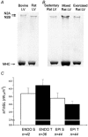
A, SDS-PAGE gel showing comparison of titin isoforms in bovine (lane 1) and sedentary rat (lane 2) left ventricle indicating that the bovine sample has an additional band (N2A) of higher molecular weight and slower mobility than the major band (N2B). This shows that the gel system can resolve different isoforms of titin and that sedentary rat left ventricle expresses only N2B myosin heavy chain (MHC). B, SDS-PAGE gel loaded with left ventricular myocardium from sedentary and exercised rat showing no difference in the expression of titin isoforms, as confirmed by a mixture of sedentary and exercised samples run in the central position. The faint band visible below N2B is a truncated form that is generally considered to arise from proteolysis (Wang, 1985). C, the mean slopes of resting tension-SL relationship (δrT/δSL) for the four groups of cells were not significantly different from each other (P > 0.05, power 43 %, n = number of cells, N = 7 sedentary and 7 trained animals).
Figure 8
Figure 8. Action potential records from representative myocytes
A, ENDO S and EPI S myocytes showing regional differences in action potential duration (APD). B, ENDO T and EPI T left ventricular myocytes; note that the regional difference in the late APD is absent. C and D, traces from previous panels emphasising the effect of training results in a lengthening of the APD in EPI cells.

Similar articles

Cited by

References

    1. Antzelevitch C, Sicouri S, Litovsky SH, Lukas A, Krishnan SC, Di Diego JM, Gintant GA, Liu D-W. Heterogeneity within the ventricular wall: electrophysiology and pharmacology of epicardial, endocardial and M cells. Circulation Research. 1991;69:1427–1449. - PubMed
    1. Anversa P, Ricci R, Olivetti G. Quantitative structural analysis of the myocardium during physiologic growth and induced cardiac hypertrophy: a review. Journal of the American College of Cardiology. 1986;7:1140–1149. - PubMed
    1. Asami S, Hirano T, Yamaguchi R, Tsurudome Y, Itoh H, Kasai H. Effects of forced and spontaneous exercise on 8-hydroxyguanosine levels in rat organs. Biochemical and Biophysical Research Communications. 1998;24:678–682. - PubMed
    1. Bryant SM, Shipsey J, Hart G. Normal regional distribution of membrane current density in the rat left ventricle is altered in catecholamine-induced hypertrophy. Cardiovascular Research. 1999;42:391–400. - PubMed
    1. Calaghan SC, White E. The role of calcium in the response of cardiac muscle to stretch. Progress in Biophysics and Molecular Biology. 1999;71:1–31. - PubMed

Publication types

MeSH terms

LinkOut - more resources