. 2002 Jun;129(2):530-9.
doi: 10.1104/pp.003970.

NPSN11 is a cell plate-associated SNARE protein that interacts with the syntaxin KNOLLE

Affiliations

NPSN11 is a cell plate-associated SNARE protein that interacts with the syntaxin KNOLLE

Haiyan Zheng et al. Plant Physiol. 2002 Jun.

Abstract

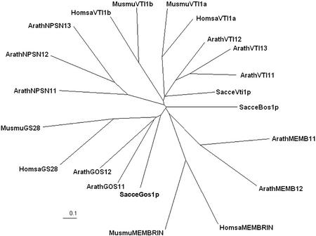
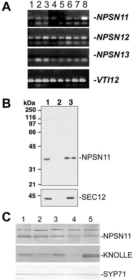
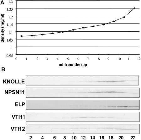
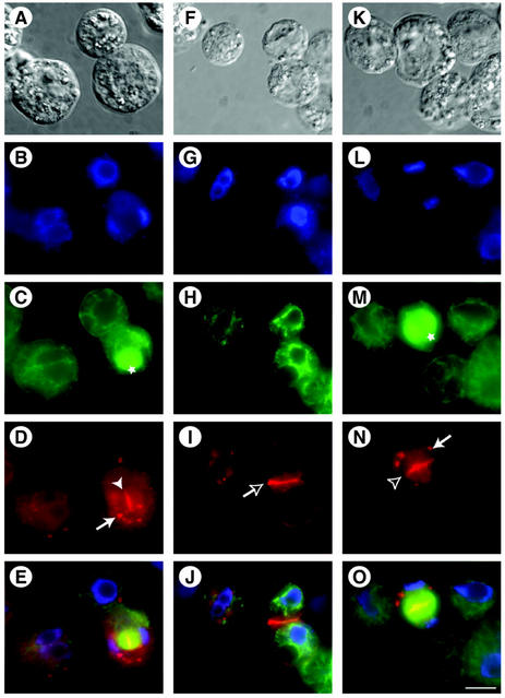
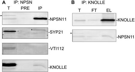
SNAREs are important components of the vesicle trafficking machinery in eukaryotic cells. In plants, SNAREs have been found to play a variety of roles in the development and physiology of the whole organism. Here, we describe the identification and characterization of a novel plant-specific SNARE, NPSN11, a member of a closely related small gene family in Arabidopsis. NSPN11 is highly expressed in actively dividing cells. In a subcellular fractionation experiment, NSPN11 cofractionates with the cytokinesis-specific syntaxin, KNOLLE, which is required for the formation of the cell plate. By immunofluorescence microscopy, NSPN11 was localized to the cell plate in dividing cells. Consistent with the localization studies, NSPN11 was found to interact with KNOLLE. Our results suggest that NPSN11 is another component of the membrane trafficking and fusion machinery involved in cell plate formation.


Figures

Figure 1
Figure 1
NPSN group SNAREs are novel to the plant kingdom. Representative SNARE protein sequences were acquired from GenBank (Arath, Arabidopsis; Sacce, budding yeast; Homsa, human; Musmu, mouse; see “Materials and Methods” for accession nos.) and aligned with the CLUSTALW algorithm. A phylogenetic tree was visualized with TreeView and was prepared for the figure with Adobe Photoshop.
Figure 2
Figure 2
NPSN11 and -12 have different tissue distribution patterns. A, RT-PCR was performed with primers specific for NPSN11, NPSN12, NPSN13, and VTI12, using total RNA prepared from the roots of a 3-week-old liquid-cultured plants (lane 1), the leaves of the same liquid-cultured plants (lane 2), flowers from mature soil-grown plants (lane 3), expanding rosette leaves (lane 4), mature rosette leaves (lane 5), green siliques (lane 6), the lower 3 cm of stem (lane 7), and the top 3 cm of the stem (lane 8). Amplified products were separated on agarose gels and visualized with ethidium bromide. Bands specific to each gene are indicated with a dash. No product could be observed with the NPSN13-specific primers. B, Extracts of Arabidopsis suspension-cultured cells were fractionated by differential centrifugation at 150,000g. Twenty micrograms of the resulting fractions of total protein (lane 1), supernatant (lane 2), and microsomal fractions (lane 3) were separated by SDS-PAGE and immunoblotted with affinity-purified NPSN11 antibodies (see “Materials and Methods”) or with the microsomal marker SEC12 (Bar-Peled and Raikhel, 1997). C, NPSN11 protein distribution. Equal amounts of total protein extracted from stems (lane 1), siliques (lane 2), roots (lane 3), leaves (lane 4), and flowers (lane 5) were separated by SDS-PAGE and blotted with antisera to NPSN11, KNOLLE, or SYP71. Bands specific for each protein are indicated with a dash.
Figure 3
Figure 3
NPSN11 T-DNA insertion mutant. A, A T-DNA insertion in the NPSN11 (At2g35190) locus was identified from the sequence-tagged database created by the SALK Institute. The insertion was confirmed and characterized as described in “Materials and Methods.” B, Protein extracts from seedlings and flowers of representative siblings of known genotype were separated by SDS-PAGE and blotted with NPSN11 antiserum to show the absence of the NPSN11 protein in the homozygous npsn11 plants.
Figure 4
Figure 4
NPSN11 and KNOLLE cofractionate on an Accudenz gradient. Total membranes prepared from 21-d-old liquid-cultured Arabidopsis roots were separated by a discontinuous Accudenz gradient. Fractions of 0.5 mL were collected from top to the bottom. The density of each fraction was plotted against the fraction number (A). Fractions were collected, and equal amounts were separated by SDS-PAGE and then blotted with the indicated antisera (B). NPSN11 cofractionated with KNOLLE but not with AtELP, the TGN marker; VTI11, which labels the PVC; or VTI12, which labels both the PVC and the TGN.
Figure 5
Figure 5
NPSN11 is localized at the cell plate during cytokinesis. Arabidopsis suspension-cultured cell protoplasts (A, F, and K) were double immunolabeled with antibodies directed against α-tubulin (α-tub) to visualize phragmoplast (marked by stars), cortical microtubules (C, H, and M), and either affinity-purified NPSN11 (D and I) or KNOLLE antisera (N). Nuclei in dividing and non-dividing cells were revealed by staining with 4′,6′-diamidino-2-phenylindole (DAPI; B, G, and L). Electronically merged images of B through D, G through I, and L through N are shown in E, J, and O, respectively. NSPN11 positive cell plate (solid arrowhead) and new plasma membrane (empty arrow) are visible in D and I, respectively. N shows the localization of KNOLLE at the cell plate (empty arrowhead). Arrows in D and N indicate NSPN11- and KNOLLE-containing intracellular organelles. Scale bar in A through O = 10 μm.
Figure 6
Figure 6
NPSN11 interacts with the syntaxin KNOLLE. A, NPSN11 protein was immunoprecipitated from total membrane proteins prepared from 5-d-old Arabidopsis suspension-cultured cells. Total protein (1/300; T), protein eluate from preimmune column (1/10; Pre) or from NPSN11-immune column (1/10; IP) was separated by SDS-PAGE. NPSN11, SYP21, VTI12, and KNOLLE were detected by western blots. Note that the 41-kD band observed with the crude NPSN11 antiserum (indicated by an arrowhead) is not immunoprecipitated. The non-specific band recognized by anti-SYP21 is indicated by an asterisk. B, Immunoprecipitation of KNOLLE from 5-d-old Arabidopsis suspension cells using rabbit anti-KNOLLE cross-linked to protein A beads. Total membrane (1/300; T), flow-through (1/300; FT), and eluted protein (1/10; EL) were separated by SDS-PAGE and detected by western blot using antisera against KNOLLE or NPSN11.

Similar articles

Cited by

References

    1. Ahmed SU, Bar-Peled M, Raikhel NV. Cloning and subcellular location of an Arabidopsis receptor-like protein that shares common features with protein-sorting receptors of eukaryotic cells. Plant Physiol. 1997;114:325–336. - PMC - PubMed
    1. Antonin W, Holroyd C, Fasshauer D, Pabst S, Fisher von Mollard G, Jahn R. A SNARE complex mediating fusion of late endosomes defines conserved properties of SNARE structure and function. EMBO J. 2000;19:6453–6464. - PMC - PubMed
    1. Assaad FF, Huet Y, Mayer U, Jürgens G. The cytokinesis gene KEULE encodes a Sec1 protein that binds the syntaxin KNOLLE. J Cell Biol. 2001;152:531–543. - PMC - PubMed
    1. Bar-Peled M, Raikhel NV. Characterization of AtSEC12 and AtSAR1: proteins likely involved in endoplasmic reticulum and Golgi transport. Plant Physiol. 1997;114:315–324. - PMC - PubMed
    1. Bassham DC, Sanderfoot AA, Kovaleva V, Zheng H, Raikhel NV. AtVPS45 complex formation at the trans-Golgi network. Mol Biol Cell. 2000;11:2251–2265. - PMC - PubMed

Publication types

MeSH terms

Associated data