. 2002 Jun;129(2):678-90.
doi: 10.1104/pp.004184.

Cytokinesis-defective mutants of Arabidopsis

Affiliations

Cytokinesis-defective mutants of Arabidopsis

Rosi Söllner et al. Plant Physiol. 2002 Jun.

Abstract

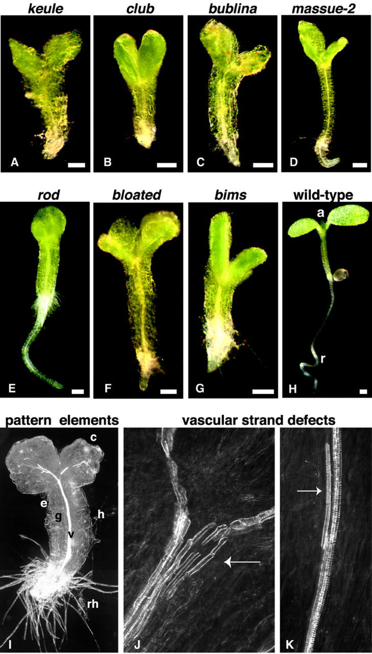
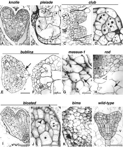
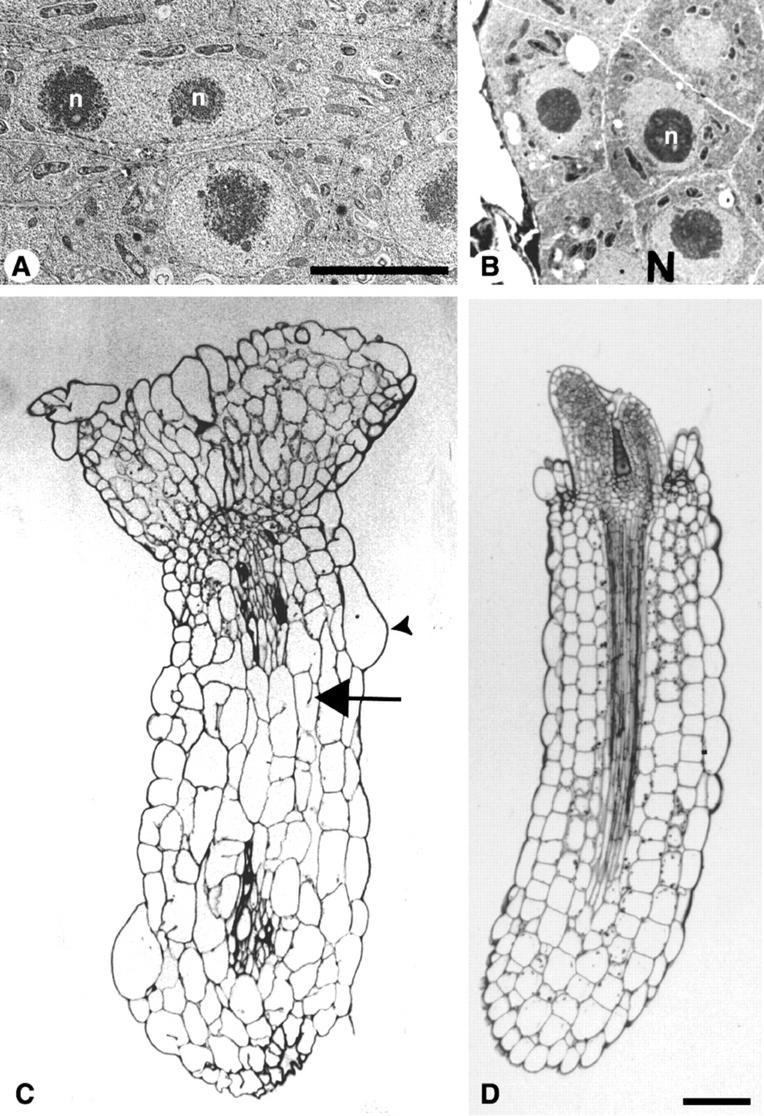
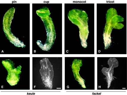
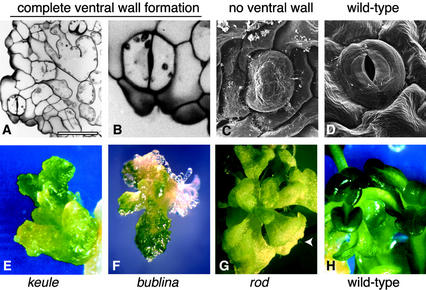
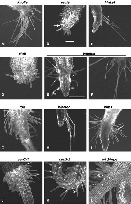
We have identified mutations in six previously uncharacterized genes of Arabidopsis, named club, bublina, massue, rod, bloated, and bims, that are required for cytokinesis. The mutants are seedling lethal, have morphological abnormalities, and are characterized by cell wall stubs, gapped walls, and multinucleate cells. In these and other respects, the new mutants are phenotypically similar to knolle, keule, hinkel, and pleiade mutants. The mutants display a gradient of stomatal phenotypes, correlating roughly with the severity of their cytokinesis defect. Similarly, the extent to which the different mutant lines were capable of growing in tissue culture correlated well with the severity of the cytokinesis defect. Phenotypic analysis of the novel and previously characterized loci indicated that the secondary consequences of a primary defect in cytokinesis include anomalies in body organization, organ number, and cellular differentiation, as well as organ fusions and perturbations of the nuclear cycle. Two of the 10 loci are required for both cytokinesis and root hair morphogenesis. The results have implications for the identification of novel cytokinesis genes and highlight the mechanistic similarity between cytokinesis and root hair morphogenesis, two processes that result in a rapid deposition of new cell walls via polarized secretion.


Figures

Figure 1
Figure 1
Bloated seedlings lacking functional root and shoot meristems. All seedlings have a rough surface layer and consist of cotyledons and a hypocotyl. Though the basal area has root-like properties, roots are absent in keule, club, bublina, and bims mutants (A, B, C, and G); stunted in massue and bloated mutants (D and F); and reduced in rod mutants (E). At the shoot apex, true leaves are absent or stunted, though these may develop upon transfer to tissue culture (see Fig. 5, F and G). The seedling phenotype is variable in all of the lines, with the exception of club and bims mutants for which the range of phenotypes is fairly narrow. The seedlings shown represent the median within the respective ranges of phenotypes, although massue mutants (D) often have considerably stronger and bloated mutants (F) considerably weaker phenotypes than those shown here. A clearing preparation shows that the apical basal body plan and the radial organization of tissue layers are generally conserved (I). The pattern elements of the seedling are shown in I and/or H; along the apical-basal axis, these include the cotyledons, hypocotyl, and root; and along the radial axis they include the epidermis, ground tissue, and vascular bundle. The vascular strands often show interruptions, misalignment, and deviation from the wild type with respect to the number and width of strands per bundle (arrow in J). In some instances, short vascular strands are found adjacent to the centrally located vascular bundles (arrow in K). Note the long root hairs at the basal end of the massue-1 mutant in I. The mutant lines shown are as indicated above each panel, and massue-1 is shown in I, bims is shown in J, and bloated is shown in K. keule allele MM125 is shown as reference in A. a, Apical meristem, first true leaf primordia; c, cotyledons; e, epidermis; g, ground tissue; h, hypocotyl; rh, root hairs; r, root; and v, vascular bundle. Bars = 200 μm.
Figure 2
Figure 2
Histological sections of embryos or seedlings revealing cell wall stubs and multinucleate cells. Histological sections were stained with toluidine blue, which stains the nuclei and cell wall, with the exception of that in G, which is stained with the cell wall-specific periodic acid schiff stain. Toluidine blue-stained nuclei appear as vacuolate structures with dark, round- or doughnut-shaped nucleoli. A, B, E, G, I, J, K, and L are embryo sections; C, D, F, and H are seedling sections. In C and D, an apical meristem is shown. B, This section depicts the basal area of a torpedo stage pleaide embryo, representing a severely affected hypocotyl and root primordium; in the same mutant, the cotyledons appear unaffected (not shown); the multinucleate cell shown encompasses more than one-half of the mutant embryo, as can be seen by comparison with the small outer cells in the same section, which suggests that the cytokinesis defects occurred early during embryogenesis; note the large nuclei with multiple nucleoli (black spots designated by arrowhead). F, Severely affected surface layer of a bublina cotyledon. H, Cotyledon cells adjacent to a true leaf primordium show the difference in cell size between expanded (lower cell with cytokinesis defects) and meristematic (upper left) cells in a rod seedling. Arrows point to cell wall stubs, small white arrow heads to multinucleate cells; larger, white arrow heads point to a cell wall gap in H and to a metaphase plate in J. In some instances, there are two juxtaposed wall stubs on either side of the cell (see black arrowhead in J), but for the most part, the cell wall stubs are anchored to the mother cell wall on one side of the cell, with no stub on the other side (see arrows in D, E, G, H, and K as well as text). Bars are 50 μm in A, C, F, I, and L; 20 μm in B, E, G, J, and K; and 10 μm in D. H and K are the same scale.
Figure 3
Figure 3
Cytokinesis-defective mutants have enlarged nuclei with multiple nucleoli, and enlarged cells. A and B are electron micrographs of embryos and C and D are histological seedling sections. A, Nucleus with two nucleoli (n), resembling a nuclear fusion, in a keule embryo. B, Wild-type nuclei (N) with single nucleoli (n) in a wild-type embryo. Note the difference in size between the nuclei in A and B, shown at the same scale. C, bublina seedling. D, Wild-type seedling, showing hypocotyl and apical meristem with the true leaf primordia; the cotyledons and root are not included due to the slightly tangential angle of the section. The bublina seedling shown in B has fewer cells per file than the wild type, but a roughly conserved number of cell files. The cells are larger than in the wild type (compare C and D, shown at the same scale). The arrowhead points to an irregularly shaped bloated surface cell, and the arrow to a cell wall stub. Bar is 5 μm in A for A and B and 100 μm in D for C and D.
Figure 4
Figure 4
Abnormal body organization in keule-like mutants. A, Pin-shaped cotyledons. B, Cup-shaped cotyledons. C, Monocotyledonous seedling. D, Tricotyledonous seedling. E, keule mutant that looks like fackel. F, Clearing preparation of keule mutant with hypocotyl deletion, with two vascular bundles (v) emerging from the basal area, as in fackel mutants. G, fackel mutant with a hypocotyl, somewhat like keule in appearance. H, Classical fackel mutant, showing central deletion and abnormal body organization. Bars = 200 μm.
Figure 5
Figure 5
A gradient of stomatal and postembryonic phenotypes. A through D, surface cells or seedling cotyledons. A and B, Histological sections. C and D, Scanning electron micrographs. Despite their heavily perturbed surface layer (A), club mutants are capable of complete ventral wall formation and form stomata with well-defined pores (B). B depicts the stomatal complex in A. By contrast, guard mother cells in keule mutants are incapable of ventral wall formation (C). E and F, Growth in tissue culture. E, keule explant on shoot-inducing medium (SIM), showing callus-like outgrowth. F, A bublina seedling transferred to SIM develops some true leaves, which show signs of being cytokinesis defective. G, rod seedling transferred to root-inducing medium shows near wild-type development, yet the leaves are irregular in shape and margins and have bloated cells. The trichomes (arrow head) have abnormal branching patterns. H, wild-type explant on SIM. Bar in A is 50 μm.
Figure 6
Figure 6
Root hair defects in keule-like mutants. Long root hairs are seen in knolle, hinkel, bublina, bloated, and bims mutants (A, C, F, G, H, and I). C and H, Long root hairs on one side of a hinkel or bloated mutant, stunted ones on the other side in contact with the agar on the plate. F, Long root hairs in bublina mutants (compare with E). E, Binucleate, bloated root hair in bublina seedling. B and D, stunted, bloated, crooked, and/or branched root hairs in keule and club mutants. Note the bulbous bases and crooked root hairs in club mutants, which resemble the defects seen in root hair mutants affected in tip growth, such as cen3 (J and K). n, Nucleus. Root hairs in rod mutants are often somewhat shorter than in the wild type (G), yet rod mutants are capable of growing long root hairs. Arrows designate branched root hair in B, bulbous bases in J, and crooked hairs in K. Bars are 100 μm. With the exception of E, all panels are the same scale as in B.

References

    1. Assaad FF. Plant cytokinesis: exploring the links. Plant Physiol. 2001a;126:509–516. - PMC - PubMed
    1. Assaad FF. Of weeds and men: what genomes teach us about plant cell biology. Curr Opin Plant Biol. 2001b;4:478–487. - PubMed
    1. Assaad FF, Huet Y, Mayer U, Jürgens G. The cytokinesis gene KEULE encodes a Sec1 protein which binds the syntaxin KNOLLE. J Cell Biol. 2001;152:531–543. - PMC - PubMed
    1. Assaad FF, Mayer U, Wanner G, Jürgens G. The KEULE gene is involved in cytokinesis in Arabidopsis. Mol Gen Genet. 1996;253:267–277. - PubMed
    1. Balasubramanian MK, McCollum D, Surana U. Tying the knot: linking cytokinesis to the nuclear cycle. J Cell Sci. 2000;113:1503–1513. - PubMed

Publication types

MeSH terms