Comparative Study
. 2002 Jun;129(2):691-705.
doi: 10.1104/pp.004010.

RASPBERRY3 gene encodes a novel protein important for embryo development

Affiliations
Comparative Study

RASPBERRY3 gene encodes a novel protein important for embryo development

Nestor R Apuya et al. Plant Physiol. 2002 Jun.

Erratum in

  • Plant Physiol 2002 Oct;130(2):1073. Harada John H [corrected to Harada John J]

Abstract

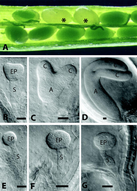
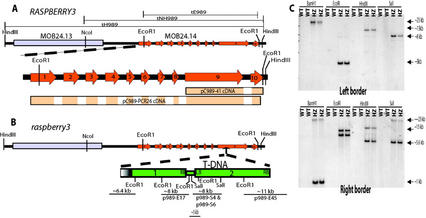
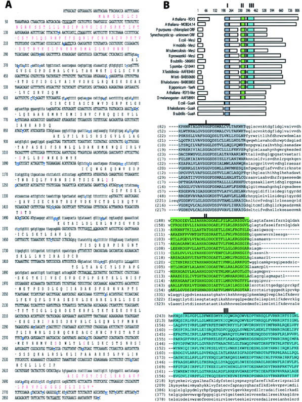
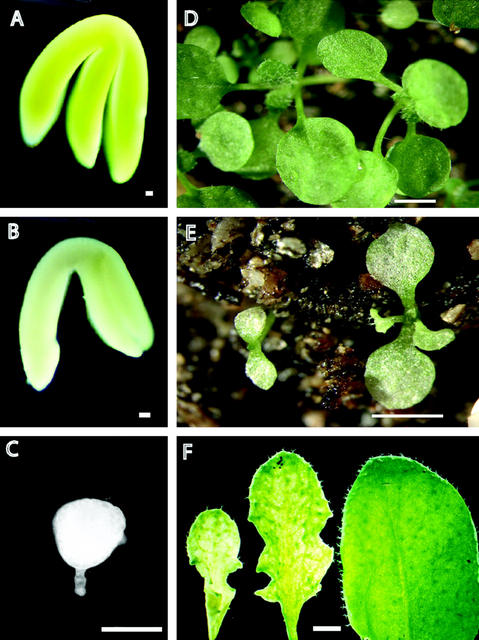
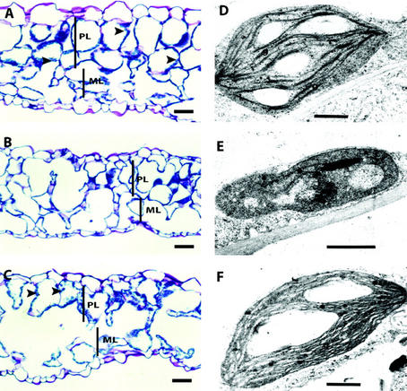
We identified a new gene that is interrupted by T-DNA in an Arabidopsis embryo mutant called raspberry3. raspberry3 has "raspberry-like" cellular protuberances with an enlarged suspensor characteristic of other raspberry embryo mutants, and is arrested morphologically at the globular stage of embryo development. The predicted RASPBERRY3 protein has domains found in proteins present in prokaryotes and algae chloroplasts. Computer prediction analysis suggests that the RASPBERRY3protein may be localized in the chloroplast. Complementation analysis supports the possibility that the RASPBERRY3 protein may be involved in chloroplast development. Our experiments demonstrate the important role of the chloroplast, directly or indirectly, in embryo morphogenesis and development.


Figures

Figure 1
Figure 1
Developmental analysis of rsy3 mutant embryos. A, Typical heterozygous siliques containing wild-type and mutant (highlighted with asterisks) seeds. Nomarski images of wild-type embryos (B–D) were taken from the same siliques from which the corresponding mutant embryos (E–G) were taken (see “Materials and Methods”). A, Axis; C, cotyledon; EP, embryo proper; S, suspensor. Bars = 25 μm.
Figure 2
Figure 2
Gene organization of RSY3 and T-DNA insertion in rsy3 mutant. A, Diagrammatic representation of a portion of the lambda genomic clone containing the RSY3 gene in chromosome 3. The predicted RSY3 gene (annotated for Columbia ecotype as MOB24.14) is expanded below the clone to highlight exons represented by solid arrows in orange and numbered accordingly. The genomic fragments tH989, tNH989, and tE989 used in the complementation analysis are outlined above the genomic clone. The cDNA clones are designated below the expanded region of RSY3 gene. Clone pC989–41 represents a partial cDNA isolated from a library, and clone pC989–41 represents the nearly full-length cDNA that were isolated using 5′- and 3′-RACE. Only the areas highlighted in colors within the rectangles represent the cDNA sequences. B, Diagrammatic representation of the T-DNA insertion in the rsy3 embryo mutant. Two T-DNAs that are arranged in concatemer are inserted in exon 9. Some of the EcoRI and SaI fragments, as revealed by plasmid rescue analysis, are highlighted with the approximate sizes written above the lines. Some of the restriction sites relevant to the DNA analysis shown in C are indicated. C, Restriction analysis of genomic DNAs isolated from wild-type (WT) and heterozygous (HZ) rsy3 individual segregants. DNAs were digested with restriction enzymes as indicated and were size separated by electrophoresis in a 1% (w/v) gel. The resulting blots were hybridized with a left or a right border probe as indicated in each panel. Restriction enzymes used are indicated. Note: Diagrams in A and B are not drawn to scale.
Figure 3
Figure 3
RSY3 genomic DNA sequence and its predicted protein. A, Portion of the genomic DNA sequence of the RSY3 gene. Introns are in lowercase letters, and the exons and the untranslated sequences are in uppercase letters. Nucleotides highlighted in blue are missing or are different from the reported sequence based on a Columbia background (see sequence of predicted gene MOB24.12 in accession AB020746). The predicted amino acids are given above the coding sequence. Amino acids highlighted in red are those that differ from the predicted amino acid sequence in the RSY3 locus of the Columbia ecotype. B, Alignment of the RSY3 protein to other proteins with similar domains. The amino acid sequences derived from the predicted coding sequences were aligned using the AlignX program of Vector NTI software. The highlighted domains (I, II, and III) were subsequently found using the AlignX Block program of the same software. Domain I, highlighted in gray, is the putative ATP-binding domain. Consensus core regions (as cited in the text) within the three domains are highlighted with a bold line above the sequences. The proteins included in the above alignment have the following accession numbers: Porphyra purpurea (AAC08269), Synechocystis sp (BAA10210), Escherichia coli (BAA77863), Heliobacillus mobilis (AAC84036), Mycobacterium tuberculosis (AAK48088), Rickettsia prowazekii (CAA14513), Bacillus subtilis (BAA05302), Xylella fastidiosia (AAF83469), Mesorhizobium loti (BAB50680), Bacillus halodurans (BAB03802), Bradyrhizobium japonicum (BAB50680), Saccharomyces pombe (CAA94698), E. coli GuaA (AAG57618), B. halodurans GuaA (Q9KF78), B. subtilis GuaA (P29727), Arabidopsis RSY3-like (AAC16077), and fruit fly (Drosophila melanogaster) predicted protein (AAF58991). The accession number for the RSY3 genomic sequence is AY077630.
Figure 4
Figure 4
The embryo-defective morphology of the rsy3 mutant can be rescued by a partial RSY3 genomic fragment. A through C, Three types of embryos produced by a heterozygous rsy3/RSY3 plant containing one copy of tE989 transgene. A, Morphologically wild-type green embryo (RSY3/RSY3;tE989). B, Partially rescued rsy3 pale-green embryo (rsy3/rsy3;tE989/tE989). C, Mutant rsy3 embryo (rsy3/rsy3). D, Seedlings generated from morphologically wild-type green embryos with genotype rsy3/RSY3; tE989. E, Seedlings generated from partially rescued rsy3 pale-green embryos with genotype rsy3/rsy3; tE989/tE989. F, Close-up view of rosette leaves taken from partially rescued rsy3 (rsy3/rsy3; tE989/tE989) plants (first two leaves from left; approximately 50 d postgermination) and from wild-type plants (right leaf; approximately 30 d postgermination). Bars = 50 μm in A through C; 3 mm in D and E; and = 2 mm in F.
Figure 5
Figure 5
Histological analysis of partially rescued rsy3 leaves. Separate sections of leaves shown in Figure 4E were taken for histological analysis (A–C) and for transmission electron microscopic (TEM) analysis of chloroplasts (D–F). Tissue section (A) and TEM of chloroplast (D) from morphologically wild-type green plants (RSY3/RSY3;tE989). Leaf tissue section (B) and TEM of a chloroplast (E) from a pale-yellow region of partially rescued rsy3 mutant plants (rsy3/rsy3; tE989/tE989). Leaf tissue section (C) and TEM of a chloroplast (F) from a green region of partially rescued rsy3 mutant plants (rsy3/rsy3; tE989/tE989). Arrowheads point to chloroplasts. PL, Palisade mesophyll layer; ML, spongy mesophyll layer; g, grana. Bars = 100 μm in A through C and = 0.15 μm in D through F.
Figure 6
Figure 6
Molecular analysis of partially rescued rsy3 plants. A, Diagram of the construct used in the partial complementation. The EcoRI fragment (see Fig. 2A) was blunt-end ligated into the NotI site of the T-DNA vector pGHS166N (see “Materials and Methods”). P2 is the mannopine synthase promoter contained within pGSH166N vector. B, Genomic DNA restriction analysis of some F2 segregants generated from the complementation cross between rsy3/RSY3 and rsy3/RSY3;tE989 lines. Two separate sets of genomic DNAs were digested with EcoRI or NotI and were size fractionated by electrophoresis in a 1% (w/v) agarose gel; the resulting blots were hybridized with the 2.5-kb EcoRI fragment of the RSY3 genomic clone (see Fig. 2A). C, RNA analysis of RSY3 transcripts in different tissues of wild-type (left panel) and in seedlings generated from partially complemented rsy3 mutant (rsy3/rsy3;tE989) plants (right panel). The length of exposure for the left panel was 3 d, and for the right panel was 1 d. D, Portion of the cDNA sequence generated from the reverse transcriptase-PCR analysis using mRNA samples from partially rescued rsy3 mutant (rsy3/rsy3;tE989) plants (see “Materials and Methods”). The series of dots (…) represents internal sequences identical to the sequence shown in Figure 3A (see also accession no. AY077630).

References

    1. Aida M, Ishida T, Fukaki H, Fujisawa H, Tasaka M. Genes involved in organ separation in Arabidopsis: an analysis of the cup-shaped cotyledon mutant. Plant Cell. 1997;9:841–857. - PMC - PubMed
    1. Albert S, Despres B, Guilleminot J, Bechtold N, Pelletier G, Delseny M, Devic M. The EMB 506 gene encodes a novel ankyrin repeat containing protein that is essential for the normal development of Arabidopsis embryos. Plant J. 1999;17:169–179. - PubMed
    1. Altmann T, Felix G, Jessop A, Kauschmann A, Uwer U, Peña-Cortés H, Willmitzer L. Ac/Ds transposon mutagenesis in Arabidopsis thaliana: mutant spectrum and frequency of Ds insertion mutants. Mol Gen Genet. 1995;247:646–652. - PubMed
    1. Altschul SF, Madden TL, Schaffer AA, Zhang J, Zhang Z, Miller W, Lipman DJ. Gapped BLAST and PSI-BLAST: a new generation of protein database search programs. Nucleic Acids Res. 1997;25:3389–3402. - PMC - PubMed
    1. Apuya NR, Yadegari R, Fischer RL, Harada JJ, Zimmerman JL, Goldberg RB. The Arabidopsis embryo mutant schlepperless has a defect in the chaperonin-60α gene. Plant Physiol. 2001;126:717–730. - PMC - PubMed

Publication types

MeSH terms

Associated data