Comparative Study
. 2002 Jun;129(2):717-32.
doi: 10.1104/pp.001016.

Evidence for a Ustilago maydis steroid 5alpha-reductase by functional expression in Arabidopsis det2-1 mutants

Affiliations
Comparative Study

Evidence for a Ustilago maydis steroid 5alpha-reductase by functional expression in Arabidopsis det2-1 mutants

Christoph W Basse et al. Plant Physiol. 2002 Jun.

Abstract

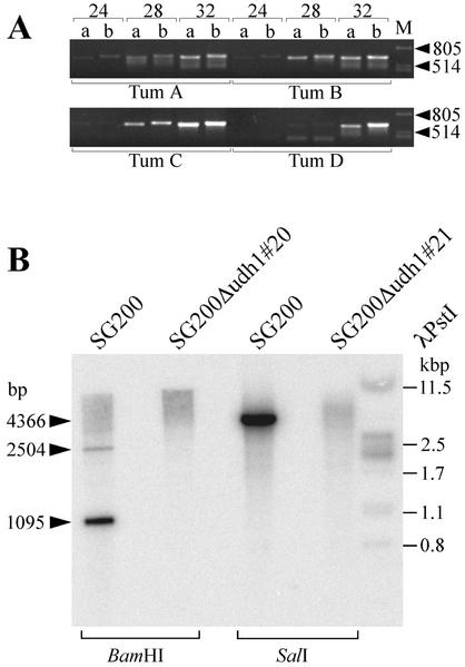
We have identified a gene (udh1) in the basidiomycete Ustilago maydis that is induced during the parasitic interaction with its host plant maize (Zea mays). udh1 encodes a protein with high similarity to mammalian and plant 5alpha-steroid reductases. Udh1 differs from those of known 5alpha-steroid reductases by six additional domains, partially predicted to be membrane-spanning. A fusion protein of Udh1 and the green fluorescent protein provided evidence for endoplasmic reticulum localization in U. maydis. The function of the Udh1 protein was demonstrated by complementing Arabidopsis det2-1 mutants, which display a dwarf phenotype due to a mutation in the 5alpha-steroid reductase encoding DET2 gene. det2-1 mutant plants expressing either the udh1 or the DET2 gene controlled by the cauliflower mosaic virus 35S promoter differed from wild-type Columbia plants by accelerated stem growth, flower and seed development and a reduction in size and number of rosette leaves. The accelerated growth phenotype of udh1 transgenic plants was stably inherited and was favored under reduced light conditions. Truncation of the N-terminal 70 amino acids of the Udh1 protein abolished the ability to restore growth in det2-1 plants. Our results demonstrate the existence of a 5alpha-steroid reductase encoding gene in fungi and suggest a common ancestor between fungal, plant, and mammalian proteins.


Figures

Figure 1
Figure 1
Nucleotide sequence and derived amino acid sequence of udh1. Shown is the DNA sequence of the 1,722-bp genomic AgeI-HindIII fragment (GenBank accession no. AF502086) containing the udh1 gene. The translation start ATG codon is underlined and the stop codon of udh1 is indicated by an asterisk. Primers used for generating udh1-eGFP fusions are indicated by arrows. The deletion in Δudh1 strains comprised the region from Phe-78 to Ala-358 (boxed). The poly(A) site is indicated at position 1,453.
Figure 2
Figure 2
Multiple alignment of Udh1 with other 5α-reductase sequences. A, Udh1, U. maydis Udh1; SP5R, putative S. pombe 5α-reductase; DET2, Arabidopsis DET2; rS5R1, rat type 1 5α-reductase. Amino acids (numbered at the borders) dominating a column are boxed and shaded. Gaps have been inserted to maximize the number of identities. Predicted transmembrane helices of Udh1 and rS5R1 are in boldface and emphasized by bars (gray for Udh1 and black for rS5R1). The highly conserved Glu residue, which is altered in the det2-1 mutant and corresponds to Glu-311 of Udh1, is marked by a dot. Conserved positions within Udh1 stretch V and the respective region of SP5R are boxed. B, Assignment of amino acids from the Udh1 sequence to matching positions from either the rS5R1 (top) or the DET2 (bottom) amino acid sequence. Amino acids are numbered from the N to C terminus.
Figure 3
Figure 3
Expression analysis of udh1. A, U. maydis strains indicated at the top were grown on CM charcoal for 2 d before RNA isolation. Five to 15 μg of RNA was loaded in each lane. B, Lane 1, RNA (15 μg) from strain FB2 grown on CM charcoal for 2 d. Lanes 2 through 9, RNA (5 μg; 2 μg in lane 9) prepared from infected maize tissue between 2 and 12 d after inoculation with a mixture of strains FB1 and FB2. The northern blots were probed with a 32P-labeled udh1 cDNA fragment (positions 1,019–1,449 in Fig. 1). A and B, Radioactive signals were quantified (boxed) and standardized (FB2 = 1) by comparison with signals obtained by hybridization with the constitutively expressed U. maydis ppi gene. Staining with methylene blue reflected the amounts of total RNA loaded. The fungal and maize ribosomal RNA bands are indicated.
Figure 4
Figure 4
Localization of Udh1. U. maydis strains CL13/pugh1#8 (column A), CL13/pugh1#10 (column B) harboring ectopic insertions of the udh1-eGFP fusion construct, and strain CB35 (Basse et al., 2000; column C), expressing the eGFP gene under the constitutive strong otef promoter, were grown in YEPS medium and assayed by differential interference contrast (DIC) light microscopy (top row), and epifluorescence to detect eGFP (medium row) and DAPI (bottom row) distribution. The bar represents 10 μm. Bottom, Schematic depiction of the udh1-eGFP fusion construct. udh1 ORF promoter and 3′ sequences are depicted by grayed arrows and bars, respectively.
Figure 5
Figure 5
Genotypic verification by CAPS analysis. A, Schematic representation of CAPS analysis. Numbers refer to MnlI restriction sites and respective fragments. The 152-bp MnlI fragment (boxed) is only contained in wild-type fragments. B, Representative example of CAPS analysis. PCR products obtained from amplification with primers det3h/det3r were restricted with MnlI and subjected to Southern analysis using the DET2 EcoRV fragment as probe (see “Materials and Methods”). Minor signals above the expected MnlI bands in the various lanes reflect incomplete digests of PCR products.
Figure 6
Figure 6
Expression analysis of transgenic Arabidopsis plants. A and B, RNA (0.3–1.8 μg) from the indicated plants (see “Materials and Methods”) and from U. maydis strain FB1 (20 μg) grown on CM charcoal for 2 d was loaded. Radioactive signals were quantified (boxed) on both membranes and standardized (det2-1/pBUB1#1 = 100) by comparison to signals obtained after subsequent hybridization with the Arabidopsis 32P-labeled actin probe (ACT2). A, RNA from wild-type Col-0 plants 62 d after sowing was included as control. The northern blot was probed with the 32P-labeled AgeI-HindIII udh1 fragment. Arrowheads indicate the full-length udh1, the truncated udh1 (udh1ΔN70), the U. maydis endogenous udh1, and the actin transcript. The actin signal in the U. maydis lane is caused by cross-hybridization with the ACT2 probe. The different sizes of the udh1 and udh1ΔN70 transcripts were also reflected by faster migration of the truncated udh1 transcript. B, RNA from two different wild-type Col-0 and det2-1 mutant plants 61 d after sowing was included as control. The northern blot was probed with the 32P-labeled DET2 EcoRV fragment. DET2 and ACT2 transcripts are indicated by arrowheads. Faintly detectable endogenous DET2 transcripts are indicated by asterisks.
Figure 7
Figure 7
Development of Arabidopsis pBUB1, pBDET2, and pBUA1 transformants. A to C, Plants from series 2, 42 d after sowing. A, Wild-type Col-0. B, Mutant det2-1. C, Mutant det2-1 transgenic for pBUB1. Arrows indicate terminal flowers that developed in Col-0 and transgenic det2-1 plants. D and E, Plants from series 3, 61 d after sowing. D, Mutant det2-1 transgenic for pBUB1 (det2-1/pBUB#1, 2, 8, and 12). Seed pods are indicated by arrows. E, Wild-type Col-0 plants. Seed pods have not yet developed. F to H, Plants from series 4, 43 d after sowing. F, Transformants det2-1/pBDET2#1, 2, 3, 6, 7, and 8 are numbered from 5 to 10. det2-1 plants carrying the pBDET2 construct (5–10) display seed pod development (arrows). Homozygous det2-1 (1–3) and heterozygous DET2/det2-1 (4) plants that escaped Basta treatment but lacked the transgene. G, Control wild-type Col-0 plants. H, pBUA1 transgenic det2-1 plants (2 and 3) are phenotypically indifferent from a det2-1 plant (1) that escaped Basta selection but lacked the transgene.
Figure 8
Figure 8
Development of T2det2-1/pBUB1 plants (series 5 and 6) compared with F1Col-0 plants. A to C, Plants from series 5, 33 d after sowing. D to F, Plants from series 6, 33 d after sowing. All presented T2det2-1/pBUB1 plants, four (B), and six (E) Col-0 plants, respectively, were analyzed with respect to the genotype and the presence (A and D) or absence (B, C, E, and F) of the transgene. Growth conditions were as specified in Table II. A and D, T2det2-1/pBUB1. B and E, Wild-type Col-0. C and F, T2det2-1/pBUB1 that displayed a dwarf phenotype and lacked the transgene. Each pot contains two plants. The numbers in D refer to the plants used for northern analysis (see Fig. 9).
Figure 9
Figure 9
Expression of the udh1 gene in differently developed transgenic Arabidopsis plants. RNA (1 μg) isolated from rosette leaves of T2det2-1/pBUB1 plants from series 6, 33 d after sowing and from U. maydis strain FB2 (4 μg) grown on CM charcoal for 2 d was loaded (see “Materials and Methods”). Northern blots were probed with the 32P-labeled AgeI-HindIII udh1 fragment, the DET2 EcoRV fragment, and the Arabidopsis 32P-labeled actin probe (ACT2) as loading control. The ratio of quantified signals (udh1/ACT2) was calculated (arrowhead). Staining with methylene blue reflected the amounts of total RNA loaded. The full-length udh1, endogenous U. maydis udh1, ACT2, and the fungal and maize ribosomal RNA bands are indicated (arrowheads). Bottom, Developmental stage of investigated plants 35 d after sowing: B, Flower bud; F, flower; S, seed pods. The numbers refer to stem lengths in centimeters.
Figure 10
Figure 10
Analysis of Δudh1 strains. A, Detection of SG200 and SG200Δudh1 strains in maize tumors. Chromosomal DNA (75 ng) isolated from tumors 10 d after inoculation with mixtures of SG200/SG200Δudh1#12 (Tum A), SG200/SG200Δudh1#15 (Tum B), SG200/SG200Δudh1#20 (Tum C), and SG200/SG200Δudh1#21 (Tum D) was used as template for PCR with the primer combinations a and b (see “Materials and Methods”). Expected sizes of PCR products were the 622-bp fragment of the hph gene flanked by udh1 5′-ORF sequences (a) and the 643-bp udh1 fragment (b). The numbers above the lanes refer to the PCR cycles performed. Phage lambda (M) DNA (500 ng) digested with PstI was used as size marker. The sizes of the respective fragments are indicated in basepairs. B, Non-stringent Southern analysis of wild-type SG200, SG200Δudh1#20, and SG200Δudh1#21 strains. Chromosomal DNA isolated from the strains indicated was restricted with either BamHI or SalI, and probed with a udh1 fragment spanning the deletion of the mutant udh1 allele. Attribution of sizes to the hybridizing bands is indicated (arrowheads). The sizes of cross-hybridizing λPstI fragments are marked on the right.

References

    1. Aichinger C. Identifizierung pflanzenabhängig-regulierter Gene in Ustilago maydis. PhD thesis. Munich, Germany: LMU München; 2000.
    1. Altmann T. A tale of dwarfs and drugs: brassinosteroids to the rescue. Trends Genet. 1998;14:490–495. - PubMed
    1. Altschul SF, Madden TL, Schäffer AA, Zhang J, Zhang Z, Miller W, Lipman DJ. Gapped BLAST and PSI-BLAST: a new generation of protein database search programs. Nucleic Acids Res. 1997;25:3389–3402. - PMC - PubMed
    1. Andersson S, Bishop RW, Russell DW. Expression cloning and regulation of steroid 5 alpha-reductase, an enzyme essential for male sexual differentiation. J Biol Chem. 1989;264:16249–16255. - PMC - PubMed
    1. Andersson S, Russell DW. Structural and biochemical properties of cloned and expressed human and rat steroid 5 alpha-reductases. Proc Natl Acad Sci USA. 1990;87:3640–3644. - PMC - PubMed

Publication types

MeSH terms

Associated data

LinkOut - more resources