. 2002 Jun 17;195(12):1515-22.
doi: 10.1084/jem.20020033.

Cytokine requirements for acute and Basal homeostatic proliferation of naive and memory CD8+ T cells

Affiliations

Cytokine requirements for acute and Basal homeostatic proliferation of naive and memory CD8+ T cells

Ananda W Goldrath et al. J Exp Med. .

Abstract

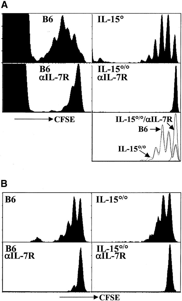
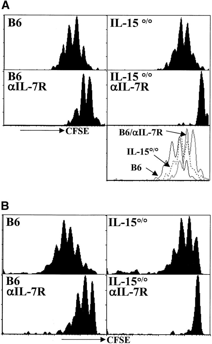
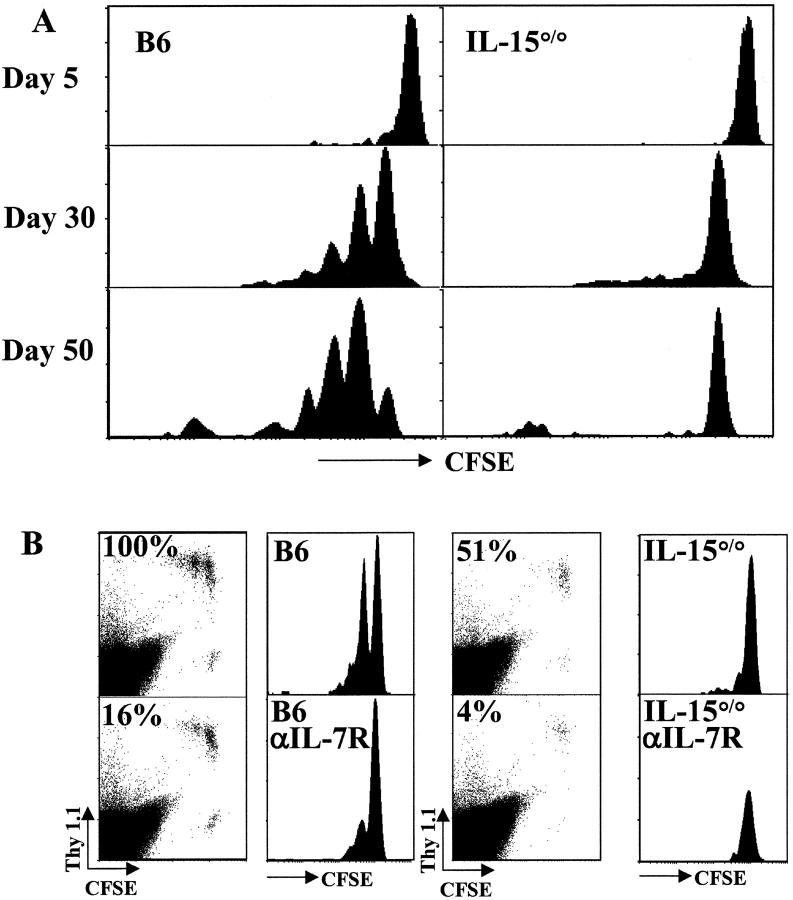
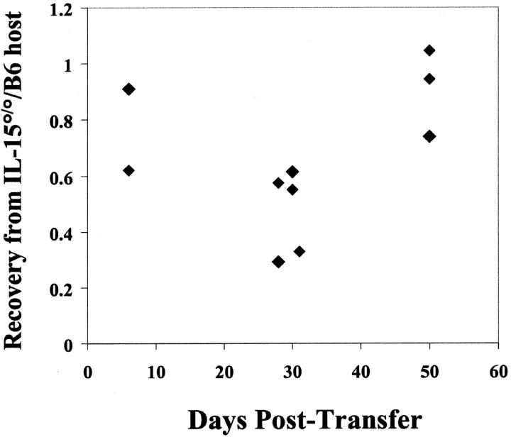
Both naive and memory T cells undergo antigen-independent proliferation after transfer into a T cell-depleted environment (acute homeostatic proliferation), whereas only memory T cells slowly divide in a full T cell compartment (basal proliferation). We show, first, that naive and memory CD8+ T cells have different cytokine requirements for acute homeostatic proliferation. Interleukin (IL)-7 receptor(R)alpha-mediated signals were obligatory for proliferation of naive T cells in lymphopenic hosts, whereas IL-15 did not influence their division. Memory T cells, on the other hand, could use either IL-7Ralpha- or IL-15-mediated signals for acute homeostatic proliferation: their proliferation was delayed when either IL-7Ralpha was blocked or IL-15 removed, but only when both signals were absent was proliferation ablated. Second, the cytokine requirements for basal and acute homeostatic proliferation of CD8+ memory T cells differ, as basal division of memory T cells was blocked completely in IL-15-deficient hosts. These data suggest a possible mechanism for the dearth of memory CD8+ T cells in IL-15- and IL-15Ralpha-deficient mice is their impaired basal proliferation. Our results show that naive and memory T lymphocytes differ in their cytokine dependence for acute homeostatic proliferation and that memory T lymphocytes have distinct requirements for proliferation in full versus empty compartments.


Figures

Figure 1.
Figure 1.
Acute homeostatic proliferation of naive CD8+ T cells requires IL-7Rα–, but not IL-15–, mediated signals. (A) Cells pooled from LN and spleen of OT-I RAG°/° mice were labeled with CFSE and transferred intravenously into B6 or IL-15°/° hosts irradiated the previous day. Indicated recipients were given 1 mg anti–IL-7Rα mAb intraperitoneally every other day beginning at the time of transfer. Data are from two experiments. Histogram plots of CFSE intensity in Vβ5+CD8+ cells (left panels from one transfer) or Vα2+CD8+ gated cells (right panels from a second transfer) are shown for representative recipients 6 d after transfer. The bottom right panel is an overlay of CFSE intensity in CD8+Vα2+ gated cells from B6 (solid line), IL-15°/° (broken line), and anti–IL-7Rα–treated IL-15°/°hosts (gray line) all from the same experiment. (B) Naive polyclonal CD44loCD122lo CD8+ T cells were sorted from LN and spleen cells from CD45.1 congenic mice, labeled with CFSE, and transferred into recipients as above. Histogram plots of CFSE intensity in CD8+CD45.1+ gated cells on day 6 after transfer are shown. Data are representative of three or more experiments.
Figure 3.
Figure 3.
CD8+ memory T cells express higher levels of IL-7Rα and IL-15Rα than naive CD8+ T cells. (A) Relative expression of IL-7Rα and IL-15Rα mRNA by naive and memory OT-I and polyclonal CD8+ T cells. Data shown are representative of duplicate of RNA samples for each condition that were each analyzed three times. Data were normalized to respective housekeeping and CD8α gene expression and plotted relative to OT-I naive cells. (B) IL-7Rα surface expression for OT-I and polyclonal naive and memory cells. Histogram overlays (left panel) represent staining for IL-7Rα for polyclonal naive (CD8+CD44lo, thin gray line), polyclonal memory (CD8+CD44hi, thick gray line), OT-I naive (OT-I RAG°/°, thin black line), or OT-I memory (CD8+ Thy1.1+, thick black line) cells or isotype staining (dashed line). Dot plots show CD44 versus IL-7Rα surface expression on gated polyclonal CD8+ and CD8+ Thy1.1+ OT-I memory T cells from the same mouse >6 mo after transfer of OT-I cells and infection with recombinant vaccinia virus.
Figure 2.
Figure 2.
Both IL-7Rα– and IL-15R–mediated signals contribute to the acute homeostatic proliferation of CD8+ memory T cells in irradiated hosts. (A) OT-I memory T cells (CD8+ Thy1.1+) were enriched, labeled with CFSE, and transferred to irradiated recipients as described for Fig. 1. Histograms of CFSE intensity for CD8+Vα2+Thy1.1+ gated cells are shown 6 d after transfer. The bottom right panel is an overlay of CFSE intensity in CD8+Thy1.1+ gated cells from B6 (solid line), IL-15°/° (broken line) and anti–IL-7Rα–treated B6 hosts (gray line). (B) Memory phenotype (CD44hiCD122hi) polyclonal CD8+ T cells were sorted from LN and spleen of CD45.1 congenic mice, CFSE labeled and transferred into recipients as above. Histogram plots of CFSE intensity of CD8+CD45.1+ gated cells are shown on day 6 after transfer. Data are representative of three experiments.
Figure 4.
Figure 4.
Basal proliferation of memory CD8+ T cells requires IL-15. (A) OT-I memory T cells (CD8+ Thy1.1+) were enriched, labeled with CFSE, and transferred to unirradiated B6 (left panels) or IL-15°/° (right panels) recipients. Proliferation of transferred memory T cells was followed as CFSE staining intensity of CD8+Thy1.1+ cells at 5, 30, and 50 d after transfer. (B) OT-I memory T cells were transferred into B6 (left panels) or IL-15°/° (right panels) hosts and untreated (top panels) or treated with anti–IL-7Rα mAb for 30 d (bottom panels). Histograms of CFSE intensity in CD8+Thy1.1+ gated cells and dot plots of live gated pooled spleen and LN cells are shown. The percentages indicated on each dot plot represent the relative recovery of transferred memory cells from spleen (mean number of recovered OT-I memory cells from each condition divided by the number recovered from the B6 control hosts). Data are representative of multiple experiments.
Figure 5.
Figure 5.
Recovery of OT-I memory T cells from B6 and IL-15°/° hosts. Results are expressed as the relative number of recovered Thy1.1+ OT-I memory T cells recovered from the spleen of IL-15°/° hosts normalized to the number of recovered memory T cells from B6 controls. Data are from multiple experiments.

Comment in

References

    1. Marrack, P., J. Bender, D. Hildeman, M. Jordan, T. Mitchell, M. Murakami, A. Sakamoto, B.C. Schaefer, B. Swanson, and J. Kappler. 2000. Homeostasis of αβ TCR+ T cells. Nat. Immunol. 1:107–111. - PubMed
    1. Freitas, A.A., and B. Rocha. 2000. Population biology of lymphocytes: the fight for survival. Annu. Rev. Immunol. 18:83–111. - PubMed
    1. Goldrath, A.W., and M.J. Bevan. 1999. Selecting and maintaining a diverse T-cell repertoire. Nature. 402:255–262. - PubMed
    1. Tough, D.F., and J. Sprent. 1994. Turnover of naive- and memory-phenotype T cells. J. Exp. Med. 179:1127–1135. - PMC - PubMed
    1. Murali-Krishna, K., L.L. Ming, S. Sambhara, F. Lemonnier, J. Altman, and R. Ahmed. 1999. Persistence of memory CD8 T cells in MHC class I deficient mice. Science. 286:1377–1381. - PubMed

Publication types