. 2002 Jun 17;195(12):1541-8.
doi: 10.1084/jem.20020369.

Interleukin 15 is required for proliferative renewal of virus-specific memory CD8 T cells

Affiliations

Interleukin 15 is required for proliferative renewal of virus-specific memory CD8 T cells

Todd C Becker et al. J Exp Med. .

Abstract

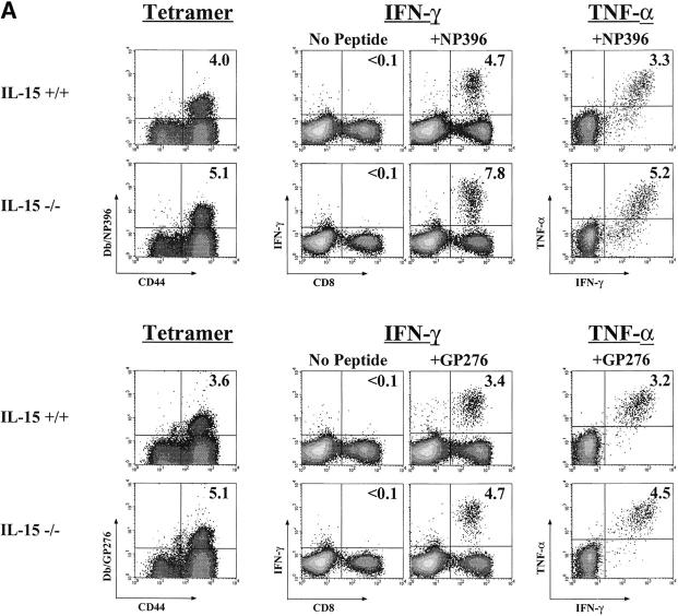
The generation and efficient maintenance of antigen-specific memory T cells is essential for long-lasting immunological protection. In this study, we examined the role of interleukin (IL)-15 in the generation and maintenance of virus-specific memory CD8 T cells using mice deficient in either IL-15 or the IL-15 receptor alpha chain. Both cytokine- and receptor-deficient mice made potent primary CD8 T cell responses to infection with lymphocytic choriomeningitis virus (LCMV), effectively cleared the virus and generated a pool of antigen-specific memory CD8 T cells that were phenotypically and functionally similar to memory CD8 T cells present in IL-15(+/+) mice. However, longitudinal analysis revealed a slow attrition of virus-specific memory CD8 T cells in the absence of IL-15 signals. This loss of CD8 T cells was due to a severe defect in the proliferative renewal of antigen-specific memory CD8 T cells in IL-15(-/-) mice. Taken together, these results show that IL-15 is not essential for the generation of memory CD8 T cells, but is required for homeostatic proliferation to maintain populations of memory cells over long periods of time.


Figures

Figure 1.
Figure 1.
IL-15−/− mice mount a substantial CD8 T cell response to LCMV infection but memory CD8 T cell numbers gradually decline. (A) Longitudinal PBMC analysis was performed by serially bleeding individual mice on the indicated days after infection with LCMV. PBMCs were stained with MHC tetra-mers of two LCMV epitopes (NP396 and GP276). Plots are gated on CD8+ T cells, and the numbers represent the number of MHC tetramer binding cells as a percentage of total PBMCs (bold) and as a percentage of CD8+ T cells (parentheses). Staining is representative of four to seven mice per group. (B) The numbers of epitope-specific cells/106 PBMCs in IL-15−/− mice are expressed as a percentage of the number/106 PBMC in control mice. Staining was performed as in A. Numbers are averages of four to seven mice. (C) At 80 d after infection, IL-15−/− and IL-15+/− littermate control mice were killed and single cell suspensions from spleens were stained with MHC tetramers as above. Absolute numbers of LCMV-specific memory CD8 T cells in the spleen were calculated. LCMV-specific CD4 responses were similar in both IL-15−/− and IL-15+/− control mice (data not shown).
Figure 1.
Figure 1.
IL-15−/− mice mount a substantial CD8 T cell response to LCMV infection but memory CD8 T cell numbers gradually decline. (A) Longitudinal PBMC analysis was performed by serially bleeding individual mice on the indicated days after infection with LCMV. PBMCs were stained with MHC tetra-mers of two LCMV epitopes (NP396 and GP276). Plots are gated on CD8+ T cells, and the numbers represent the number of MHC tetramer binding cells as a percentage of total PBMCs (bold) and as a percentage of CD8+ T cells (parentheses). Staining is representative of four to seven mice per group. (B) The numbers of epitope-specific cells/106 PBMCs in IL-15−/− mice are expressed as a percentage of the number/106 PBMC in control mice. Staining was performed as in A. Numbers are averages of four to seven mice. (C) At 80 d after infection, IL-15−/− and IL-15+/− littermate control mice were killed and single cell suspensions from spleens were stained with MHC tetramers as above. Absolute numbers of LCMV-specific memory CD8 T cells in the spleen were calculated. LCMV-specific CD4 responses were similar in both IL-15−/− and IL-15+/− control mice (data not shown).
Figure 2.
Figure 2.
IL-15Rα2/− mice generate a potent LCMV-specific CD8 T cell response but memory CD8 T cell numbers are reduced relative to controls at day 70 after infection. (A) NP396-specific CD8 T cells from the PBMCs were stained as in Fig. 1. Staining is representative of five mice per group. (B) Total numbers of memory LCMV-specific CD8 T cells were determined in the spleen and liver by tetramer staining as described in Fig. 1 C. Numbers indicate the average of three to four mice per group. LCMV-specific CD4 responses were similar in both IL-15Rα2/− and IL-15Rα1/− control mice (data not shown).
Figure 2.
Figure 2.
IL-15Rα2/− mice generate a potent LCMV-specific CD8 T cell response but memory CD8 T cell numbers are reduced relative to controls at day 70 after infection. (A) NP396-specific CD8 T cells from the PBMCs were stained as in Fig. 1. Staining is representative of five mice per group. (B) Total numbers of memory LCMV-specific CD8 T cells were determined in the spleen and liver by tetramer staining as described in Fig. 1 C. Numbers indicate the average of three to four mice per group. LCMV-specific CD4 responses were similar in both IL-15Rα2/− and IL-15Rα1/− control mice (data not shown).
Figure 3.
Figure 3.
Antigen-specific memory CD8 T cells in IL-15−/− mice are functionally and phenotypically similar to memory CD8 T cells in IL-15+/+ mice. (A) Splenocytes from IL-15−/− or IL-15+/+ control mice were prepared 40 d after infection. Cells were stimulated in vitro for 5 h with NP396 or GP276 peptides, then intracellular cytokine staining was performed for IFN-γ and TNF-α. The percentage of CD8 T cells producing either cytokine is essentially equivalent to the percentage of tetramer-binding cells in all cases. Tetramer staining and IFN-γ/TNF-α dual staining is shown gated on CD8 T cells. Numbers in the top right represent the percentage of CD8 T cells (B) LCMV-specific GP33 tetramer+ CD8 T cells from the spleens of IL-15−/− mice or IL-15+/+ controls were stained for CD44 and CD122 at 40 d after infection. The dark line indicates GP33 tetramer+ cells from a IL-15+/+ mouse while the dashed line represents GP33 tetramer+ CD8 T cells from an IL-15−/− mouse. Naive controls are shown in light gray; all CD8s from a naive mouse for the CD44 plot and CD44loCD8s for the CD122 plot.
Figure 3.
Figure 3.
Antigen-specific memory CD8 T cells in IL-15−/− mice are functionally and phenotypically similar to memory CD8 T cells in IL-15+/+ mice. (A) Splenocytes from IL-15−/− or IL-15+/+ control mice were prepared 40 d after infection. Cells were stimulated in vitro for 5 h with NP396 or GP276 peptides, then intracellular cytokine staining was performed for IFN-γ and TNF-α. The percentage of CD8 T cells producing either cytokine is essentially equivalent to the percentage of tetramer-binding cells in all cases. Tetramer staining and IFN-γ/TNF-α dual staining is shown gated on CD8 T cells. Numbers in the top right represent the percentage of CD8 T cells (B) LCMV-specific GP33 tetramer+ CD8 T cells from the spleens of IL-15−/− mice or IL-15+/+ controls were stained for CD44 and CD122 at 40 d after infection. The dark line indicates GP33 tetramer+ cells from a IL-15+/+ mouse while the dashed line represents GP33 tetramer+ CD8 T cells from an IL-15−/− mouse. Naive controls are shown in light gray; all CD8s from a naive mouse for the CD44 plot and CD44loCD8s for the CD122 plot.
Figure 4.
Figure 4.
Memory CD8 T cells in IL-15−/− mice generate a potent recall response. IL-15+/− and IL-15−/− mice were immunized with the Armstrong strain of LCMV. 3 mo later, mice were challenged with the virulent LCMV clone 13 strain intravenously. PBMCs were stained using MHC class I tetramers at days 0, 3, and 8 after rechallenge. Plots are gated on CD8+ T cells, and the numbers indicate the percentages of MHC tetramer positive cells as a percentage of total PBMCs (top number) and as a percentage of CD8+ T cells (bottom number).
Figure 5.
Figure 5.
Memory CD8 T cells do not undergo homeostatic proliferation, but undivided cells are maintained in an IL-15−/− environment. (A) Splenocytes from LCMV immune mice were labeled with CFSE and transferred into naive recipients. 30 d after transfer, spleens were removed from recipient animals and cells were stained using MHC class I tetramers to identify antigen specific memory CD8 T cells. Transferred antigen-specific cells proliferated in IL-15+/+ but not IL-15−/− mice over a 30-d period. In the right column, the same experiment was performed using splenocytes from immune IL-15−/− mice and similar results were observed. Homeostatic proliferation of CD4 T cells occurred and was normal in all environments (data not shown) (B) Labeled cells recovered from the recipient spleens at 30 d after transfer were quantified. In addition, the number of undivided cells (i.e., zero division CFSE peak) and the number of divided cells (sum of cells in divisions 1–4) are graphed. Results are similar for transfers of memory cells from +/+ mice (left panel) and IL-15−/− mice (right).
Figure 5.
Figure 5.
Memory CD8 T cells do not undergo homeostatic proliferation, but undivided cells are maintained in an IL-15−/− environment. (A) Splenocytes from LCMV immune mice were labeled with CFSE and transferred into naive recipients. 30 d after transfer, spleens were removed from recipient animals and cells were stained using MHC class I tetramers to identify antigen specific memory CD8 T cells. Transferred antigen-specific cells proliferated in IL-15+/+ but not IL-15−/− mice over a 30-d period. In the right column, the same experiment was performed using splenocytes from immune IL-15−/− mice and similar results were observed. Homeostatic proliferation of CD4 T cells occurred and was normal in all environments (data not shown) (B) Labeled cells recovered from the recipient spleens at 30 d after transfer were quantified. In addition, the number of undivided cells (i.e., zero division CFSE peak) and the number of divided cells (sum of cells in divisions 1–4) are graphed. Results are similar for transfers of memory cells from +/+ mice (left panel) and IL-15−/− mice (right).

Comment in

Similar articles

Cited by

References

    1. Ahmed, R., and D. Gray. 1996. Immunological memory and protective immunity: understanding their relation. Science. 272:54–60. - PubMed
    1. Bachmann, M.F., M. Barner, A. Viola, and M. Kopf. 1999. Distinct kinetics of cytokine production and cytolysis in effector and memory T cells after viral infection. Eur. J. Immunol. 29:291–299. - PubMed
    1. Harrington, L.E., M. Galvan, L.G. Baum, J.D. Altman, and R. Ahmed. 2000. Differentiating between memory and effector CD8 T cells by altered expression of cell surface O-glycans. J. Exp. Med. 191:1241–1246. - PMC - PubMed
    1. Veiga-Fernandes, H., U. Walter, C. Bourgeois, A. McLean, and B. Rocha. 2000. Response of naive and memory CD8+ T cells to antigen stimulation in vivo. Nat. Immunol. 1:47–53. - PubMed
    1. Kaech, S.M., and R. Ahmed. 2001. Memory CD8+ T cell differentiation: initial antigen encounter triggers a developmental program in naive cells. Nat. Immunol. 2:415–422. - PMC - PubMed

Publication types