. 2002 Jun 17;195(12):1549-63.
doi: 10.1084/jem.20011700.

Mesenchymal stem cells in perichondrium express activated leukocyte cell adhesion molecule and participate in bone marrow formation

Affiliations

Mesenchymal stem cells in perichondrium express activated leukocyte cell adhesion molecule and participate in bone marrow formation

Fumio Arai et al. J Exp Med. .

Abstract

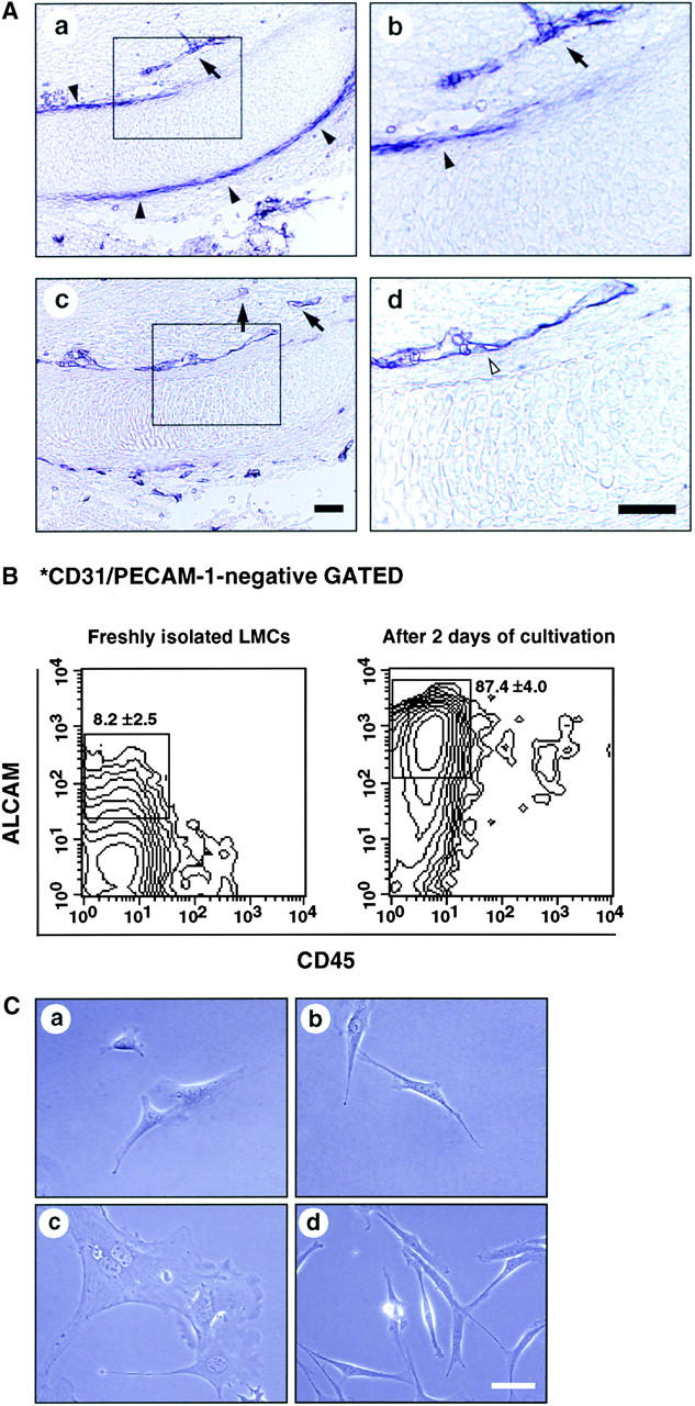
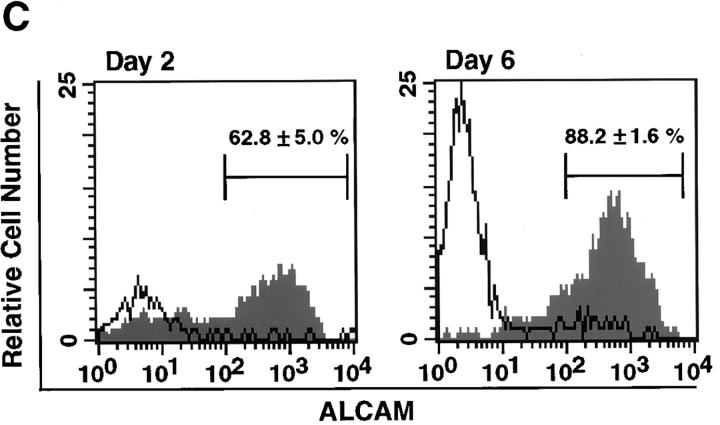
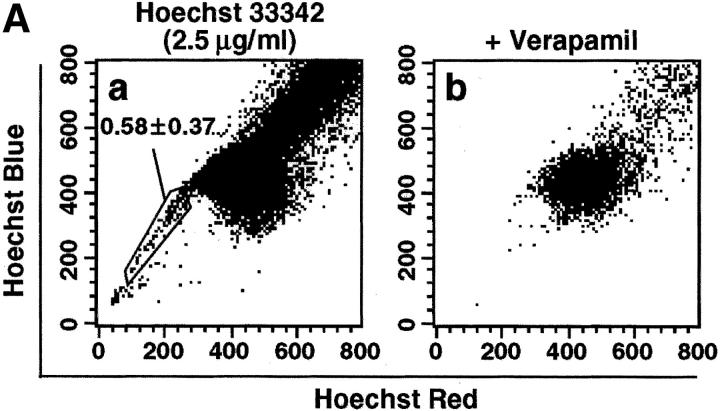
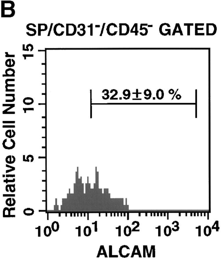
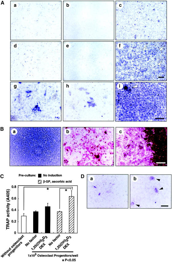
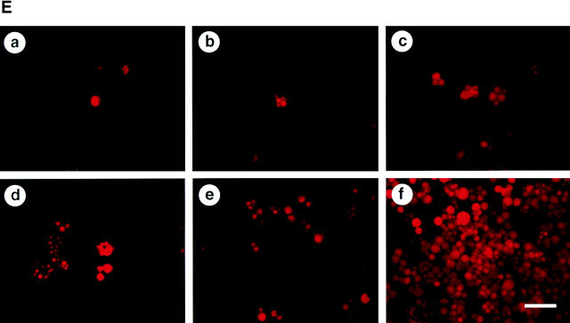
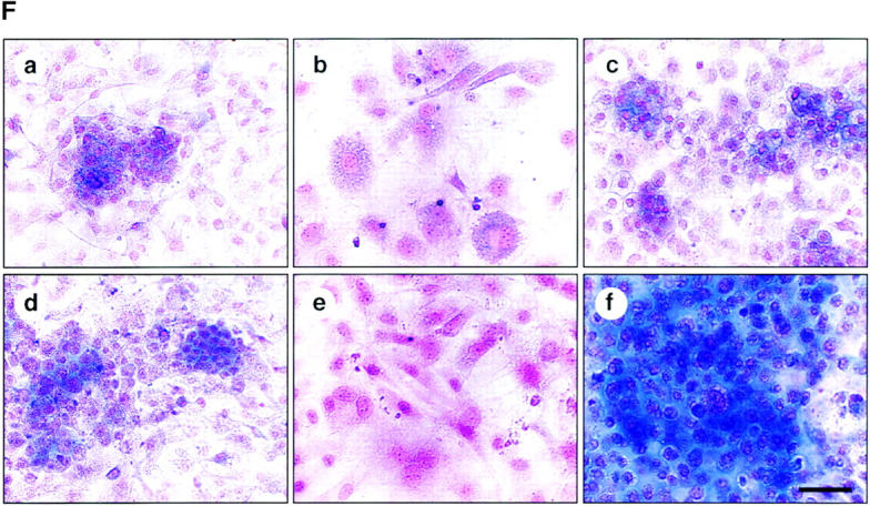
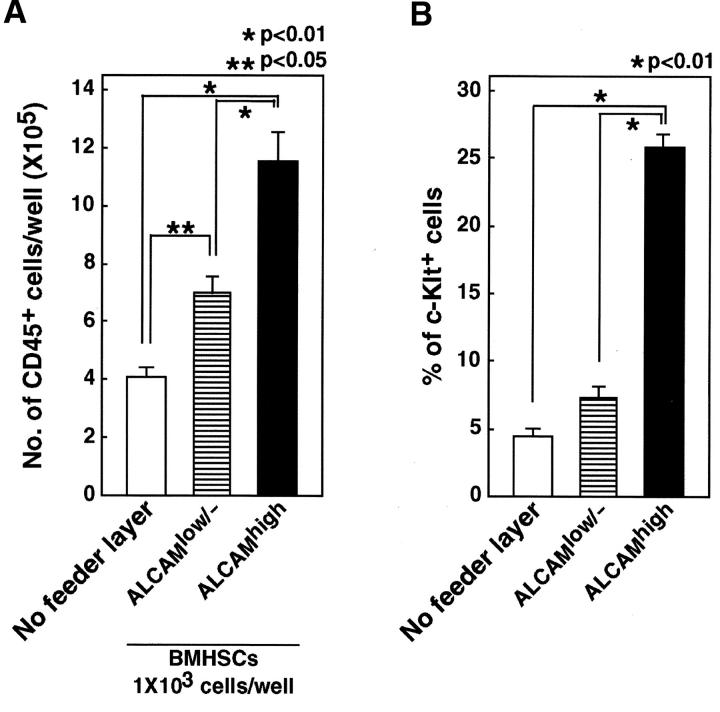
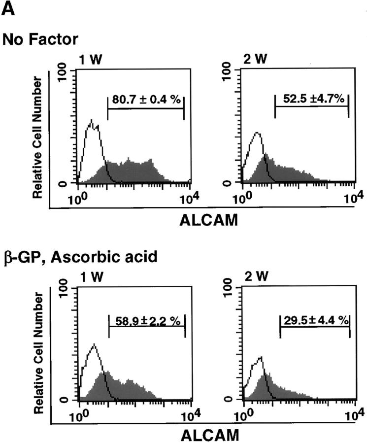
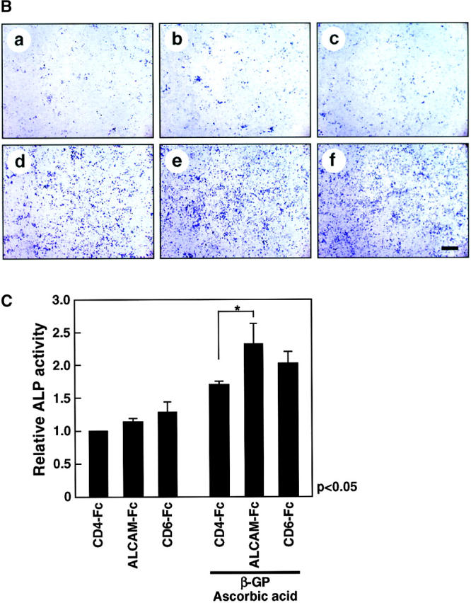
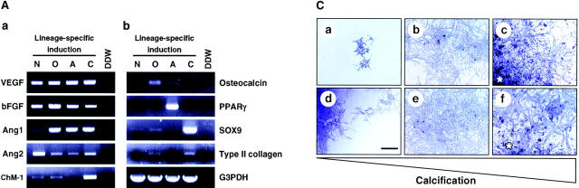
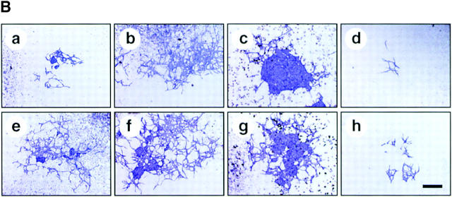
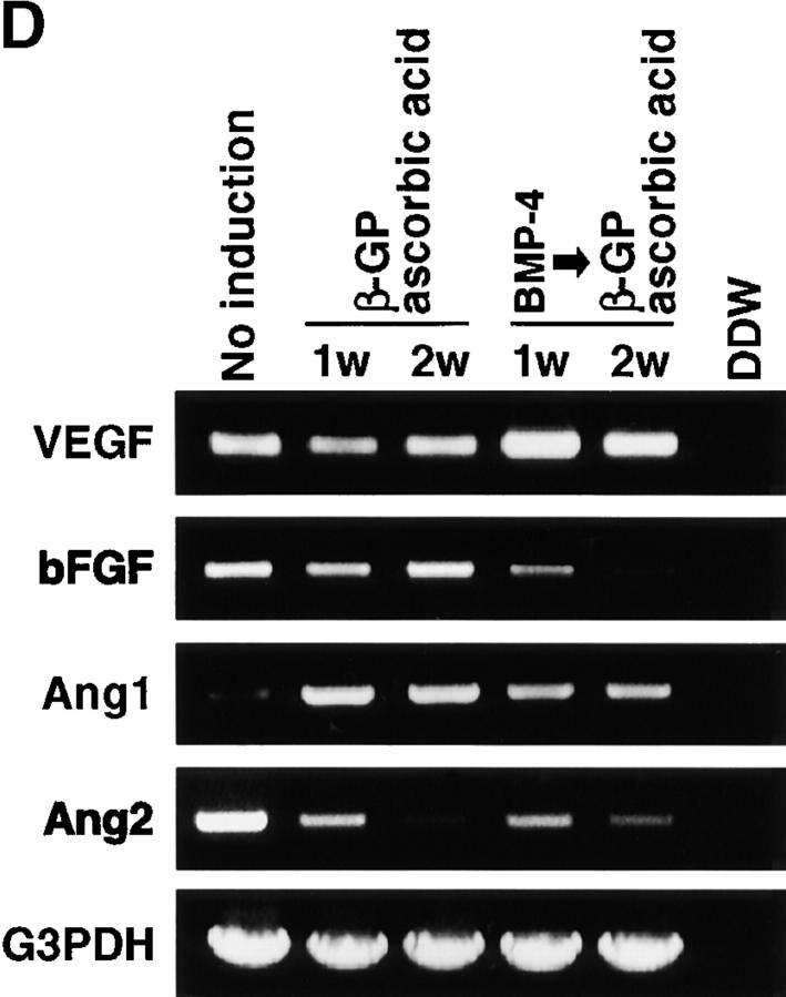
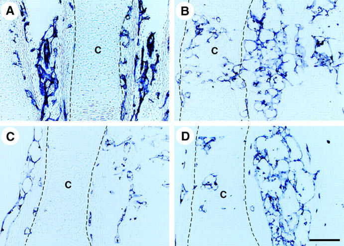
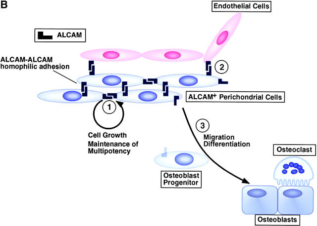
Perichondrium in fetal limb is composed of undifferentiated mesenchymal cells. However, the multipotency of cells in this region and the role of perichondrium in bone marrow formation are not well understood. In this report, we purified and characterized perichondrial cells using a monoclonal antibody against activated leukocyte cell adhesion molecule (ALCAM) and investigated the role of perichondrial cells in hematopoietic bone marrow formation. ALCAM is expressed on hematopoietic cells, endothelial cells, bone marrow stromal cells, and mesenchymal stem cells and mediates homophilic (ALCAM-ALCAM)/heterophilic (ALCAM-CD6) cell adhesion. Here we show by immunohistochemical staining that ALCAM is expressed in perichondrium. ALCAM+ perichondrial cells isolated by FACS exhibit the characteristics of mesenchymal stem cells. ALCAM+ cells can differentiate into osteoblasts, adipocytes, chondrocytes, and stromal cells, which can support osteoclastogenesis, hematopoiesis, and angiogenesis. Furthermore, the addition of ALCAM-Fc or CD6-Fc to the metatarsal culture, the invasion of the blood vessels to a cartilage was inhibited. Our findings indicate that ALCAM+ perichondrial cells participate in vascular invasion by recruiting osteoclasts and vessels. These findings suggest that perichondrium might serve as a stem cell reservoir and play an important role in the early development of a bone and bone marrow.


Figures

Figure 1.
Figure 1.
Perichondrial cells express ALCAM. (A) Longitudinal sections of E13.5 forelimb were stained with anti-ALCAM mAb (a and b) and anti–PECAM-1 mAb (c and d). ALCAM+ cells (black arrowheads) are seen in the perichondrium region. PECAM-1+ endothelial cells (white arrowheads) were detected surrounding cartilage and ALCAM-expressing perichondrium. ALCAM+ endothelial cells distributed toward the perichondrium (allows). b and d are higher magnifications of enclosed boxes in a and c, respectively. Scale bar, 100 μm. (B) LMCs derived from E13.5 limb were stained with anti-CD45, anti–PECAM-1, and anti-ALCAM mAbs and then fractionated by FACS®. PECAM-1 cells were gated and examined for expression of CD45 and ALCAM by contour blot. The percentages of ALCAM+ cells are representative of triplicate experiments. (C) Morphology of sorted ALCAMhigh (a and b) and ALCAMlow/− cells (c and d). (a and c) 12 h after plating. (b and d) 4 d after plating. Scale bar, 50 μm.
Figure 2.
Figure 2.
Characterization of perichondrial cells. (A) Comparison of proliferation between freshly isolated ALCAM+ and ALCAM cells. ALCAM+ and ALCAM cells were sorted from freshly isolated LMCs and plated into 48-well plates at 1.5 × 10⁴ cells per well. Cell numbers were determined on days 1–4. (B) Effects of ALCAM-Fc and CD6-Fc on the cell proliferation. ALCAM+ cells were sorted from freshly prepared LMCs, and cultured for 5 d in the presence of various concentrations of Fc-chimeric proteins. The number of expanded cells was counted on days 0.5, 3, and 5. (a) The ratio of cell growth. The cell number of the 0.5 d of culture in the presence of control-Fc was set at 1.0. (b) The relative cell numbers on each day of culture. The cell number in cultivation with control-Fc in each culture period was set as 1.0. The data shown represent the mean ±SD. (C) ALCAM cells were isolated from freshly prepared LMCs, and the expression of ALCAM was examined on days 2 and 6 of cultivation. The percentages of ALCAM+ cells in each panel are representative of triplicate experiments. (D) Flow cytometric characterization of ALCAMhigh cells. The expression of cell surface markers indicated on panels (a–l) was examined by FACS®.
Figure 2.
Figure 2.
Characterization of perichondrial cells. (A) Comparison of proliferation between freshly isolated ALCAM+ and ALCAM cells. ALCAM+ and ALCAM cells were sorted from freshly isolated LMCs and plated into 48-well plates at 1.5 × 10⁴ cells per well. Cell numbers were determined on days 1–4. (B) Effects of ALCAM-Fc and CD6-Fc on the cell proliferation. ALCAM+ cells were sorted from freshly prepared LMCs, and cultured for 5 d in the presence of various concentrations of Fc-chimeric proteins. The number of expanded cells was counted on days 0.5, 3, and 5. (a) The ratio of cell growth. The cell number of the 0.5 d of culture in the presence of control-Fc was set at 1.0. (b) The relative cell numbers on each day of culture. The cell number in cultivation with control-Fc in each culture period was set as 1.0. The data shown represent the mean ±SD. (C) ALCAM cells were isolated from freshly prepared LMCs, and the expression of ALCAM was examined on days 2 and 6 of cultivation. The percentages of ALCAM+ cells in each panel are representative of triplicate experiments. (D) Flow cytometric characterization of ALCAMhigh cells. The expression of cell surface markers indicated on panels (a–l) was examined by FACS®.
Figure 3.
Figure 3.
Limb SP cells express ALCAM. (A) LMCs were stained with Hoechst 33342 and analyzed for expression of ALCAM, PECAM-1, and CD45 in the SP cell fraction. (a) Hoechst 33342 staining and emission pattern of LMCs. SP cells were indicated in the enclosed region. (b) Hoechst 33342 staining and emission pattern of LMCs in the presence of verapamil. (B) A fluorescence histogram shows the ALCAM staining profile of the SP gated with the PECAM-1CD45 fraction. The percentages of ALCAM+ cells are representative of triplicate experiments.
Figure 3.
Figure 3.
Limb SP cells express ALCAM. (A) LMCs were stained with Hoechst 33342 and analyzed for expression of ALCAM, PECAM-1, and CD45 in the SP cell fraction. (a) Hoechst 33342 staining and emission pattern of LMCs. SP cells were indicated in the enclosed region. (b) Hoechst 33342 staining and emission pattern of LMCs in the presence of verapamil. (B) A fluorescence histogram shows the ALCAM staining profile of the SP gated with the PECAM-1CD45 fraction. The percentages of ALCAM+ cells are representative of triplicate experiments.
Figure 4.
Figure 4.
Osteoblasts, adipocytes, and chondrocyte differentiation. (A) Total LMCs (a, d, and g), ALCAMlow/− cells (b, e, and h), and ALCAMhigh cells (c, f, and i) were induced (d–i) or not-induced (a–c) to undergo osteogenic differentiation. After 2 wk, ALP staining was performed. Panels g–i show high power fields of d–f, respectively. Scale bars, 1 mm (a–f), 200 μm (g–i). (B) The region of alizarin red positive calcium deposition in ALCAMhigh cells, in which osteogenic differentiation was induced for 2 wk. (a) no factor; (b and c) β-GP and ascorbic acid. (b) The center of culture plate. (c) The corner of the culture plate. (C) TRAP activities were measured on day 7 of cocultivation of BM derived osteoclast precursor cells and ALCAMhigh cells, which were induced (hatched bar) or not induced (solid bar) to undergo osteogenic differentiation in the presence or absence of 1,25-(OH)2D3 and Dex. The data shown represents the mean ±SD. (D) Results of TRAP staining of cocultured BM-derived osteoclast precursor cells and ALCAMhigh cells, in which osteogenic differentiation was induced. TRAP-positive multinucleated cells are indicated by arrowheads. (a) no factor; (b) in the presence of 1,25-(OH)2D3 and Dex. Scale bar, 100 μm. (E) Total LMCs (a and d) ALCAMlow/− cells (b and e), and ALCAMhigh cells (c and f) were induced to undergo adipogenic differentiation (as described in Materials and Methods) and stained with Nile red. (a–c) control medium; (d–f) MDI medium. Scale bar, 200 μm. (F) Alcian blue staining after chondrogenic induction. Total LMCs (a and d), ALCAMlow/− cells (b and e), and ALCAMhigh cells (c and f) were induced to undergo chondrogenic differentiation as described in Materials and Methods. (a–c) no factor; (d–f) in the presence of BMP-2. Scale bar, 100 μm.
Figure 4.
Figure 4.
Osteoblasts, adipocytes, and chondrocyte differentiation. (A) Total LMCs (a, d, and g), ALCAMlow/− cells (b, e, and h), and ALCAMhigh cells (c, f, and i) were induced (d–i) or not-induced (a–c) to undergo osteogenic differentiation. After 2 wk, ALP staining was performed. Panels g–i show high power fields of d–f, respectively. Scale bars, 1 mm (a–f), 200 μm (g–i). (B) The region of alizarin red positive calcium deposition in ALCAMhigh cells, in which osteogenic differentiation was induced for 2 wk. (a) no factor; (b and c) β-GP and ascorbic acid. (b) The center of culture plate. (c) The corner of the culture plate. (C) TRAP activities were measured on day 7 of cocultivation of BM derived osteoclast precursor cells and ALCAMhigh cells, which were induced (hatched bar) or not induced (solid bar) to undergo osteogenic differentiation in the presence or absence of 1,25-(OH)2D3 and Dex. The data shown represents the mean ±SD. (D) Results of TRAP staining of cocultured BM-derived osteoclast precursor cells and ALCAMhigh cells, in which osteogenic differentiation was induced. TRAP-positive multinucleated cells are indicated by arrowheads. (a) no factor; (b) in the presence of 1,25-(OH)2D3 and Dex. Scale bar, 100 μm. (E) Total LMCs (a and d) ALCAMlow/− cells (b and e), and ALCAMhigh cells (c and f) were induced to undergo adipogenic differentiation (as described in Materials and Methods) and stained with Nile red. (a–c) control medium; (d–f) MDI medium. Scale bar, 200 μm. (F) Alcian blue staining after chondrogenic induction. Total LMCs (a and d), ALCAMlow/− cells (b and e), and ALCAMhigh cells (c and f) were induced to undergo chondrogenic differentiation as described in Materials and Methods. (a–c) no factor; (d–f) in the presence of BMP-2. Scale bar, 100 μm.
Figure 6.
Figure 6.
ALCAMhigh cells support hematopoiesis. Total of 103 Lin-c-Kit+Sca-1+ cells were cultured without feeder layer or cocultured with ALCAMhigh or ALCAMlow/− cells in the presence of SCF, IL-6, IL-7, and EPO. After 8 d of cultivation, the number of CD45+ cell was scored (A), and expression of c-Kit was analyzed by FACS® (B). The data shown represents the mean ±SD.
Figure 5.
Figure 5.
ALCAM-mediated homophilic adhesion plays a role in osteogenic differentiation. (A) Changes in expression of ALCAM were examined by FACS® after 1 wk (1 W) and 2 wk (2 W) of osteoblastic differentiation. The percentages of ALCAM+ cells in each panel represent triplicate experiments. (B) Osteogenic differentiation of ALCAMhigh cells was induced in the presence of control-Fc (a and d), ALCAM-Fc (b and e), or CD6-Fc (c and f). After 2 wk of cultivation, all cells were stained by ALP. (a–c) No factor; (d–f) in the presence of β-GP and ascorbic acid. Scale bar, 1 mm. (C) Relative ALP-activities were measured on day 14 of cultivation. ALP-activity from in the presence of control-Fc alone was set at 1.0. The data shown represents the mean ±SD.
Figure 5.
Figure 5.
ALCAM-mediated homophilic adhesion plays a role in osteogenic differentiation. (A) Changes in expression of ALCAM were examined by FACS® after 1 wk (1 W) and 2 wk (2 W) of osteoblastic differentiation. The percentages of ALCAM+ cells in each panel represent triplicate experiments. (B) Osteogenic differentiation of ALCAMhigh cells was induced in the presence of control-Fc (a and d), ALCAM-Fc (b and e), or CD6-Fc (c and f). After 2 wk of cultivation, all cells were stained by ALP. (a–c) No factor; (d–f) in the presence of β-GP and ascorbic acid. Scale bar, 1 mm. (C) Relative ALP-activities were measured on day 14 of cultivation. ALP-activity from in the presence of control-Fc alone was set at 1.0. The data shown represents the mean ±SD.
Figure 7.
Figure 7.
ALCAMhigh cells support growth and differentiation of endothelial cells. (A) Expression of VEGF, bFGF, Ang1, Ang2, and ChM-1 was analyzed by RT-PCR after induction of mesenchymal lineages (a). The expression of mesenchymal lineage markers (b). N, not induced; O, osteogenic; A, adipogenic; and C, chondrogenic induction. (B) Cocultivation of E9.5 P-Sp–derived cells and ALCAMhigh cells in the presence (e–h) or absence (a–d) of VEGF. The cells of feeder layers were not induced (a and e) or induced to undergo osteoblast (b and f) adipocyte (c and g), and chondrogenic (d and h) induction. After 10 d of cultivation, endothelial cells were verified by PECAM-1 reactivity. Scale bar, 500 μm. (C) Differences in angiogenesis supportive activity between calcified or not-calcified osteogenic differentiation. ALCAMhigh cells were precultured in β-GP and ascorbic acid for 1 wk (not-calcified condition, b and e) or rh-BMP-4 for 1 d, followed by treatment with β-GP and ascorbic acid for an additional 6 d (calcified condition, c and f). P-Sp–derived cells were cocultured on each feeder layer in the presence (c–f) or absence (a–c) of VEGF. After 10 d of cocultivation, cells were fixed and stained anti–PECAM-1 mAb. Asterisks indicate a region where calcium deposition appears to occur. Scale bar, 500 μm. (D) Expression of VEGF, bFGF, Ang1, and Ang2 in calcified or not calcified osteogenic induction analyzed by RT-PCR.
Figure 7.
Figure 7.
ALCAMhigh cells support growth and differentiation of endothelial cells. (A) Expression of VEGF, bFGF, Ang1, Ang2, and ChM-1 was analyzed by RT-PCR after induction of mesenchymal lineages (a). The expression of mesenchymal lineage markers (b). N, not induced; O, osteogenic; A, adipogenic; and C, chondrogenic induction. (B) Cocultivation of E9.5 P-Sp–derived cells and ALCAMhigh cells in the presence (e–h) or absence (a–d) of VEGF. The cells of feeder layers were not induced (a and e) or induced to undergo osteoblast (b and f) adipocyte (c and g), and chondrogenic (d and h) induction. After 10 d of cultivation, endothelial cells were verified by PECAM-1 reactivity. Scale bar, 500 μm. (C) Differences in angiogenesis supportive activity between calcified or not-calcified osteogenic differentiation. ALCAMhigh cells were precultured in β-GP and ascorbic acid for 1 wk (not-calcified condition, b and e) or rh-BMP-4 for 1 d, followed by treatment with β-GP and ascorbic acid for an additional 6 d (calcified condition, c and f). P-Sp–derived cells were cocultured on each feeder layer in the presence (c–f) or absence (a–c) of VEGF. After 10 d of cocultivation, cells were fixed and stained anti–PECAM-1 mAb. Asterisks indicate a region where calcium deposition appears to occur. Scale bar, 500 μm. (D) Expression of VEGF, bFGF, Ang1, and Ang2 in calcified or not calcified osteogenic induction analyzed by RT-PCR.
Figure 8.
Figure 8.
ALCAM-Fc and CD6-Fc inhibit vascular invasion into cartilage. PECAM-1 immunohistochemical staining of metatarsal culture sections. E17.5 mouse three middle metatarsals of hind limb were dissected and cultured for 5 d in 10% FCS/BGJb medium in the presence of Fc-chimeric protein (A) before cultivation. (B) control-Fc. (C) CD6-Fc. (D) ALCAM-Fc. The dotted line indicates the margin of cartilage. Scale bar, 100 μm. Representative data from independent three experiments are shown.
Figure 9.
Figure 9.
Model of multipotency of ALCAM+ perichondrial cells and the function of ALCAM in the perichondrium. (A) ALCAM+ perichondrial cells differentiate into osteo-, adipo-, chondro-, and stromal cell lineages and support osteoclastogenesis, hematopoiesis, and angiogenesis. VEGF, bFGF, and Ang1, which are expressed by stromal cells, osteoblasts, adipocytes, and hypertrophic chondrocytes, promote angiogenesis. ChM-1, which is expressed by immature chondrocytes, inhibits angiogenesis. Osteoblasts support osteoclastogenesis by the expression of RANKL and M-CSF. (B, 1) ALCAM-mediated homophilic cell-cell adhesion supports cell growth and maintenance of multipotency. (2) ALCAM-mediated cell adhesion between perichondrial cells and endothelial cells attracts vessels to cartilage. (3) Loss of ALCAM–ALCAM interaction or downregulation of ALCAM assists osteogenic differentiation and may facilitate migration of osteoprogenitor cells into cartilage.
Figure 9.
Figure 9.
Model of multipotency of ALCAM+ perichondrial cells and the function of ALCAM in the perichondrium. (A) ALCAM+ perichondrial cells differentiate into osteo-, adipo-, chondro-, and stromal cell lineages and support osteoclastogenesis, hematopoiesis, and angiogenesis. VEGF, bFGF, and Ang1, which are expressed by stromal cells, osteoblasts, adipocytes, and hypertrophic chondrocytes, promote angiogenesis. ChM-1, which is expressed by immature chondrocytes, inhibits angiogenesis. Osteoblasts support osteoclastogenesis by the expression of RANKL and M-CSF. (B, 1) ALCAM-mediated homophilic cell-cell adhesion supports cell growth and maintenance of multipotency. (2) ALCAM-mediated cell adhesion between perichondrial cells and endothelial cells attracts vessels to cartilage. (3) Loss of ALCAM–ALCAM interaction or downregulation of ALCAM assists osteogenic differentiation and may facilitate migration of osteoprogenitor cells into cartilage.

Similar articles

Cited by

References

    1. Hinchcliffe, J.R., and D.R. Johnson. 1990. The development of the vertebrate limb. Clarendon Press, Oxford. 1–300.
    1. Horton, W.A. 1993. Cartilage Morphology. Extracellular Matrix and Heritable Disorders of Connective Tissue. P.M. Royce and B. Steinman, editors. Alan R. Liss, New York. pp. 73–84.
    1. Caplan, A.I., and D.G. Pechak. 1987. The cellular and molecular embryology of bone formation. Bone and Mineral Research. W.A. Peck, editor, Elsevier, New York. pp. 117–183.
    1. Baron, R.E. 1996. Anatomy and ultrastructure of bone. Primer on the Metabolic Bone Diseases and Disorders of Mineral Metabolism, third edition. M.J. Favus, editor, Lippincott-Raven, New York. pp. 3–10.
    1. Pittenger, M.F., A.M. Mackay, S.C. Beck, R.K. Jaiswal, R. Douglas, J.D. Mosca, M.A. Moorman, D.W. Simonetti, S. Creig, and D.R. Marshak. 1999. Multilineage potential of adult human mesenchymal stem cells. Science. 284:143–147. - PubMed

Publication types

Substances