. 2002 Jun;109(12):1541-50.
doi: 10.1172/JCI15420.

Rab proteins mediate Golgi transport of caveola-internalized glycosphingolipids and correct lipid trafficking in Niemann-Pick C cells

Affiliations

Rab proteins mediate Golgi transport of caveola-internalized glycosphingolipids and correct lipid trafficking in Niemann-Pick C cells

Amit Choudhury et al. J Clin Invest. 2002 Jun.

Abstract

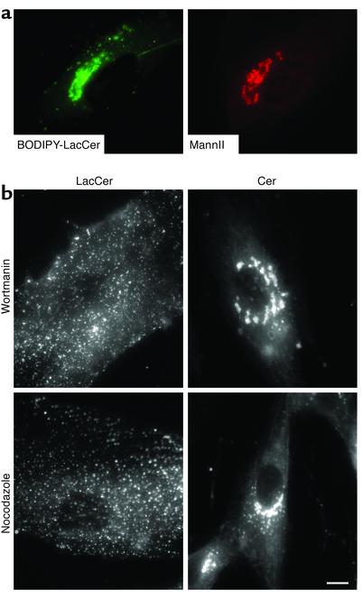
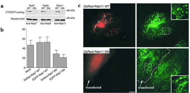
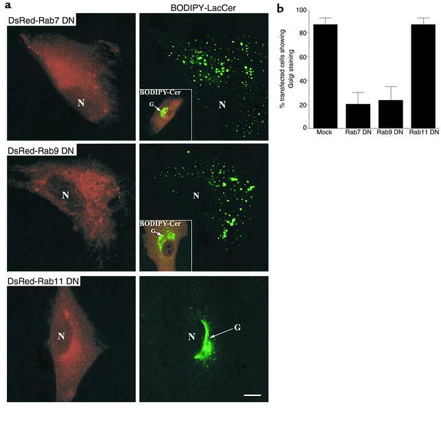
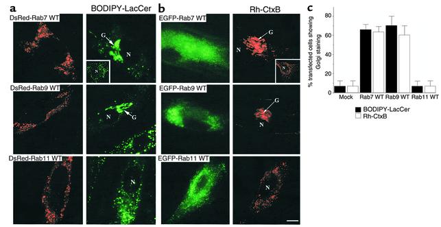
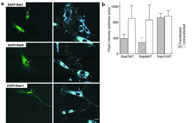
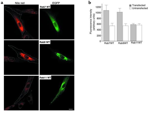
We recently showed that human skin fibroblasts internalize fluorescent analogues of the glycosphingolipids lactosylceramide and globoside almost exclusively by a clathrin-independent mechanism involving caveolae. In contrast, a sphingomyelin analogue is internalized approximately equally via clathrin-dependent and caveolar routes. Here, we further characterized the caveolar pathway for glycosphingolipids, showing that Golgi targeting of sphingolipids internalized via caveolae required microtubules and phosphoinositol 3-kinases and was inhibited in cells expressing dominant-negative Rab7 and Rab9 constructs. In addition, overexpression of wild-type Rab7 or Rab9 (but not Rab11) in Niemann-Pick type C (NP-C) lipid storage disease fibroblasts resulted in correction of lipid trafficking defects, including restoration of Golgi targeting of fluorescent lactosylceramide and endogenous GM(1) ganglioside, and a dramatic reduction in intracellular cholesterol stores. Our results demonstrate a role for Rab7 and Rab9 in the Golgi targeting of glycosphingolipids and suggest a new therapeutic approach for restoring normal lipid trafficking in NP-C cells.


Figures

Figure 1
Figure 1
Fluorescent LacCer is transported from the PM to the Golgi apparatus in normal HSFs and is dependent on microtubules and activity of PI3Ks. (a) Cells grown on etched grid coverslips were incubated with 2.5 μM BODIPY-LacCer for 30 minutes at 4°C in HMEM, washed, further incubated for 45 minutes at 37°C, and then back-exchanged at 10°C to remove any fluorescent lipid present at the PM. Live cell imaging (left panel) revealed a perinuclear distribution of the LacCer analogue. Cells were then fixed and immunolabeled with an antibody against the Golgi marker mannosidase II, and the same field was rephotographed for mannosidase II staining (MannII; right panel). (b) Cells were preincubated with wortmannin or nocodazole and subsequently pulse-labeled with BODIPY-LacCer as above. Golgi targeting of LacCer was inhibited in 80% (nocodazole), 50% (wortmannin), 70% (LY294002, not shown), or 5% (untreated) of the cells (n = 20 for each). No disruption of Golgi morphology was detected for the indicated inhibitors using BODIPY-Cer as a vital stain for this organelle. Bar, 10 μM.
Figure 2
Figure 2
Characterization of Rab–fluorescent protein constructs. (a) GTP overlay assay and immunoblot analysis of DsRed-Rab fusion proteins. HSFs were transfected with DsRed-Rab7, -Rab9, or -Rab11 (WT or DN) constructs, and 48 hours later cell lysates were prepared. Lysate samples were separated by SDS-PAGE and transferred to nitrocellulose membranes. The blots were incubated for 2 hours with [α-32P]GTP and exposed to x-ray film. The GTP blots were then stripped of GTP and were independently incubated with rabbit anti-Rab7, -Rab9, or -Rab11 monoclonal antibodies. Bands were visualized after secondary antibody incubations by chemiluminescence. (b) Effect of Rab7 WT and mutant protein overexpression on [125I]EGF degradation in HeLa cells. Transfected or mock-transfected cells were incubated with [125I]EGF for 10 minutes at 37°C, washed, and incubated for another 30 minutes at 37°C. TCA-soluble counts in cell lysates and extracellular medium were used to calculate EGF degradation (see Methods). Data are expressed as mean ± SD of three independent experiments. (c) Intracellular localization of Tfn in HeLa cells. Cells overexpressing DsRed-Rab11 WT or DN fusion proteins were incubated with FITC-Tfn for 45 minutes at 37°C. Samples were then washed, acid-stripped to remove fluorescent Tfn from the cell surface, and observed under the fluorescence microscope. In cells overexpressing WT DsRed-Rab11, Tfn colocalized with Rab11 fluorescent protein in punctate vesicular structures, while in cells overexpressing the DN construct, Tfn was found in tubular structures. These distributions are readily seen in the higher-magnification insets (right panels). Bar, 10 μM.
Figure 3
Figure 3
Golgi targeting of BODIPY-LacCer is blocked in HSFs expressing DN mutants of Rab7 or Rab9, but not Rab11. (a) Normal HSFs were transfected with plasmids encoding DsRed fusion proteins of DN Rab7, Rab9, or Rab11. Forty-eight hours later, the samples were pulse-labeled with BODIPY-LacCer, and Golgi targeting was assessed by fluorescence microscopy. Golgi morphology was not significantly altered by overexpression of the DN Rab proteins as assessed by BO-DIPY-Cer, seen in insets showing overlay images of DN Rab–expressing cells (red) and Cer (green). N, nucleus; G, Golgi apparatus. Bar, 10 μm. (b) Golgi targeting of BODIPY-LacCer in HSFs overexpressing DN DsRed-Rab7, -Rab9, or -Rab11 proteins. Transfected cells were identified by DsRed fluorescence and then scored as Golgi-positive (e.g., Figure 3a, bottom right panel) or Golgi-negative (e.g., Figure 3a, top panels). Bar labeled “Mock” represents cells transfected with empty vector. Values are expressed as mean of three independent experiments (n = 10) for each condition.
Figure 4
Figure 4
Overexpression of WT Rab7 or Rab9 (but not Rab11) restores BODIPY-LacCer and fluorescent CtxB targeting to the Golgi apparatus of NP-C fibroblasts. Cells were transfected with plasmids encoding (a) DsRed fusion proteins or (b) EGFP fusion proteins of WT Rab7, Rab9, or Rab11. Forty-eight hours later, the samples were pulse-labeled with (a) BODIPY-LacCer or (b) Rh-CtxB. Golgi targeting of LacCer and CtxB was assessed by fluorescence microscopy. (a and b) Top rows: Transfected cells overexpressing WT Rab7 showed intense Golgi staining of LacCer (green) and CtxB (red), with little punctate cytoplasmic staining. In contrast, untransfected cells showed punctate cytoplasmic staining with either LacCer or CtxB (see insets). Middle rows: Similar results to those obtained with WT Rab7 were seen in cells overexpressing WT Rab9. Bottom rows: Little or no Golgi targeting of fluorescent LacCer or CtxB was observed in transfected cells overexpressing WT Rab11. Bar, 10 μm. (c) Golgi-labeling by BODIPY-LacCer or CtxB in NP-C cells overexpressing WT Rab’s was quantified as described in Figure 3b legend.
Figure 5
Figure 5
Overexpression of WT Rab7 or Rab9 (but not Rab11) in NP-C cells reduced the accumulation of intracellular cholesterol. (a) Cells were transfected with plasmids expressing EGFP fusion proteins of WT Rab7, Rab9, or Rab11 and cultured in EMEM plus 10% FBS. Forty-eight hours later, the distribution of free cholesterol was examined by filipin staining (blue fluorescence). Transfected cells identified by green fluorescence are outlined in the filipin images. Bar, 25 μm. (b) Quantitation of filipin fluorescence in NP-C cells overexpressing WT Rab7, Rab9, or Rab11 proteins. Total filipin fluorescence of individual transfected or adjacent untransfected cells was calculated by image analysis. Values shown are mean ± SD from a typical experiment (n = 50). Similar results were obtained in each of three independent experiments.
Figure 6
Figure 6
Overexpression of WT Rab7 or Rab9 (but not Rab11) in NP-C cells increased the accumulation of neutral lipids. (a) Cells were transfected with plasmids expressing EGFP fusion proteins of WT Rab7, Rab9, or Rab11 as in Figure 5. Forty-eight hours later, samples were fixed and stained for the presence of cholesterol esters and other neutral lipids using Nile Red (34). Transfected cells identified by green fluorescence are outlined on the Nile Red images. Bar, 25 μm. (b) Quantitation of Nile Red fluorescence in NP-C cells overexpressing WT Rab7, Rab9, or Rab11 proteins. Total Nile Red fluorescence of individual transfected or adjacent untransfected cells was calculated by image analysis. Values are mean ± SD from a typical experiment (n = 30). Similar results were obtained in each of three independent experiments.

References

    1. Green DR. Apoptosissphingomyelin hydrolysis. The flip side. J Cell Biol. 2000;150:F5–F7. - PMC - PubMed
    1. Kolesnick RN, Goni FM, Alonso A. Compartmentalization of ceramide signaling: physical foundations and biological effects. J Cell Physiol. 2000;184:285–300. - PubMed
    1. van Meer G, Holthuis JC. Sphingolipid transport in eukaryotic cells. Biochim Biophys Acta. 2000; 1486:145–170. - PubMed
    1. Brown DA, London E. Structure and function of sphingolipid- and cholesterol-rich membrane rafts. J Biol Chem. 2000;275:17221–17224. - PubMed
    1. Hoekstra D, van IJzendoorn SC. Lipid trafficking and sorting: how cholesterol is filling gaps. Curr Opin Cell Biol. 2000;12:496–502. - PubMed

Publication types

MeSH terms