. 2002 Jun;109(12):1551-9.
doi: 10.1172/JCI15234.

Blockade of TGF-beta inhibits mammary tumor cell viability, migration, and metastases

Affiliations

Blockade of TGF-beta inhibits mammary tumor cell viability, migration, and metastases

Rebecca S Muraoka et al. J Clin Invest. 2002 Jun.

Abstract

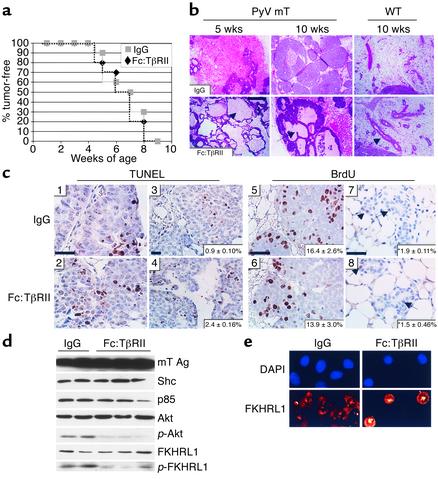
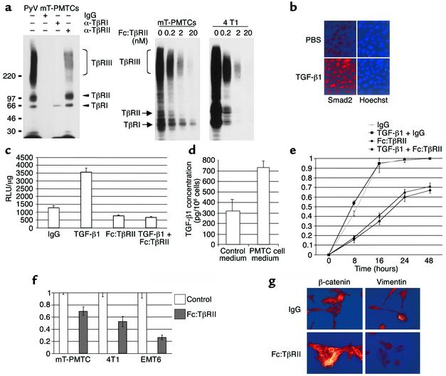
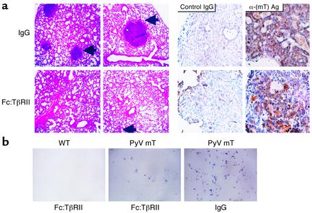
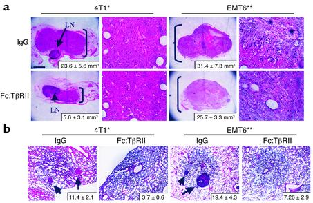
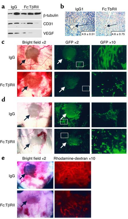
TGF-betas are potent inhibitors of epithelial cell proliferation. However, in established carcinomas, autocrine/paracrine TGF-beta interactions can enhance tumor cell viability and progression. Thus, we studied the effect of a soluble Fc:TGF-beta type II receptor fusion protein (Fc:TbetaRII) on transgenic and transplantable models of breast cancer metastases. Systemic administration of Fc:TbetaRII did not alter primary mammary tumor latency in MMTV-Polyomavirus middle T antigen transgenic mice. However, Fc:TbetaRII increased apoptosis in primary tumors, while reducing tumor cell motility, intravasation, and lung metastases. These effects correlated with inhibition of Akt activity and FKHRL1 phosphorylation. Fc:TbetaRII also inhibited metastases from transplanted 4T1 and EMT-6 mammary tumors in syngeneic BALB/c mice. Tumor microvessel density in a mouse dorsal skin window chamber was unaffected by Fc:TbetaRII. Therefore, blockade of TGF-beta signaling may reduce tumor cell viability and migratory potential and represents a testable therapeutic approach against metastatic carcinomas.


Figures

Figure 1
Figure 1
Tumor morphology, rate of apoptosis, and Akt signaling are altered by treatment with Fc:TβRII. (a) MMTV/PyV mT mice treated with Fc:TβRII or normal mouse IgG were examined twice weekly from 21 to 110 days of age. The initial observation of tumor onset is represented. (b) Histologic sections of tumors from 35- or 70-day-old transgenic or wild-type mice. Arrowheads indicate dilated ducts filled with secretory products. (c) TUNEL analysis (panels 1–4) of tumors harvested from 35- or 70-day-old mice treated with Fc:TβRII or IgG. n = 6 per condition. Quantification of percentage of apoptotic nuclei (bottom right corner of each panel) was calculated using the following equation: (number of TUNEL-positive nuclei in ×400 field) / (number of total nuclei in ×400 field). BrdU incorporation analysis (panels 5–8) of tumors harvested from 70-day-old MMTV/PyV mT or wild-type mice. Arrowheads in panels 7 and 8 indicate BrdU-positive nuclei. Quantification of percentage of BrdU-positive nuclei (bottom right corner of each panel) was calculated using the following equation: (number of BrdU-positive nuclei in ×400 field) / (number of total nuclei in ×400 field). *P = 0.15. Scale bars = 25 μm. (d) Tumor extracts harvested from 110-day-old transgenic mice were subjected to Western blot analysis using Ab’s against the mT Ag, Shc, p85, Akt, p-Akt, FKHRL1, and p-FKHRL. (e) PMTCs were incubated with or without 20 nM Fc:TβRII for 6 hours and stained with a FKHRL1 Ab followed by staining with Cy3-conjugated anti-rabbit Ab. Nuclei were counterstained with DAPI.
Figure 2
Figure 2
Fc:TβRII inhibits autocrine TGF-β signaling in PyV mT PMTCs. (a) Left panel: PMTCs were affinity labeled with 125I-TGF-β1, resolved directly (lane 1) or immunoprecipitated with the indicated TβR antibodies or IgG. Right panel: PMTCs and 4T1 cells were labeled with 125I-TGFβ1 in the presence of the indicated concentrations of Fc:TβRII. (b) Immunofluorescent detection of Smad2 in PMTCs treated with PBS or TGF-β1. (c) PMTCs transfected with a 3TP-Lux were treated with Fc:TβRII, TGF-β1, or both. The results are presented as relative light units (RLUs)/μg of total protein.The values represent the average of 3 experiments performed in duplicate, ± SE. (d) PMTCs or PMECs were cultured in serum-free medium for 24 hours. Conditioned medium was collected and analyzed for TGF-β1 using ELISA. Results are shown as an average of 5 experiments, analyzed in triplicate. (e) PMTCs were grown to confluency and wounded. The results are presented as the percentage of the total area of the original wound enclosed by cells and represent the average ± SD obtained from five experiments, analyzed in triplicate. (f) Transwell assays were performed using PMTCs, 4T1 cells, or EMT6 cells. The number of cells migrating to the lower side of the filter in controls was given the value of 1, such that migration of cells is represented as a fraction of control. Values shown are the average (± SE) of triplicate transwells in three experiments. (g) Immunocytochemical detection of β-catenin or vimentin in PMTCs cultured in the presence of the indicated factors. Representative photographs are shown.
Figure 3
Figure 3
Fc:TβRII decreases PyV mT mouse tumor cell intravasation and lung metastases. (a) Hematoxylin and eosin–stained lung sections (left panels) or mT Ag immunohistochemical analysis (right panels) of lungs harvested from 110-day-old PyV mT mice. Arrows point to lung metastases. Immunohistochemistry was performed using normal mouse IgG or pAb 762 against middle T antigen [α-(mT) Ag]. (b) Photomicrographs of hematoxylin-stained tumor cell colonies harvested from the blood of 110-day-old wild-type or PyV mT mice treated with normal mouse IgG or Fc:TβRII.
Figure 4
Figure 4
Fc:TβRII inhibits 4T1 and EMT6 primary tumor size and lung metastases. Tumor cells (0.5 × 10⁵ 4T1 or EMT6) were injected into the mammary fat pad of BALB/c mice. Mice were treated twice weekly with Fc:TβRII. (a) After 10 days, primary tumors were resected. Arrows indicate the lymph nodes of the number 4 mammary gland. The average tumor volume (n = 8 per condition), calculated by the formula vol-ume = width2 × length/2, is indicated in the lower right corner. *P = 0.011; **P = 0.05. (b) Lungs were collected at 8 weeks and examined for lung surface metastases. Arrows indicate metastases. The average number of metastases per mouse is shown in the bottom right corner of each panel (n = 8 per condition). *P = 0.026; **P = 0.034.
Figure 5
Figure 5
Fc:TβRII alters tumor cell density and invasiveness but not tumor angiogenesis. (a) Tumor lysates from PyV mT mice were subjected to Western blot analysis using Ab’s against β-tubulin, CD31, or VEGF. The VEGF isoform shown is mouse VEGF-A120, migrating at 14 kDa. (b) Immunohistochemical detection of CD31 in tumors harvested from 110-day-old MMTV/PyV mT mice treated with normal mouse IgG or Fc:TβRII. The average number of vessels per ×400 field is indicated in the lower-right corner of each panel. (n = 10 random tumor fields from three mice per condition.) Dorsal skin window analysis of in vivo tumor angiogenesis. One hundred 4T1-GFP cells were implanted into dorsal window chambers in BALB/c mice along with Fc:TβRII-, IgG-, or PBS-releasing pellets on day 0. Arrows indicate location of the pellet. On day 15, mice were administered rhodamine-conjugated dextran. Digital photomicrographs were taken on days 5 (c), 10 (d), and 15 (e) under green field (to visualize 4T1-GFP tumor cells), red field (to visualize the rhodamine-labeled vasculature), and bright field. Relative tumor density was calculated using the following equation: (average number of fluorescent pixels per sample in Fc:TβRII group)/(average number of fluorescent pixels per sample in IgG control group and PBS control group combined). The relative vascular density was determined using the following equation: (number of fluorescent pixels in test sample)/(number of fluorescent pixels in IgG and PBS controls).

Comment in

References

    1. Hanahan D, Weinberg RA. The hallmarks of cancer. Cell. 2000;100:57–70. - PubMed
    1. Massague J. TGF-beta signal transduction. Annu Rev Biochem. 1998;67:753–791. - PubMed
    1. Derynck R, Akhurst RJ, Balmain A. TGF-beta signaling in tumor suppression and cancer progression. Nat Genet. 2001;29:117–129. - PubMed
    1. Pierce DF, Jr, et al. Inhibition of mammary duct development but not alveolar outgrowth during pregnancy in transgenic mice expressing active TGF-beta 1. Genes Dev. 1993;7:2308–2317. - PubMed
    1. Jhappan C, et al. Targeting expression of a transforming growth factor beta 1 transgene to the pregnant mammary gland inhibits alveolar development and lactation. EMBO J. 1993;12:1835–1845. - PMC - PubMed

Publication types

MeSH terms

Substances