. 2002 Jun;109(12):1571-8.
doi: 10.1172/JCI14308.

Regulation of seizure spreading by neuroserpin and tissue-type plasminogen activator is plasminogen-independent

Affiliations

Regulation of seizure spreading by neuroserpin and tissue-type plasminogen activator is plasminogen-independent

Manuel Yepes et al. J Clin Invest. 2002 Jun.

Abstract

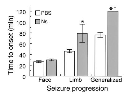
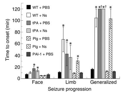
Tissue-type plasminogen activator (tPA) is a highly specific serine proteinase expressed in the CNS during events that require neuronal plasticity. In this study we demonstrate that endogenous tPA mediates the progression of kainic acid-induced (KA-induced) seizures by promoting the synchronization of neuronal activity required for seizure spreading, and that, unlike KA-induced cell death, this activity is plasminogen-independent. Specifically, seizure induction by KA injection into the amygdala induces tPA activity and cell death in both hippocampi, and unilateral treatment of rats with neuroserpin, a natural inhibitor of tPA in the brain, enhances neuronal survival in both hippocampi. Inhibition of tPA within the hippocampus by neuroserpin treatment does not prevent seizure onset but instead markedly delays the progression of seizure activity in both rats and wild-type mice. In tPA-deficient mice, seizure progression is significantly delayed, and neuroserpin treatment does not further delay seizure spreading. In contrast, plasminogen-deficient mice show a pattern of seizure spreading and a response to neuroserpin that is similar to that of wild-type animals. These findings indicate that tPA acts on a substrate other than plasminogen and that the effects of neuroserpin on seizure progression and neuronal cell survival are mediated through the inhibition of tPA.


Figures

Figure 1
Figure 1
Model of KA-induced seizures in the limbic system. A diagram of a coronal section through the rat hippocampi and amygdaloid nuclei is shown. KA was injected into the amygdaloid nuclei (a). Dashed arrows show the propagation of the abnormal electrical activity, originating in the amygdala, first to the ipsilateral (b) and then to the contralateral (c) hippocampus through the corpus callosum (d). Neuroserpin or PBS was injected into the hippocampus ipsilateral to the amygdala injected with KA (b).
Figure 2
Figure 2
Kinetic analysis of tPA activity and neuroserpin antigen in the limbic system during seizure. Seizures were induced by KA injection into the amygdala as described in Methods, and the animals were euthanized at 10, 30, and 60 minutes after injection. Cryostat sections were stained with DAPI to visualize nuclei and then analyzed either for in situ tPA activity by plasminogen-casein zymography with internally quenched fluorescent casein (red) or for neuroserpin antigen by immunofluorescence (green). The top panel of each part shows tPA activity, and the bottom panel of each part shows neuroserpin antigen (αNs). (ac) Ten minutes after KA injection; (df) 30 minutes after injection; (gi) 60 minutes after injection. (jl) Top panels: 60 minutes after KA injection, with anti-tPA antibodies (αtPA) in the overlay; bottom panels: normal rat brain with no KA injection. a, d, g, and j show the basolateral amygdala ipsilateral (Ipsi) to the injection site; b, e, h, and k show the ipsilateral hippocampus; c, f, i, and l show the contralateral (Contra) hippocampus. Original magnification, ×100.
Figure 3
Figure 3
Quantitative analysis of neuronal loss in the hippocampus 24 hours after seizure. KA and neuroserpin treatments were as described in Methods. The animals were euthanized 24 hours after KA injection, tissue sections were stained with H&E, and surviving neurons in the CA1-CA3 and dentate gyrus (DG) layers of the hippocampus were counted microscopically and compared with control sections from untreated animals. (a) The results of this analysis (n = 3 for each condition). *P < 0.05 relative to the same cell layer from PBS-treated animals. I, ipsilateral; C, contralateral. (b) An H&E stain of a normal rat hippocampus at the junction of the CA2 and CA3 layers. (c) The same region 24 hours after KA injection. Original magnification, ×200. The open arrows in c show examples of cells counted as dead, and the filled arrows show examples of cells counted as alive.
Figure 4
Figure 4
Clinical analysis of seizure behavior in rats. KA-treated rats were injected with neuroserpin (Ns) or PBS as described in Methods, and seizure behavior was recorded for 2 hours. Convulsive behavior was classified in three categories: 1, seizures affecting one side of the face or neck; 2, convulsion involving limbs only on one side; and 3, generalized bilateral seizure activity involving four limbs. For each condition, n = 6. *P < 0.05 relative to PBS-treated animals. None of the animals generalized during the experiment.
Figure 5
Figure 5
EEG recording in rats with KA-induced seizures. Monopolar electrodes were positioned into the ipsilateral or contralateral hippocampus as described in Methods. a and c are ipsilateral (Ipsi) and b and d are contralateral (Contra) to the KA injection. (a and b) PBS-treated animals; (c and d) neuroserpin-treated (Ns) animals. In a and b, the large black arrows indicate ictal activity (seizure), the large white arrows indicate interictal activity, and the dashed black arrows indicate the depression of the electrical activity immediately following seizure. In c, interictal activity is observed throughout the entire trace, and in d, no interictal activity is seen. Animals were monitored for 120 minutes (n = 3 for each condition).
Figure 6
Figure 6
Analysis of tPA activity in brain extracts. (a) SDS-PAGE zymography of brain extracts. Lane 1 is a mixture of human tPA and a rat kidney extract as a marker for rat urokinase plasminogen activator (uPA). Lane 2 is empty, and lanes 3–10 are hippocampal extracts 1 hour after KA injection. Lanes 3, 5, 7, and 9 are ipsilateral (I) and lanes 4, 6, 8, and 10 are contralateral (C) to the injection. Lanes 3 and 4 are from sham-operated animals that did not receive KA (Sham), and all other lanes show animals that received KA. Lanes 5 and 6 show PBS-treated animals, lanes 7 and 8 are from neuroserpin-treated animals (Ns), and lanes 9 and 10 are from animals that underwent corpus callosotomy (Call). (b) Quantitative analysis of PA activity from SDS-PAGE zymography of brain extracts 1 hour after KA injection. The results represent the average fold increase in tPA activity in the hippocampus ipsilateral (Ipsi) and contralateral (Contra) to the KA injection, relative to normal base-line tPA activity in sham-operated animals. PBS and Ns represent animals treated with PBS or neuroserpin, respectively (n = 5 for each condition), and Call represents brain extracts of animals that underwent corpus callosotomy (n = 2). *P < 0.05 relative to PBS-treated animals. AU, arbitrary units.
Figure 7
Figure 7
Clinical analysis of seizure behavior in mice. KA-treated mice were injected with neuroserpin (Ns) or PBS as described in Methods, and seizure behavior was recorded for 2 hours. Convulsive behavior was classified as in Figure 4. WT, wild-type mice; tPA, tPA–/– mice; Plg, Plg–/– mice; PAI-1, PAI-1–/– mice. *P < 0.05 relative to PBS-treated wild-type animals. None of the animals generalized at this time. For each condition, n = 5–10.

Comment in

References

    1. Carroll PM, Tsirka SE, Richards WG, Frohman MA, Strickland S. The mouse tissue-plasminogen activator gene 5′ flanking region directs appropriate expression in development and a seizure-enhanced response in the CNS. Development. 1994;120:3173–3183. - PubMed
    1. Qian Z, Gilbert ME, Colicos MA, Kandel ER, Kuhl D. Tissue-plasminogen activator is induced as an immediate-early gene during seizure, kindling and long-term potentiation. Nature. 1993;361:453–457. - PubMed
    1. Seeds NW, Williams BL, Bickford PC. Tissue-plasminogen activator induction in Purkinje neurons after cerebellar motor learning. Science. 1995;270:1992–1994. - PubMed
    1. Bugge TH, et al. Loss of fibrinogen rescues mice from the pleiotropic effects of plasminogen deficiency. Cell. 1996;87:709–719. - PubMed
    1. Tsirka SE, Gualandris A, Amaral DG, Strickland S. Excitotoxin-induced neuronal degeneration and seizure are mediated by tissue-plasminogen activator. Nature. 1995;377:340–344. - PubMed

Publication types

MeSH terms