. 2002 Jun;109(12):1625-33.
doi: 10.1172/JCI14873.

Estrogen alters thresholds for B cell apoptosis and activation

Affiliations

Estrogen alters thresholds for B cell apoptosis and activation

Christine M Grimaldi et al. J Clin Invest. 2002 Jun.

Abstract

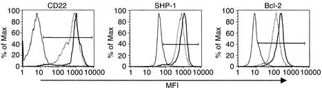
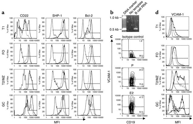
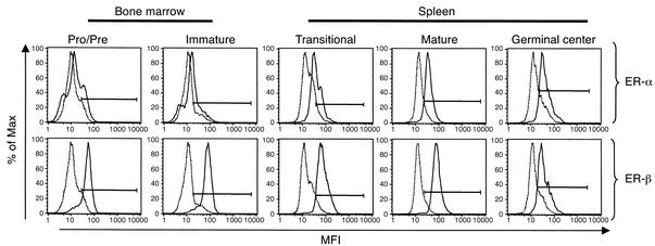
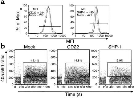
Estrogen is thought to contribute to the increased frequency of autoimmune disorders occurring in females, but a molecular basis for its effects on autoimmunity remains to be elucidated. We have shown previously that estrogen leads to the survival and activation of autoreactive cells in the naive repertoire. To identify the molecular pathways involved in B cell tolerance, we sought to identify genes that are differentially regulated by estrogen in mouse B cells. Several genes involved in B cell activation and survival, including cd22, shp-1, bcl-2, and vcam-1, were upregulated by estrogen in B cells. We found that overexpression of CD22 and SHP-1 in B cells decreased B cell receptor signaling. Estrogen receptors alpha and beta are expressed on B cells and are functional, since they can directly upregulate expression of CD22, SHP-1, and Bcl-2. Estrogen treatment protected isolated primary B cells from B cell receptor-mediated apoptosis. These results suggest that estrogen induces a genetic program that alters survival and activation of B cells in a B cell-autonomous fashion and thus skews the naive immune system toward autoreactivity.


Figures

Figure 1
Figure 1
Expression profile of E2-regulated molecules in B cells. Mean fluorescence intensity of CD22, SHP-1, and Bcl-2 was determined by flow cytometry. Representative histograms of CD19-gated events are shown. The marker depicts positive staining above the isotype-matched control. A broken line represents the profile for the P-treated mouse, a solid line represents the profile for the E2-treated mouse, and a thin solid line represents the isotype-matched control.
Figure 2
Figure 2
(a) Analysis of CD22, SHP-1, and Bcl-2 B cell subsets. Splenocytes from E2-treated (bold line) and P-treated (dashed line) mice (n = 5–10 per group) were analyzed by flow cytometry using Ab’s that recognize CD19 and B cell stage subset-specific markers. Representative histograms are shown, and the isotype-matched controls are represented by a thin solid line. Levels of CD21 or CD1 were used to distinguish between transitional T1 (T1; CD21lo/CD1lo), follicular (FO; CD21int/CD1int), and transitional T2 plus marginal zone (T2/MZ; CD21hi/CD1hi) B cell populations, and GL7 was used to identify germinal center (GC; GL7+) B cells. The marker depicts positive staining above the isotype-matched control. (b) Analysis of VCAM-1 in B cells. RT-PCR was performed on RNA extracted from sorted B220+, CD5 B cells, and the predicted 500-bp product for vcam-1 is shown. (c) A representative contour plot of CD19-gated events demonstrating expansion of VCAM-1+ B cells in the spleens is shown. (d) Representative histograms of stage-specific expression of VCAM-1 in E2-treated (bold line) and P-treated (dashed line) mice (n = 5) are shown, and values are presented in Table 1. The isotype-matched controls are represented by a thin solid line.
Figure 3
Figure 3
Detection of ERs in B cell subsets. Bone marrow cells and splenocytes were isolated, and cell suspensions from three mice were pooled together and stained with Ab’s to detect ER-α or ER-β plus Ab’s to detect developmental surface markers. Bone marrow pro/pre cells were identified as B220lo/sIgM and immature as B220lo/sIgM. Splenic transitional B cells were identified as B220lo/CD24hi, mature as B220int/CD24lo, and germinal center as CD19+/GL7+. The experiment was performed three times on a total of 13 mice, and representative histograms of stage-specific expression are shown. Specific staining is represented by a solid line, and the isotype-matched control is represented by a dashed line.
Figure 4
Figure 4
(a) Expression profiles of CD22- and SHP-1–transfected cells. Levels of CD22 and SHP-1 were determined by flow cytometry and histograms of mock (dashed line) and CD22- or SHP-1–transfected (solid line) cells are shown. CD22- and SHP-1–transfected cells display approximately a 20% increase in CD22 and SHP-1 levels. (b) Calcium influx in CD22- and SHP-1–transfected K46μ cells. The K46μ cell line was transfected with plasmids that express CD22 or SHP-1 and with one-tenth the concentration of a plasmid that expresses human CD4 to determine transfection efficiency. Forty-eight hours after transfection, cells were loaded with Indo-1 AM, and calcium influx was measured using 2.5–20 μg/ml of anti-IgM F(ab′)2. Representative dot plots depicting the percentage of cells responding to 10 μg/ml of anti-IgM F(ab′)2 are shown, and similar results were obtained for all doses examined. These experiments were repeated three times, and similar results were obtained.
Figure 5
Figure 5
IgM-mediated apoptosis in E2-treated splenic B cells. Levels of activated caspase 3 were measured by flow cytometry to identify (a) B220+/CD24hi (transitional) and (b) B220+/CD24lo (mature) cells undergoing apoptosis. Representative histograms of splenocytes from P- and E2-treated mice incubated with anti-IgM (solid line) or without anti-IgM (dashed line) are shown. For anti-IgM–treated splenocytes, 26,661 (P) and 12,075 (E2) transitional B cell–gated events and 139,416 (P) and 127,235 (E2) mature B cell–gated events were examined, respectively. For untreated splenocytes, 20,912 (P) and 10,437 (E2) transitional B cell–gated events and 57,600 (P) and 64,489 (E2) mature B cell–gated events were examined, respectively. The experiment was performed on B cells from five mice in each treatment group, and similar results were observed for all mice within a group. The percentage of transitional B cells from E2-treated mice undergoing apoptosis was significantly reduced (P < 0.02) compared with P-treated mice. There was no significant reduction in the percentage of mature B cells undergoing apoptosis. (c) Splenic B cells isolated from ovariectomized mice BALB/c mice (n = 5) were incubated with E2 or the E2 antagonist ICI 182,780 5 hours before the addition of anti-IgM. Representative histograms from one of three experiments are shown. For B cells incubated with anti-IgM plus ICI 182,780, 2,382 lymphocyte-gated events were examined; for B cells incubated with anti-IgM plus E2, 4,161 lymphocyte-gated events were examined; for B cells incubated with ICI 182,780 alone, 6,426 lymphocyte-gated events were examined; and for B cells incubated with E2 alone, 7109 lymphocyte-gated events were examined. Anti–IgM-induced apoptosis was reduced in cells pretreated with E2 (P < 0.05).

References

    1. Smithson G, Couse JF, Lubahn DB, Korach KS, Kincade PW. The role of estrogen receptors and androgen receptors in sex steroid regulation of B lymphopoiesis. J Immunol. 1998;161:27–34. - PubMed
    1. Butterworth M, McClellan B, Allansmith M. Influence of sex in immunoglobulin levels. Nature. 1967;214:1224–1225. - PubMed
    1. Schuurs AH, Verheul HA. Effects of gender and sex steroids on the immune response. J Steroid Biochem. 1990;35:157–172. - PubMed
    1. Grossman CJ, Roselle GA, Mendenhall CL. Sex steroid regulation of autoimmunity. J Steroid Biochem Mol Biol. 1991;40:649–659. - PubMed
    1. Ansar AS, Penhale WJ, Talal N. Sex hormones, immune responses, and autoimmune diseases. Mechanisms of sex hormone action. Am J Pathol. 1985;121:531–551. - PMC - PubMed

Publication types

MeSH terms