. 2002 Jul;11(7):1779-87.
doi: 10.1110/ps.0202702.

Structural basis for cyclodextrins' suppression of human growth hormone aggregation

Affiliations

Structural basis for cyclodextrins' suppression of human growth hormone aggregation

Daniel Erik Otzen et al. Protein Sci. 2002 Jul.

Abstract

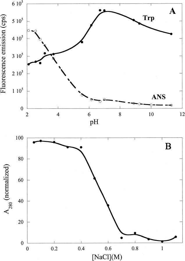
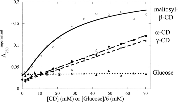
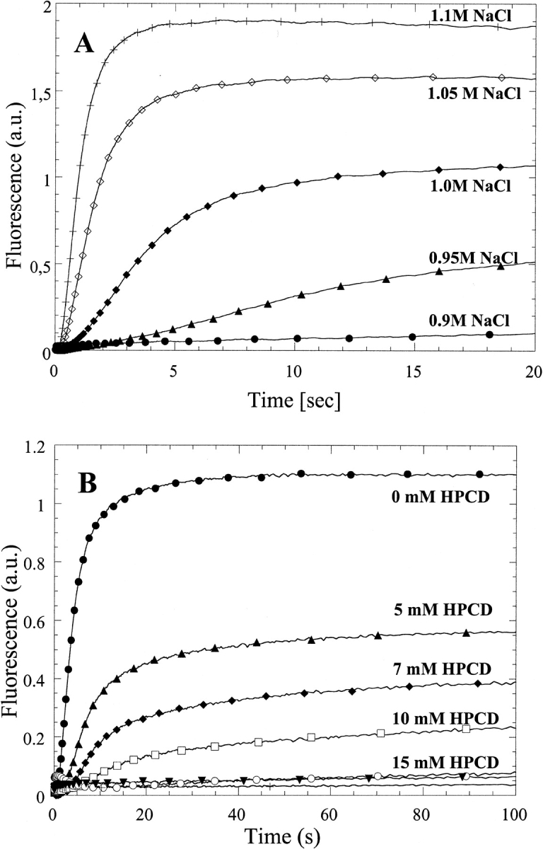
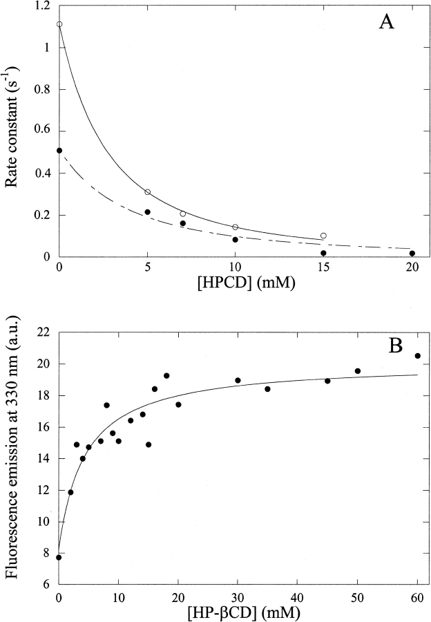
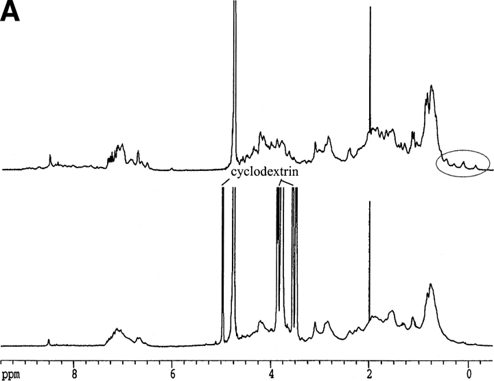
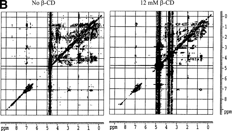
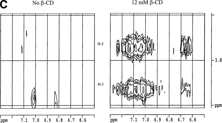
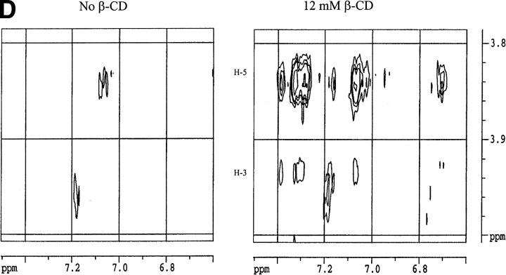
Many therapeutic proteins require storage at room temperature for extended periods of time. This can lead to aggregation and loss of function. Cyclodextrins (CDs) have been shown to function as aggregation suppressors for a wide range of proteins. Their potency is often ascribed to their affinity for aromatic amino acids, whose surface exposure would otherwise lead to protein association. However, no detailed structural studies are available. Here we investigate the interactions between human growth hormone (hGH) and different CDs at low pH. Although hGH aggregates readily at pH 2.5 in 1 M NaCl to form amorphous aggregates, the presence of 25 to 50 mM of various beta-CD derivatives is sufficient to completely avoid this. alpha- and gamma-CD are considerably less effective. Stopped-flow data on the aggregation reaction in the presence of beta-CD are analyzed according to a minimalist association model to yield an apparent hGH-beta-CD dissociation constant of approximately 6 mM. This value is very similar to that obtained by simple fluorescence-based titration of hGH with beta-CD. Nuclear magnetic resonance studies indicate that beta-CD leads to a more unfolded conformation of hGH at low pH and predominantly binds to the aromatic side-chains. This indicates that aromatic amino acids are important components of regions of residual structure that may form nuclei for aggregation.


Figures

Fig. 1.
Fig. 1.
(A) Binding of the fluorescent probe 1,8-anilino-naphthalene-sulfonic acid (ANS) to human growth hormone (hGH) as a function of pH in the absence of salt. The increase in ANS fluorescence at low pH, together with the accompanying decrease in Trp fluorescence, supports the accumulation of a partially folded A-state. The solid lines are smoothing curves intended to guide the eye. (B) NaCl- aggregation of hGH at pH 2.5. 22 μM hGH were incubated with 0 to 1.1 M NaCl at 25°C for 1 h and spun down at 14,000 rpm for 15 min, and the A280 of the supernatant was measured. The solid lines are smoothing curves.
Fig. 2.
Fig. 2.
Solubilization of hGH by various cyclodextrins (CDs) at pH 2.5 in 1 M NaCl. Glucose is included as a control to show that the reaction is not a simple solvent effect but requires the presence of a CD cavity. The concentration of glucose is sixfold higher than indicated on the X-axis to facilitate comparison with the CDs.
Fig. 3.
Fig. 3.
(A) Increase in apparent fluorescence on mixing hGH with increasing concentrations of NaCl at pH 2.5. (B) As in A but in 1.0 M NaCl and 0 to 25 mM hydroxy-propyl-β-CD (HPCD).
Fig. 4.
Fig. 4.
(A) The fast rate of aggregation of hGH (empty circles) and the reciprocal of the lag time (filled circles) at pH 2.5 in 1 M NaCl fitted to Equation 1. This yields an apparent dissociation constant of 5.6 ± 0.2 and 7.8 ± 1.0 mM, respectively. (B) Titration of 20 μM hGH with hydroxy-propyl-β-CD (HP-βCD) in 25 mM Gly (pH 2.5), followed by tryptophan fluorescence (excitation, 295 nm; emission, 330 nm). The data are fitted to a simple binding curve to yield an apparent dissociation constant of 4.6 ± 1.2 mM.
Fig. 5.
Fig. 5.
(A) One-dimensional nuclear magnetic resonance spectra of hGH at pD 1.7 alone (top spectrum) and with 12 mM β-CD (lower spectrum). Note the disppearance of the methyl peaks around 0 ppm. (B) Two-dimensional NOESY spectrum of hGH at pD 1.7 alone (left) and with 12 mM β-CD (right), highlighting the cross-peaks between β-CD and hGH. (C) Close-up of the region of interaction between hGH and β-CD from B (right) and the region in the absence of β-CD (left). (D) As C but at pD 6.8.
Fig. 5.
Fig. 5.
(A) One-dimensional nuclear magnetic resonance spectra of hGH at pD 1.7 alone (top spectrum) and with 12 mM β-CD (lower spectrum). Note the disppearance of the methyl peaks around 0 ppm. (B) Two-dimensional NOESY spectrum of hGH at pD 1.7 alone (left) and with 12 mM β-CD (right), highlighting the cross-peaks between β-CD and hGH. (C) Close-up of the region of interaction between hGH and β-CD from B (right) and the region in the absence of β-CD (left). (D) As C but at pD 6.8.

Similar articles

Cited by

References

    1. Abildgaard, F., Jorgensen, A.M.M., Led, J.J., Christensen, T., Jensen, E.B., Junker, F., and Dalboge, H. 1992. Characterization of tertiary interactions in a folded protein by NMR methods: Studies of pH-induced structural changes in human growth hormone. Biochemistry 31 8587–8596. - PubMed
    1. Banga, A.K. and Mitra, R. 1993. Minimization of shaking-induced formation of insoluble aggregates of insulin by cyclodextrins. J. Drug. Target. 1 341–345. - PubMed
    1. Bartels, C., Xia, T.-H., Billeter, M., Günter, P., and Wüthrich, K. 1995. The program XEASY for computer-supported NMR spectral of biological macromolecules. J. Biomol. NMR 5 1–10. - PubMed
    1. Branchu, S., Forbes, R.T., York, P., Petrén, S., Nyqvist, H., and Camber, O. 1999. Hydroxypropyl-β-cyclodextrin inhibits spray-drying–induced inactivation of β-galactosidase. J. Pharm. Sci. 88 905–911. - PubMed
    1. Brems, D.N. 1988. Solubility of different conformers of bovine growth hormone. Biochemistry 27 4541–4546.

Publication types

LinkOut - more resources