. 2002 Jun 25;99(13):9061-6.
doi: 10.1073/pnas.132092099. Epub 2002 Jun 17.

Reconstitution in yeast of the Arabidopsis SOS signaling pathway for Na+ homeostasis

Affiliations

Reconstitution in yeast of the Arabidopsis SOS signaling pathway for Na+ homeostasis

Francisco J Quintero et al. Proc Natl Acad Sci U S A. .

Abstract

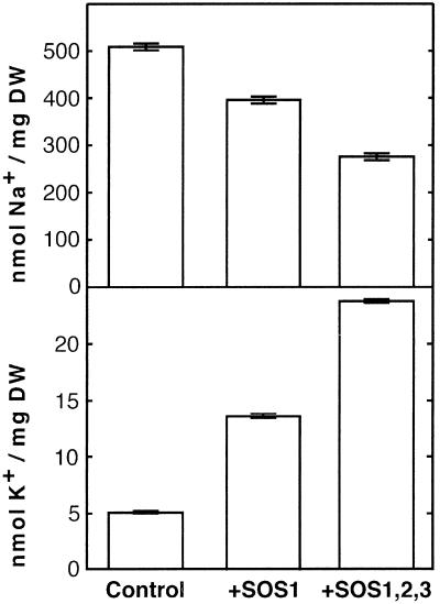
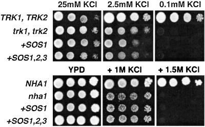
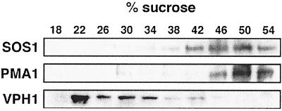
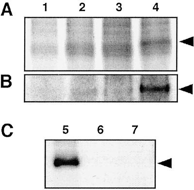
The Arabidopsis thaliana SOS1 protein is a putative Na+/H+ antiporter that functions in Na+ extrusion and is essential for the NaCl tolerance of plants. sos1 mutant plants share phenotypic similarities with mutants lacking the protein kinase SOS2 and the Ca2+ sensor SOS3. To investigate whether the three SOS proteins function in the same response pathway, we have reconstituted the SOS system in yeast cells. Expression of SOS1 improved the Na+ tolerance of yeast mutants lacking endogenous Na+ transporters. Coexpression of SOS2 and SOS3 dramatically increased SOS1-dependent Na+ tolerance, whereas SOS2 or SOS3 individually had no effect. The SOS2/SOS3 kinase complex promoted the phosphorylation of SOS1. A constitutively active form of SOS2 phosphorylated SOS1 in vitro independently of SOS3, but could not fully substitute for the SOS2/SOS3 kinase complex for activation of SOS1 in vivo. Further, we show that SOS3 recruits SOS2 to the plasma membrane. Although sos1 mutant plants display defective K+ uptake at low external concentrations, neither the unmodified nor the SOS2/SOS3-activated SOS1 protein showed K+ transport capacity in vivo, suggesting that the role of SOS1 on K+ uptake is indirect. Our results provide an example of functional reconstitution of a plant response pathway in a heterologous system and demonstrate that the SOS1 ion transporter, the SOS2 protein kinase, and its associated Ca2+ sensor SOS3 constitute a functional module. We propose a model in which SOS3 activates and directs SOS2 to the plasma membrane for the stimulatory phosphorylation of the Na+ transporter SOS1.


Figures

Figure 1
Figure 1
SOS2 and SOS3 increase the NaCl tolerance of yeast expressing the SOS1 ion transporter. AXT3K cells transformed with an empty vector (control) or expressing the indicated combination of Arabidopsis genes were grown overnight in liquid AP medium with 1 mM KCl. Five microliters of serial decimal dilutions were spotted onto plates of the same medium or supplemented with 70 mM NaCl. Plates were incubated at 28°C and photographed after 4 days. Plasmids used for expression of the SOS proteins were: pSOS1–1 for SOS1; pFL2T for SOS2; pFL3T for SOS3; pFL32T for SOS2 and SOS3; and pFL2ΔT for SOS2T/DΔ308.
Figure 2
Figure 2
Expression of SOS genes reduces Na+ accumulation and improves K+ status. Cells of strain AXT3K transformed with an empty vector (control), or expressing SOS1 (+SOS1) or transformed with plasmids for the simultaneous expression of SOS1, SOS2, and SOS3 (+SOS1,2,3) were grown in liquid AP medium with 1 mM KCl and 50 mM NaCl. When cultures reached OD550 ≈ 0.2, cells were collected by filtration, and their Na+ and K+ contents were determined. Units are nmol of ion per mg dry weight of cell samples. Data shown are the average and SE of ion contents of three independent cultures of each strain.
Figure 3
Figure 3
Activated SOS1 does not complement yeast mutants deficient in K+ transport. Cells of strains WΔ3 (trk1, trk2) and ANT3 (nha1) were transformed with SOS1 (+SOS1) or SOS1, SOS2, and SOS3 (+SOS1,2,3). Transformants of WΔ3 and ANT3 were grown overnight in AP medium supplemented with 25 mM KCl or YPD, respectively. Five microliters of serial decimal dilutions were spotted onto plates of the corresponding medium supplemented with the indicated KCl concentrations. Cells of strains W303 (TRK1, TRK2) and G19 (NHA1) were used as control as indicated. Plates were incubated at 28°C and photographed after 4 days.
Figure 4
Figure 4
Subcellular localization of SOS1. Total membrane extracts from AXT3K cells expressing SOS1 (plasmid pSOS1–2His) were fractionated on a 10-step sucrose gradient (18–54% wt/wt). Samples (25 μg protein) were resolved by SDS/PAGE and blotted. Western blots show the distribution of markers for the plasma membrane, PMA1, the vacuolar membrane, VPH1, and the SOS1 protein.
Figure 5
Figure 5
Interaction of SOS2 and SOS3 at the plasma membrane. Complementation of the cdc25-2 mutation through SOS2–SOS3 interaction. Cells of genotype cdc25–2 were transformed with the hSos:SOS2 translational fusion in plasmid pSRS2–1 (SOS2) or with a similar fusion using the SOS2T/DΔ308 variant in plasmid pSRS2–2 (SOS2T/DΔ308). These cells were subsequently transformed with plasmids containing the genes SOS1 and SOS3 as indicated for each lane. Plasmid pADNS–SosF (hSosF) was used as positive control. Transformants were grown on YPD plates at 23°C or 37°C. Only cells containing both the SOS3 gene and the hSos:SOS2 chimera grew at 37°C, as did control hSosF-transformed cells.
Figure 6
Figure 6
The SOS2/SOS3 kinase complex phosphorylates SOS1. Crude membranes extracts (10 μg protein) containing SOS1 (A) or purified SOS1:His6x protein (100 ng; B) were incubated with 20 μg of protein extracts from AXT3K cells carrying an empty plasmid (lane 1), or plasmid pFL2T for expression of SOS2 (lane 2), plasmid pFL3T for expression of SOS3 (lane 3), or plasmid pFL32T for expression of both SOS2 and SOS3 (lane 4). (C) Purified SOS1:His6x protein (100 ng) was incubated with 100 ng of purified GST:SOS2T/DΔ308 fusion protein (lane 5). Control samples without the GST:SOS2T/DΔ308 protein (lane 6) or the SOS1:His6x protein (lane 7) are also shown. All reactions were carried on kinase buffer for 30 min. Aliquots of kinase reactions were resolved by SDS/PAGE and exposed to x-ray films. Additional lanes with 100 ng of purified SOS1:His6x were included for protein staining and assessment of correspondence with phosphorylated bands (not shown). Arrows indicate the 127-kDa bands pertaining to phosphorylated SOS1.

References

    1. Hasegawa P M, Bressan R A, Zhu J-K, Bohnert H J. Annu Rev Plant Physiol Plant Mol Biol. 2000;51:463–499. - PubMed
    1. Blumwald E, Aharon G S, Apse M P. Biochim Biophys Acta. 2000;1465:140–151. - PubMed
    1. Shi H, Ishitani M, Kim C, Zhu J-K. Proc Natl Acad Sci USA. 2000;97:6896–6901. - PMC - PubMed
    1. Shi S, Quintero F J, Pardo J M, Zhu J-K. Plant Cell. 2002;14:465–477. - PMC - PubMed
    1. Zhu J-K. Plant Physiol. 2000;124:941–948. - PMC - PubMed

Publication types