

Objective comparison of quantitative imaging modalities without the use of a gold standard
- PMID: 12071615
- PMCID: PMC3150581
- DOI: 10.1109/TMI.2002.1009380
Objective comparison of quantitative imaging modalities without the use of a gold standard
Abstract
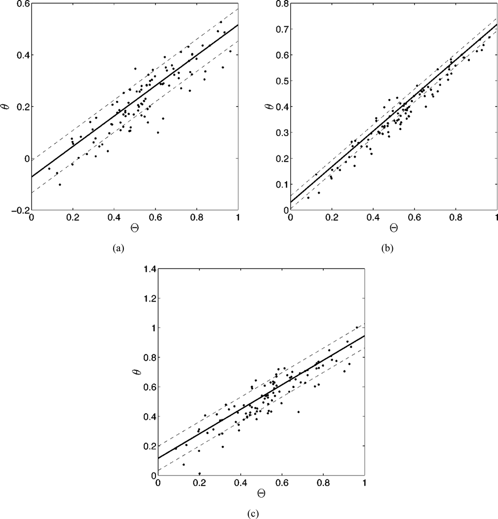
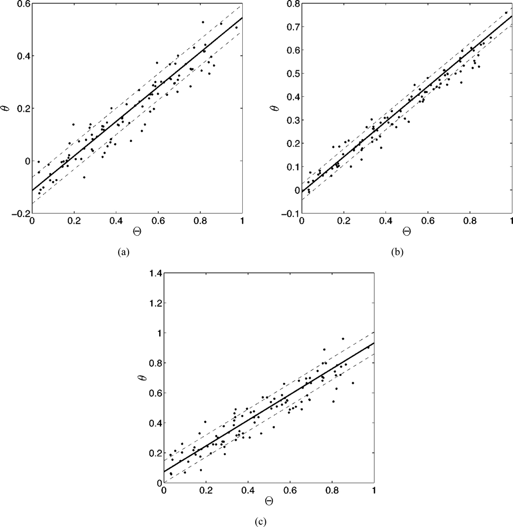
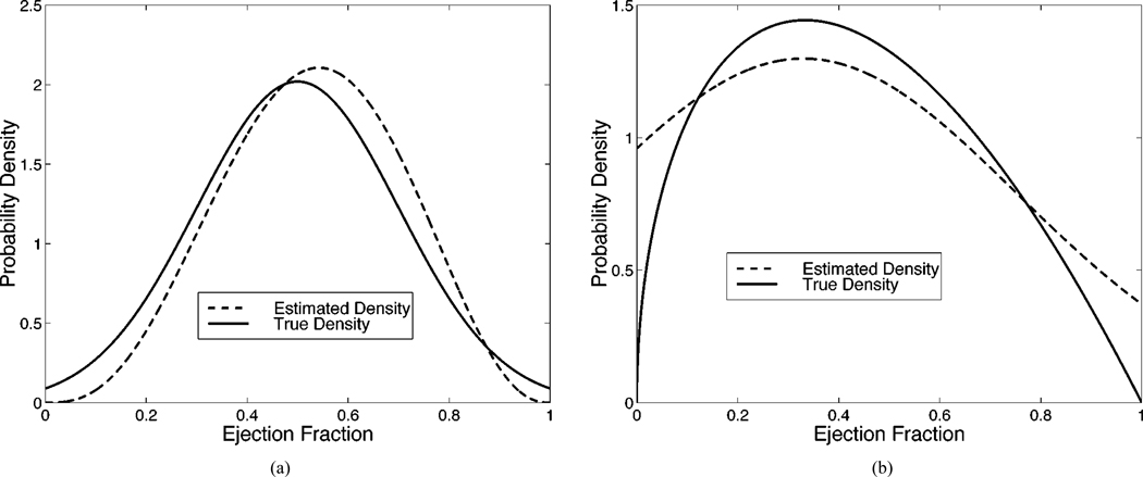
Imaging is often used for the purpose of estimating the value of some parameter of interest. For example, a cardiologist may measure the ejection fraction (EF) of the heart in order to know how much blood is being pumped out of the heart on each stroke. In clinical practice, however, it is difficult to evaluate an estimation method because the gold standard is not known, e.g., a cardiologist does not know the true EF of a patient. Thus, researchers have often evaluated an estimation method by plotting its results against the results of another (more accepted) estimation method, which amounts to using one set of estimates as the pseudogold standard. In this paper, we present a maximum-likelihood approach for evaluating and comparing different estimation methods without the use of a gold standard with specific emphasis on the problem of evaluating EF estimation methods. Results of numerous simulation studies will be presented and indicate that the method can precisely and accurately estimate the parameters of a regression line without a gold standard, i.e., without the x axis.
Figures
References
-
- Barrett HH. Objective assessment of image quality: Effects of quantum noise and object variability. J. Opt. Soc. Amer. A. 1990;vol. 7(no. 7):1266–1278. - PubMed
-
- Barrett HH, Denny JL, Wagner RF, Myers KJ. Objective assessment of image quality. II. Fisher information, Fourier crosstalk and figures of merit for task performance. J. Opt. Soc. Amer. A. 1995;vol. 12(no. 5):834–852. - PubMed
-
- Barrett HH, Abbey CK, Clarkson E. Objective assessment of image quality: III. ROC metrics, ideal observers and likelihood-generating functions. J. Opt. Soc. Amer. A. 1998;vol. 15(no. 6):1520–1535. - PubMed
-
- Achtert A, King MA, Dahlberg ST, Pretorius PH, LaCroix KH, Tsui BMW. An investigation of the estimation of ejection fractions and cardiac volumes by a quantitative gated spect software package in simulated spect images. J. Nucl. Cardiol. 1998 Mar-Aug;vol. 5:144–152. - PubMed
-
- Vanhove C, Franken PR. Left ventricular ejection fraction and volumes from gated blood pool tomography: Comparison between two automatic algorithms that work in three-dimensional space. J. Nucl. Cardiol. 2001 Jul-Aug;vol. 8:466–471. - PubMed
