. 2002 Jul;76(14):6944-56.
doi: 10.1128/jvi.76.14.6944-6956.2002.

Template requirements for de novo RNA synthesis by hepatitis C virus nonstructural protein 5B polymerase on the viral X RNA

Affiliations

Template requirements for de novo RNA synthesis by hepatitis C virus nonstructural protein 5B polymerase on the viral X RNA

Meehyein Kim et al. J Virol. 2002 Jul.

Abstract

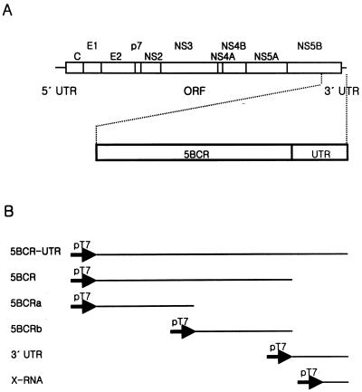
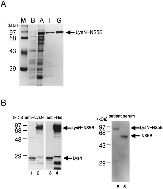
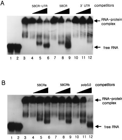
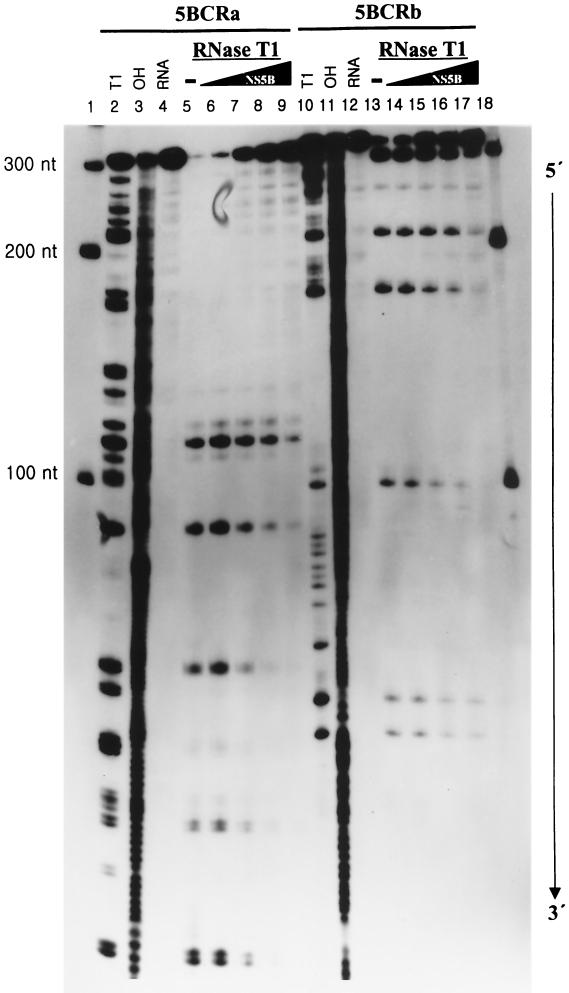
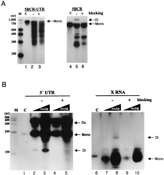
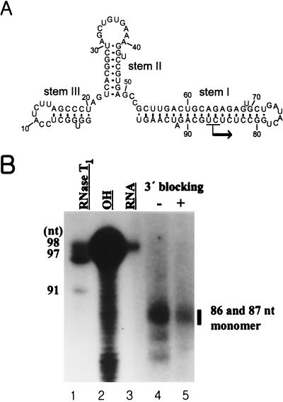
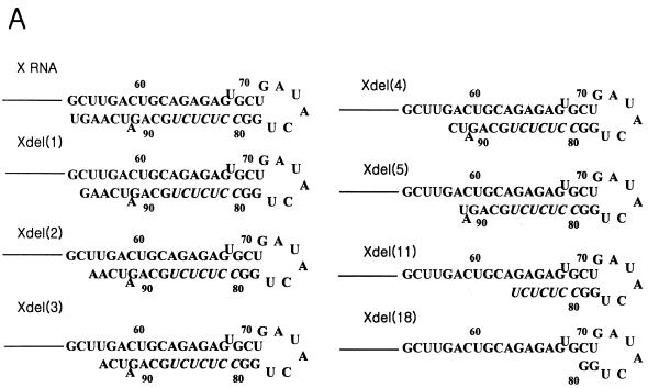
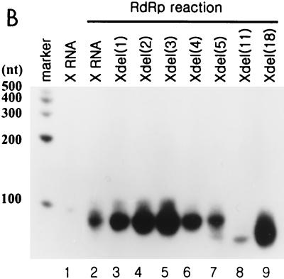
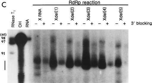
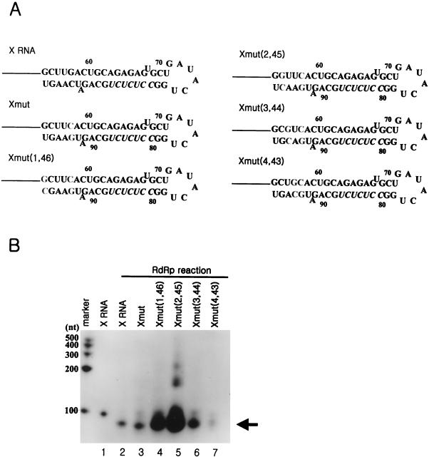
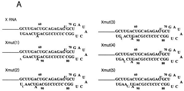
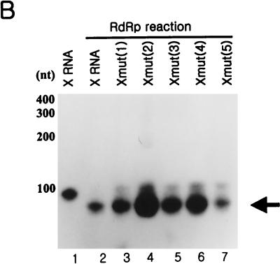
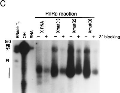
The hepatitis C virus (HCV)-encoded NS5B protein is an RNA-dependent RNA polymerase which plays a substantial role in viral replication. We expressed and purified the recombinant NS5B of an HCV genotype 3a from Esherichia coli, and we investigated its ability to bind to the viral RNA and its enzymatic activity. The results presented here demonstrate that NS5B interacts strongly with the coding region of positive-strand RNA, although not in a sequence-specific manner. It was also determined that more than two molecules of polymerase bound sequentially to this region with the direction 3' to 5'. Also, we attempted to determine the initiation site(s) of de novo synthesis by NS5B on X RNA, which contains the last 98 nucleotides of HCV positive-strand RNA. The initiation site(s) on X RNA was localized in the pyrimidine-rich region of stem I. However, when more than five of the nucleotides of stem I in X RNA were deleted from the 3' end, RNA synthesis initiated at another site of the specific ribonucleotide. Our study also showed that the efficiency of RNA synthesis, which was directed by X RNA, was maximized by the GC base pair at the penultimate position from the 3' end of the stem. These results will provide some clues to understanding the mechanism of HCV genomic RNA replication in terms of viral RNA-NS5B interaction and the initiation of de novo RNA synthesis.


Figures

FIG. 1.
FIG. 1.
HCV genome structure and RNA templates for in vitro assay. (A) The HCV genome organization is presented with 5′ and 3′ UTRs (solid lines) and the ORF (open box). The polyprotein cleavage products are indicated. A detailed view of the RNA template domains containing a part of NS5B and the 3′ UTR is drawn below the full viral genome. 5BCR, one-third of NS5B from the 3′ terminus (nt 8760 to 9405 of the genome); UTR, 3′ UTR (nt 9406 to 9588 of the genome). (B) Summary of template constructs used in this study. All of the RNAs were transcribed directly from the PCR products which were amplified from plasmid pSP3a using the specific primers. The solid arrows indicate the promoter for the T7 RNA polymerase.
FIG. 2.
FIG. 2.
Expression of recombinant HCV NS5B protein in E. coli. (A) The HCV LysN-NS5B protein eluted from an ion-exchange and a gel filtration column was subjected to sodium dodecyl sulfate-10% polyacrylamide gel electrophoresis and visualized by Coomassie blue staining. Lanes: M, size marker; B, cell extract before induction; A, cell extract after induction with IPTG; I, eluted fractions from an ion-exchange column; G, eluted fraction from a gel filtration column. (B) Western blot analysis of the purified proteins was performed with various antibodies. Lanes 1 and 3, LysN protein from a metal affinity column detected with rabbit anti-LysN antibody and anti-His antibody, respectively; lanes 2 and 4, partially purified LysN-NS5B protein from an ion-exchange column with rabbit anti-LysN antibody and anti-His antibody, respectively; lane 5, finally purified LysN-NS5B protein expressed in E. coli with patient serum; lane 6, purified NS5B protein generated from a baculovirus expression system in the insect cells as a positive control. Both of the proteins in lanes 5 and 6 were purified from a gel filtration column.
FIG. 3.
FIG. 3.
Gel mobility shift assay of HCV NS5B protein and the viral RNA. The binding assay was conducted with HCV NS5B protein and the 5BCR RNA as a probe by increasing the amounts of competitor RNAs. (A) The competitor RNAs were the unlabeled 5BCR-UTR, 5BCR, and 3′ UTR RNAs. (B) The competitor RNAs were the 5BCRa, 5BCRb, and homopolymer of poly(U) RNAs. Lanes 1, labeled 5BCR RNA as a marker for free RNA; lanes 2, probe RNA with LysN; lanes 3, probe RNA with NS5B. In the mixture of NS5B and the probe RNA, the unlabeled competitor RNAs of 1- (lanes 4, 7, and 10), 5- (lanes 5, 8, and 11), and 10-fold (lanes 6, 9, and 12) molar excess to the probe RNA were added. The products of the RNA-protein complex and free RNA are indicated at the right of each gel.
FIG. 4.
FIG. 4.
Footprinting analysis of 5BCRa and 5BCRb RNAs with NS5B. The 3′-end-labeled RNAs (20 fmol) were incubated at 30°C for 15 min in the absence (lanes 4 and 12) or in the presence (lanes 5 and 13) of RNase T1. The probe RNA was incubated with the NS5B protein by increasing its amount (lanes 6 and 14, 50 fmol; lanes 7 and 15, 100 fmol; lanes 8 and 16, 200 fmol; lanes 9 and 17, 400 fmol of protein) and then digested by RNase T1. The RNA fragments were then resolved on an 8 M urea-5% polyacrylamide gel. Lanes 1 and 18, RNA size markers; lanes 2 and 10, RNase T1-digested products under denaturing conditions; lanes 3 and 11, fragments generated by partial alkaline hydrolysis. The numbers on the left side of the gel indicate the length of the RNA marker.
FIG. 5.
FIG. 5.
Analysis of RdRp reaction products from viral RNAs as templates. (A) The RdRp reaction on 5BCR-UTR RNA and 5BCR RNA. The template of each product is indicated at the top of the panel. The reaction was performed with 0.1 pmol of RNA template and 0.1 to 0.3 pmol of NS5B by adding a recombinant NTP mixture of 500 μM GTP, 250 μM ATP, 250 μM UTP, 100 μM CTP, and 0.5 μCi of CTP in the 15-μl reaction volume. Lane M, size marker; lanes 1 and 4, internally labeled input templates; lanes 2 and 5, RdRp products without (−) blocking the 3′-OH group of the template RNA; lanes 3 and 6, RdRp products with (+) blocking of the 3′-OH group of the template RNAs. Mono, the monomer RNA; Di, the dimer RNA. (B) RdRp reaction on 3′ UTR RNA and X RNA. The template of each product is indicated at the top of the gel. The RdRp reaction was performed with 12 pmol of RNA template by increasing the amounts of NS5B from 1 (lanes 2, 4, 7, and 9) to 3 pmol (lanes 3, 5, 8, and 10). Other contents for RNA synthesis are given in Materials and Methods. Lane M, size marker; lanes 1 and 6, internally labeled input template; lanes 2, 3, 7, and 8, labeled products without (−) blocking the 3′-OH group of the template RNAs; lanes 4, 5, 9, and 10, labeled products with (+) blocking of the 3′-OH group of the template RNAs. Mono, the monomer RNA; Di, the dimer RNA; Du, the duplex product by hybridization of the monomer and the template RNAs.
FIG. 6.
FIG. 6.
Determination of the initiation sites of RNA synthesis on X RNA by HCV NS5B. (A) Secondary structure of X RNA. The major initiation sites of RNA synthesis are indicated by the arrow on stem I. (B) An 8 M urea-5% polyacrylamide gel electrophoresis autoradiogram showing the RNA product of HCV NS5B using the X RNA template. The bar indicates the major RdRp products on the gel. The numbers on the left give the length of the 5′-labeled X RNA partially digested by RNase T1 (lane 1). The numbers on the right give the lengths of the RNA synthesis products of NS5B. Lane 2, partial alkaline hydrolysis of the 5′-labeled X RNA; lane 3, template X RNA labeled internally with [α-32P]CTP as a marker; lanes 4 and 5, RdRp product on the X RNA template before (−) and after (+) the 3′ end was blocked.
FIG. 7.
FIG. 7.
RdRp reaction on the deletion mutants of X RNA by HCV NS5B. (A) Sequences of stem I of the deletion mutant RNAs. The nucleotides in italics indicate the pyrimidine-rich region in stem I. (B) RdRp reaction products of wild-type X RNA and its deletion mutants on a 7 M urea-8% polyacrylamide gel. The length of the marker RNA is indicated at the left of the gel. Lane 1, template X RNA with internally incorporated 32P; lanes 2 to 9, RdRp products of X, Xdel (1), Xdel (2), Xdel (3), Xdel (4), Xdel (5), Xdel (11), and Xdel (18) RNAs, respectively. (C) An 8 M urea-5% polyacrylamide gel electrophoresis autoradiogram showing the RNA products of HCV NS5B using X RNA and the deletion mutant RNA templates before (−) or after (+) treatment with NaIO4. RNase T1, 5′-labeled X RNA partially digested by RNase T1; OH, partial alkaline hydrolysis of 5′-labeled X RNA; RNA, template X RNA labeled internally with [α-32P]CTP as a marker. The numbers on the left give the length of the RNA, and the bar at the left of the gel indicates the pyrimidine-rich region of X RNA.
FIG. 7.
FIG. 7.
RdRp reaction on the deletion mutants of X RNA by HCV NS5B. (A) Sequences of stem I of the deletion mutant RNAs. The nucleotides in italics indicate the pyrimidine-rich region in stem I. (B) RdRp reaction products of wild-type X RNA and its deletion mutants on a 7 M urea-8% polyacrylamide gel. The length of the marker RNA is indicated at the left of the gel. Lane 1, template X RNA with internally incorporated 32P; lanes 2 to 9, RdRp products of X, Xdel (1), Xdel (2), Xdel (3), Xdel (4), Xdel (5), Xdel (11), and Xdel (18) RNAs, respectively. (C) An 8 M urea-5% polyacrylamide gel electrophoresis autoradiogram showing the RNA products of HCV NS5B using X RNA and the deletion mutant RNA templates before (−) or after (+) treatment with NaIO4. RNase T1, 5′-labeled X RNA partially digested by RNase T1; OH, partial alkaline hydrolysis of 5′-labeled X RNA; RNA, template X RNA labeled internally with [α-32P]CTP as a marker. The numbers on the left give the length of the RNA, and the bar at the left of the gel indicates the pyrimidine-rich region of X RNA.
FIG. 8.
FIG. 8.
effect of the position of the GC base pair at stem I on the RdRp reaction. (A) Sequence identities of the mutant RNAs which were generated by shifting the GC base pair from the end to the inside of stem I. The nucleotides in boldface indicate the mutated sequences. Xmut RNA, as a control RNA template, was produced by reversing the GC base pair at the fifth position of the terminus of stem I. Xmut (1,46) RNA was generated from Xmut RNA by regenerating the GC base pair at the end of stem I; Xmut (2,45) RNA was generated from Xmut RNA by regenerating the GC base pair at the second position from the end of stem I; Xmut (3,44) RNA was generated from Xmut RNA by regenerating the GC base pair at the third position from the end of stem I; Xmut (4,43) RNA was generated from Xmut RNA by regenerating the GC base pair at the fourth position from the end of stem I. (B) RdRp reaction products using the mutant RNA templates which contain the GC base pair at different sites of stem I. The length of the marker RNA is indicated at the left of the gel. The monomer RNA produced by de novo synthesis is indicated by the arrow. Lane 1, internally labeled X RNA template; lane 2, RdRp product directed by X RNA; lanes 3 to 7, RdRp products of Xmut, Xmut (1,46), Xmut (2,45), Xmut (3,44), and Xmut (4,43) RNAs, respectively.
FIG. 9.
FIG. 9.
RdRp reaction of the point mutants of X RNA with HCV NS5B. (A) Nucleotide sequences in stem I of the mutant RNAs. The characters in boldface represent the elements for rendering the bulge structure. The nucleotides in italics indicate the pyrimidine-rich region in stem I. (B) Products of RdRp reaction on wild-type X RNA and its point mutants on a 7 M urea-8% polyacrylamide gel. The length of the marker RNA is indicated at the left of the gel. Lane 1, template X RNA internally labeled with 32P; lanes 2 to 7, RdRp products of X, Xmut (1), Xmut (2), Xmut (3), Xmut (4), and Xmut (5) RNAs, respectively. (C) An 8 M urea-5% polyacrylamide sequencing gel electrophoresis autoradiogram showing the de novo synthesis products of X RNA and the point mutant RNAs before (−) or after (+) treatment with NaIO4. The template RNAs for directing RNA synthesis by NS5B are indicated at the top of the gel. RNase T1, 5′-labeled X RNA partially digested by RNase T1; OH, partial alkaline hydrolysis of the 5′-labeled X RNA; RNA, template X RNA labeled internally with [α-32P]CTP as a marker. The numbers on the left give the length of the RNA, and the bar at the left of the gel indicates the pyrimidine-rich region of X RNA.
FIG. 9.
FIG. 9.
RdRp reaction of the point mutants of X RNA with HCV NS5B. (A) Nucleotide sequences in stem I of the mutant RNAs. The characters in boldface represent the elements for rendering the bulge structure. The nucleotides in italics indicate the pyrimidine-rich region in stem I. (B) Products of RdRp reaction on wild-type X RNA and its point mutants on a 7 M urea-8% polyacrylamide gel. The length of the marker RNA is indicated at the left of the gel. Lane 1, template X RNA internally labeled with 32P; lanes 2 to 7, RdRp products of X, Xmut (1), Xmut (2), Xmut (3), Xmut (4), and Xmut (5) RNAs, respectively. (C) An 8 M urea-5% polyacrylamide sequencing gel electrophoresis autoradiogram showing the de novo synthesis products of X RNA and the point mutant RNAs before (−) or after (+) treatment with NaIO4. The template RNAs for directing RNA synthesis by NS5B are indicated at the top of the gel. RNase T1, 5′-labeled X RNA partially digested by RNase T1; OH, partial alkaline hydrolysis of the 5′-labeled X RNA; RNA, template X RNA labeled internally with [α-32P]CTP as a marker. The numbers on the left give the length of the RNA, and the bar at the left of the gel indicates the pyrimidine-rich region of X RNA.

Similar articles

Cited by

References

    1. Arnold, J. J., and C. E. Cameron. 1999. Poliovirus RNA-dependent RNA polymerase (3Dpol) is sufficient for template switching in vitro. J. Biol. Chem. 274:2706-2716. - PubMed
    1. Banerjee, R., and A. Dasgupta. 2001. Specific interaction of hepatitis C virus protease/helicase NS3 with the 3′-terminal sequences of viral positive- and negative-strand RNA. J. Virol. 75:1708-1721. - PMC - PubMed
    1. Behrens, S. E., L. Tomei, and R. De Francesco. 1996. Identification and properties of the RNA-dependent RNA polymerase of hepatitis C virus. EMBO J. 15:12-22. - PMC - PubMed
    1. Cheng, J. C., M. F. Chang, and S. C. Chang. 1999. Specific interaction between the hepatitis C virus NS5B RNA polymerase and the 3′ end of the viral RNA. J. Virol. 73:7044-7049. - PMC - PubMed
    1. Choo, Q. L., G. Kuo, A. J. Weiner, L. R. Overby, D. W. Bradley, and M. Houghton. 1989. Isolation of a cDNA clone derived from a blood-borne non-A, non-B viral hepatitis genome. Science 244:359-362. - PubMed

MeSH terms

LinkOut - more resources