. 2002 Jul;76(14):7228-38.
doi: 10.1128/jvi.76.14.7228-7238.2002.

Characterization of varicella-zoster virus gene 21 and 29 proteins in infected cells

Affiliations

Characterization of varicella-zoster virus gene 21 and 29 proteins in infected cells

Randall J Cohrs et al. J Virol. 2002 Jul.

Abstract

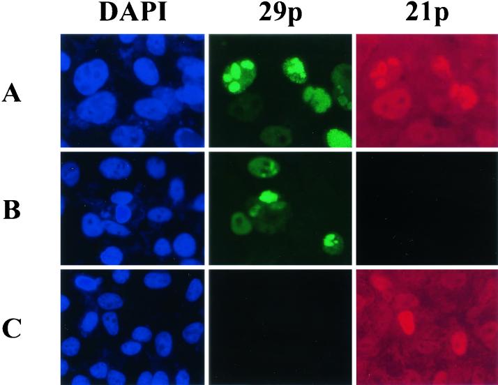
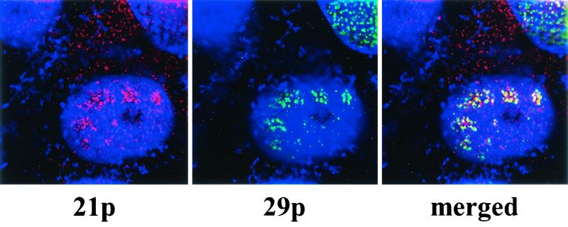
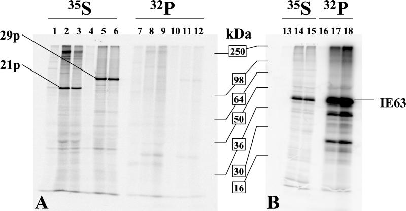
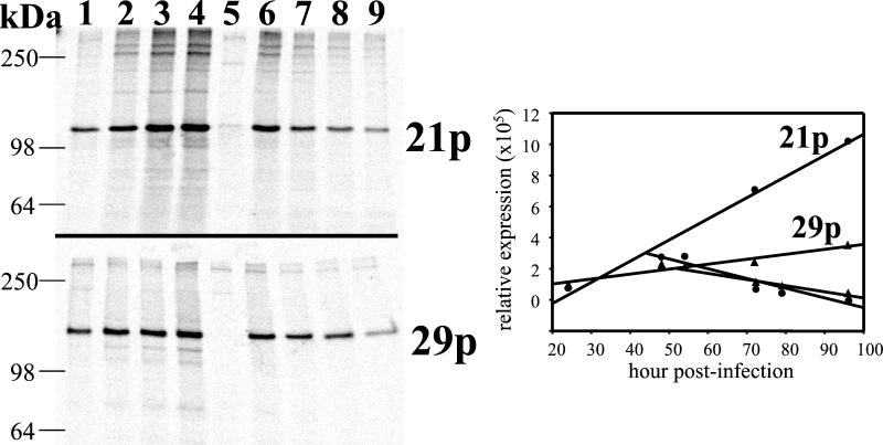
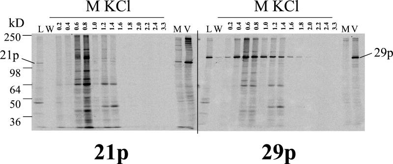
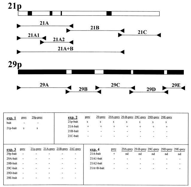
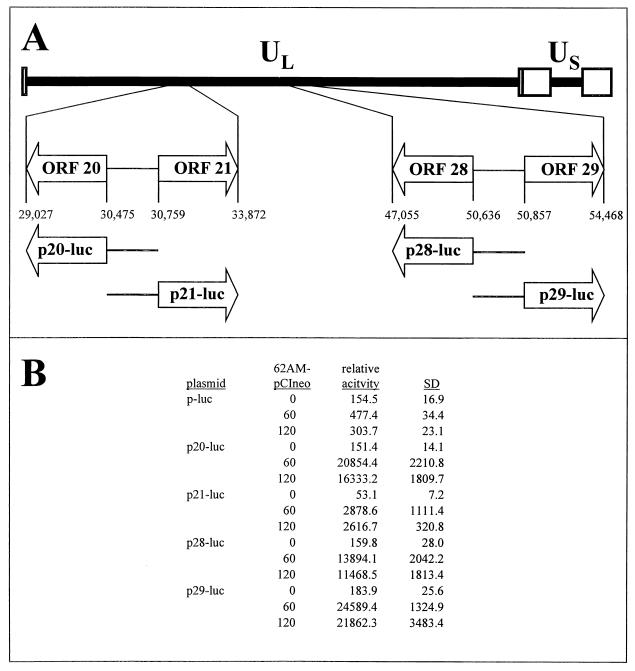
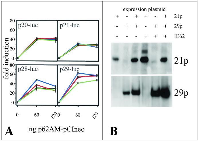
Varicella-zoster virus (VZV) transcription is limited in latently infected human ganglia. Note that much of the transcriptional capacity of the virus genome has not been analyzed in detail; to date, only VZV genes mapping to open reading frames (ORFs) 4, 21, 29, 62, and 63 have been detected. ORF 62 encodes the major immediate-early virus transcription transactivator IE62, ORF 29 encodes the major virus DNA binding protein, and ORF 21 encodes a protein associated with the developing virus nucleocapsid. We analyzed the cellular location of proteins encoded by ORF 21 (21p) and ORF 29 (29p), their phosphorylation state during productive infection, and their ability form a protein-protein complex. The locations of both 21p and 29p within infected cells mimic those of their herpes simplex virus type 1 (HSV-1) homologues (UL37 and ICP8); however, unlike these homologues, 21p is not phosphorylated and neither 21p nor 29p exhibits a protein-protein interaction. Transient transfection assays to determine the effect of 21p and 29p on transcription from VZV gene 20, 21, 28, and 29 promoters revealed no significant activation of transcription by 21p or 29p from any of the VZV gene promoters tested, and 21p did not significantly modulate the ability of IE62 to activate gene transcription. A modest increase in IE62-induced activation of gene 28 and 29 promoters was seen in the presence of 29p; however, IE62-induced activation of gene 28 and 29 promoters was reduced in the presence of 21p. A Saccharomyces cerevisiae two-hybrid analysis of 21p indicated that the protein can activate transcription when tethered within a responsive promoter. Together, the data reveal that while VZV gene 21 and HSV-1 UL37 share homology at the nucleic acid level, these proteins differ functionally.


Figures

FIG. 1.
FIG. 1.
Intracellular locations of VZV gene 21 protein (21p) and VZV gene 29 protein (29p) viewed by transmission UV microscopy. VZV-infected Vero cells were stained for cellular DNA in the nucleus (blue) with DAPI, FITC-tagged 29p (green), and Cy3-tagged 21p (red) as described in Materials and Methods. 29p and 21p are seen in punctuate intranuclear regions (row A), with 21p also present in the cytoplasm. Staining specificity was confirmed by the omission of anti-21p antibody (row B) and anti-29p antibody (row C).
FIG. 2.
FIG. 2.
Intracellular location of VZV 21p and 29p viewed by confocal UV microscopy. VZV-infected Vero cells were stained for DNA with DAPI (blue), Cy3-tagged 21p (red), and FITC-tagged 29p (green) as described in Materials and Methods. Note 21p in the nucleus and cytoplasm of infected cells (left panel) and 29p exclusively in the nucleus (middle panel). The right panel shows intranuclear colocalization of both 21p and 29p (yellow).
FIG. 3.
FIG. 3.
21p and 29p are not significantly phosphorylated in vivo. At 48 h postinfection, VZV-infected (lanes 2, 3, 5, 6, 8, 9, 11, 12, 14, 15, 17, and 18) and uninfected (lanes 1, 4, 7, 10, 13, and 16) MeWo cells were labeled with [35S]methionine (lanes 1 to 6 and 13 to 15) or 32P (lanes 7 to 12 and 16 to 18) for 6 h (lanes 2, 5, 8, 11, 14, and 17) or 22 h (lanes 1, 3, 4, 6, 7, 9, 10, 12, 13, 15, 16, and 18). At the end of the labeling period, protein extracts were prepared and immunoprecipitated as described in Materials and Methods with antisera directed against 21p (lanes 1 to 3 and 7 to 9), 29p (lanes 4 to 6 and 10 to 12), or IE63 (lanes 13 to 18). 21p and 29p were resolved by SDS-8% PAGE (panel A) and IE63 by SDS-12% PAGE (panel B). Phosphorimaging of immunoprecipitated 21p, 29p, and IE63 (40.5 kDa) demonstrated that all three proteins were synthesized during both the 6-h and 22-h labeling periods, but only IE63 was significantly phosphorylated. Two smaller phosphorylated proteins (lanes 17 and 18) are likely to be IE63 degradation products.
FIG. 4.
FIG. 4.
Pulse-chase labeling of 21p and 29p during productive VZV infection. At 24 h (lane 1), 48 h (lane 2), 72 h (lane 3), and 96 h (lane 4) postinfection, cells were labeled with [35S]methionine for 3 h and processed for immunoprecipitation of 21p or 29p. Lane 5 shows the results for uninfected cells labeled for 3 h beginning at 48 h after mock infection. The results depicted in lanes 6 to 9 indicate the stability of 21p and 29p. At 48 h postinfection, cultures were labeled with [35S]methionine for 3 h and chased with excess unlabeled methionine for an additional 6 h (lane 6), 24 h (lane 7), 31 h (lane 8), or 48 h (lane 9). Protein extracts were prepared and processed by immunoprecipitation to detect 21p or 29p. Both 21p and 29p were detected in all infected cells, but 21p and 29p did not coimmunoprecipitate in any sample. Quantitation of the immunoprecipitated proteins showed that the steady-state rates of synthesis and degradation for both 21p and 29p remained constant during the 96-h labeling period.
FIG. 5.
FIG. 5.
DNA column elution profile of 21p and 29p. High-salt nuclear protein extract was prepared from [35S]methionine-labeled VZV-infected cells and loaded (lanes L) onto a DNA-agarose gel. After extensive washing (lanes W), proteins were eluted with increasing molar concentrations of KCl. The presence of 21p and 29p in each dialyzed and concentrated fraction was determined by immunoprecipitation, SDS-PAGE, and phosphorimaging. [35S]methionine-labeled protein extracts from mock (lanes M)- and VZV (lanes V)-infected cells controlled for the location of 21p and 29p. The maximum yield of 29p was obtained in the 0.6 M KCl fraction, whereas 21p was not detected in any fraction.
FIG. 6.
FIG. 6.
Yeast two-hybrid analysis of 21p and 29p. Full-length ORFs 21p and 29p were inserted into pHybLex/Zeo (bait) or pYESTrp2 (prey) yeast expression plasmids. Oligonucleotide primers (between arrowheads) as listed in Table 1 were used to amplify regions within 21p and 29p, generating subclones of 21p and 29p for insertion into the bait and prey plasmids. The PCR primers were selected to be outside regions of homology between 21p and 29p and their analogous HSV-1 proteins (black boxes). The relative positions and nomenclatures for the subcloned fragments of 21p and 29p are listed below the respective proteins. The results of four independent yeast bait-prey transformation experiments (exp.) are shown. −, no growth on YC-WHUK+zeo plates and no β-Gal induction; +, growth on YC-WHUK+zeo plates and β-Gal induction; s, slow growth on YC-WHUK+zeo plates and slight β-Gal induction; nd, not done. The results indicate that full-length 21p contains slight intrinsic transcription activation capabilities, most likely located within the 21pA fragment of the protein. No 21p/29p interaction was demonstrated.
FIG. 7.
FIG. 7.
Cloning of gene 20, 21, 28, and 29 promoter reporter plasmids and their activation by IE62. (A) The 125-kbp VZV genome is composed of unique long (UL) and unique short (US) DNA segments, each bound by inverted repeat DNA sequences. The opposing ORFs 20 and 21, along with ORFs 28 and 29, are located within the UL segment (open boxes). The ORF 20/21 and ORF 28/29 intergenic regions were inserted into a promoterless luciferase reporter vector such that each region governed transcription of the luciferase reporter gene. (B) The resulting plasmids (p20-luc, p21-luc, p28-luc, and p29-luc) were used in transient transfection assays to determine intrinsic promoter activity and their response to induction by IE62. Data from at least duplicate experiments are presented as relative activity levels (luciferase activity normalized to β-Gal activity) to indicate the magnitude of promoter activity. Compared to the promoterless plasmid (p-luc), gene 20, 21, 28, and 29 promoters have no intrinsic activity but are induced by IE62. SD, standard deviation.
FIG. 8.
FIG. 8.
Effect of 21p and 29p on the transcriptional activation of VZV gene 20, 21, 28, and 29 promoters by IE62. In panel A, transient transfection experiments were performed to determine the effect of the presence of 21p (red lines), 29p (blue lines), or both proteins (green lines) on promoter activation (severalfold induction measured as relative activity levels normalized to 0 ng of 62AM-pCIneo) by IE62 (black lines). The presence of 21p has no effect on the induction of the promoter of gene 20, 21, 28, or 29 by IE62. The presence of 29p enhances activation of gene 28 and 29 promoters at low concentrations of IE62. The 29p-associated modulation of IE62 induction is diminished by 21p (green lines). In panel B, Western blot analysis of transfected cells showed that 21p and 29p were translated in the presence and in the absence of IE62.

References

    1. Abendroth, A., and A. Arvin. 1999. Varicella-zoster virus immune evasion. Immunol. Rev. 168:143-156. - PubMed
    1. Albright, A. G., and F. J. Jenkins. 1993. The herpes simplex virus UL37 protein is phosphorylated in infected cells. J. Virol. 67:4842-4847. - PMC - PubMed
    1. Annunziato, P. W., O. Lungu, and C. Panagiotidis. 2001. Varicella zoster virus in human and rat tissue specimens. Arch. Virol. Suppl. 17:135-142. - PubMed
    1. Ben Ze'ev, A., R. Abulafia, and S. Bratosin. 1983. Herpes simplex virus and protein transport are associated with the cytoskeletal framework and the nuclear matrix in infected BSC-1 cells. Virology 129:501-507. - PubMed
    1. Blom, N., S. Gammeltoft, and S. Brunak. 1999. Sequence and structure-based prediction of eukaryotic protein phosphorylation sites. J. Mol. Biol. 294:1351-1362. - PubMed

Publication types

MeSH terms

LinkOut - more resources