. 2002 Jul;76(14):7276-92.
doi: 10.1128/jvi.76.14.7276-7292.2002.

Identification of the neoplastically transformed cells in Marek's disease herpesvirus-induced lymphomas: recognition by the monoclonal antibody AV37

Affiliations

Identification of the neoplastically transformed cells in Marek's disease herpesvirus-induced lymphomas: recognition by the monoclonal antibody AV37

Shane C Burgess et al. J Virol. 2002 Jul.

Abstract

Understanding the interactions between herpesviruses and their host cells and also the interactions between neoplastically transformed cells and the host immune system is fundamental to understanding the mechanisms of herpesvirus oncology. However, this has been difficult as no animal models of herpesvirus-induced oncogenesis in the natural host exist in which neoplastically transformed cells are also definitively identified and may be studied in vivo. Marek's disease (MD) herpesvirus (MDV) of poultry, although a recognized natural oncogenic virus causing T-cell lymphomas, is no exception. In this work, we identify for the first time the neoplastically transformed cells in MD as the CD4(+) major histocompatibility complex (MHC) class I(hi), MHC class II(hi), interleukin-2 receptor alpha-chain-positive, CD28(lo/-), phosphoprotein 38-negative (pp38(-)), glycoprotein B-negative (gB(-)), alphabeta T-cell-receptor-positive (TCR(+)) cells which uniquely overexpress a novel host-encoded extracellular antigen that is also expressed by MDV-transformed cell lines and recognized by the monoclonal antibody (MAb) AV37. Normal uninfected leukocytes and MD lymphoma cells were isolated directly ex vivo and examined by flow cytometry with MAb recognizing AV37, known leukocyte antigens, and MDV antigens pp38 and gB. CD28 mRNA was examined by PCR. Cell cycle distribution and in vitro survival were compared for each lymphoma cell population. We demonstrate for the first time that the antigen recognized by AV37 is expressed at very low levels by small minorities of uninfected leukocytes, whereas particular MD lymphoma cells uniquely express extremely high levels of the AV37 antigen; the AV37(hi) MD lymphoma cells fulfill the accepted criteria for neoplastic transformation in vivo (protection from cell death despite hyperproliferation, presence in all MD lymphomas, and not supportive of MDV production); the lymphoma environment is essential for AV37(+) MD lymphoma cell survival; pp38 is an antigen expressed during MDV-productive infection and is not expressed by neoplastically transformed cells in vivo; AV37(+) MD lymphoma cells have the putative immune evasion mechanism of CD28 down-regulation; AV37(hi) peripheral blood leukocytes appear early after MDV infection in both MD-resistant and -susceptible chickens; and analysis of TCR variable beta chain gene family expression suggests that MD lymphomas have polyclonal origins. Identification of the neoplastically transformed cells in MD facilitates a detailed understanding of MD pathogenesis and also improves the utility of MD as a general model for herpesvirus oncology.


Figures

FIG. 1.
FIG. 1.
The AV37 antigen is expressed at high levels by MD lymphoma cells but not by uninfected leukocytes or leukocytes after mitogen activation. (A) The MAb AV37 recognizes a host-encoded antigen expressed at low levels (AV37lo) by a small proportion of uninfected PBL, splenocytes, thymocytes, bursacytes, BM cells, and E14 chorioallantoic blood leukocytes. The flow cytometry histogram overlay presented is of PBL stained with an isotype-matched control MAb (arrow 1) and AV37 (arrow 2). (B) AV37lo PBL (arrow 1) can be magnetically sorted from AV37 uninfected PBL (arrow 2). Results from magnetically sorted E14 chorioallantoic leukocytes are similar. (C) The antigen recognized by AV37 is grossly overexpressed (AV37hi) by proportions of MD lymphoma cells. The flow cytometry histogram overlay presented is of a nerve lymphoma (Table 2, bird 1) immunostained with an isotype-matched control MAb (arrow 1) or AV37 (arrow 2). (D) Mitogen and CM activation of cultured uninfected splenocytes results only in small increases in both the proportion of cells recognized by AV37 and the level of AV37 antigen expression; the high level of AV37 antigen expression observed on lymphoma cells does not occur. All data on the figure are from male splenocytes, as described in Materials and Methods. Arrow 1, 0 h, 1.31% AV37lo cells; arrow 2, 24 h, 20% CM, 2.34% AV37lo cells; arrow 3, 72 h, 20% CM, 6.13% AV37lo cells. (E) The increases in AV37lo cell percentages in the cultures are not in proportion to the percentage increases in lymphoblastoid cells in the cultures. The percentage of lymphoblasts in the male splenocytes (as described above) after 24 h of culture with 20% CM; FSC versus SSC dot plots of untreated (i) and mitogen-treated (ii) cells. (F) The percentages of AV37lo cells in the cultures are proportional to the amount of CM used (72 h of culture, male splenocytes are shown as described above).
FIG. 2.
FIG. 2.
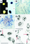
(A) Photomicrographs of uninfected PBL and E14 leukocytes recognized by the MAb AV37 which are lymphocytoid (i and ii) or thrombocytoid (iii and iv). Figures shown are of dark-field (i and iii) and light-field (ii and iv) microscopy of uninfected adult PBL after magnetic sorting with AV37. Magnification, ×920. Bar = 15 μm. (B) Immunohistochemistry of the heart from an MDV-infected chicken (Table 2, bird 5). MD lymphomas form multiple foci, each focus is composed of mainly CD4+ T cells (red) associated with fewer CVI-ChnL-68.1+ (myeloid) cells (blue). Magnification, ×23. Bar = 100 μm. The myeloid cells mainly surround, although a few are present within, each developing MD lymphoma focus. (C) Single representative focus from the heart section (shown in panel B). CD4+ cells are red, and CVI-ChnL-68.1+ cells are blue. Magnification, ×368. Bar = 100 μm. (D to F) Electron microscopy of classical changes of PCD after ovarian MD lymphoma cells were incubated with 10−4 M DEX. Magnification, ×11,960. Bars = 15 μm. (D) Normal lymphoma cells are shown after 0 h of incubation. (E) After 34 h of incubation, there is a loss of cytoplasmic processes, rounding, increased cytoplasmic density, and nuclear condensation (arrow) typical of the early stages of PCD. (F) After 48 h of incubation, apoptotic bodies with condensed nuclear remnants (arrows) typical of the late stages of PCD are present.
FIG. 3.
FIG. 3.
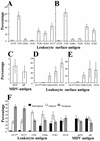
Histograms of flow cytometry data of antigen expression by cell populations in MD lymphomas (Table 2, birds 1 to 14) (means ± standard errors of the mean). (A) The majority of MD lymphoma cells are CD4+ and either TCR2+ or TCR3+; few are AV20+, CD8α+, or TCR1+. (B) The lymphoma cells recognized by AV37, regardless of expression level of the antigen (AV37+), have phenotype distributions similar to those of lymphoma cells as a whole. (C) Few lymphoma cells are (MDV antigen) pp38+ or gB+. Large proportions of AV20+, CD8α+, or TCR1+ lymphoma cells, but few CD4+ lymphoma cells and no cells expressing high levels of the AV37 antigen (AV37hi), are pp38+ (D) or gB+ (E). (F) DNA ploidy of MD lymphoma cell populations (mean ± standard error of the mean). Few AV37hi and AV37+ cells are subdiploid, relatively few are diploid, and most were tetraploid. Few CD4+ cells are subdiploid, and most are diploid. Many AV20+, CD8α+, and TCR1+ cells are subdiploid, and most pp38+ and gB+ cells are subdiploid.
FIG. 4.
FIG. 4.
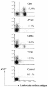
Flow cytometry dot plots of AV37 antigen expression relative to leukocyte surface antigen expression by ovarian MD lymphoma cells (Table 2, bird 2). High-level AV37 antigen expression is exhibited by CD4+ MD lymphoma cells but not by AV20+, CD8α+, or TCR1+ MD lymphoma cells. Leukocyte antigens are indicated in the top right of each dot plot. The percentages shown are percentages of cells coexpressing the leukocyte antigen and the AV37 antigen.
FIG. 5.
FIG. 5.
Confluent but not subconfluent CHCC-OU2 cell cultures are supportive of MDV production after MDV infection. On the flow cytometry dot plots, the x axes show antigen expression and the y axes show MDV infection status and cell culture conditions. Uninfected confluent and subconfluent CHCC-OU2 cultures were identical and are represented by the same dot plots.
FIG. 6.
FIG. 6.
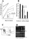
Protection from cell death in vitro and immune accessory molecule expression by MD lymphoma cell subpopulations. (A) All MD lymphoma cell subpopulations die at a similar rate when cultured in vitro. Proportions of ovarian MD lymphoma cell populations with subdiploid DNA incubated either with or without 10−4 M DEX are shown. DEX increased the proportion of all cell types with subdiploid DNA. At 48 h, AV20+, CD8α+, and TCR1+ cells could not be identified in cultures. (B) MHC class I and II, CD28, and IL-2Rα expression levels measured by flow cytometry (means ± standard errors of the means) of any cells recognized by the AV37 MAb (AV37+) and AV37 lymphoma cell populations. AV37+ MD lymphoma cells expressed more MHC class I and class II and IL-2Rα than did AV37 cells. AV37+ lymphoma cells had decreased CD28 expression. (C) Flow cytometry dot plots showing decreased (nerve lymphoma) or virtually absent (ovarian lymphoma) CD28 expression by AV37+ MD lymphoma cells. (D) RT-PCR for CD28 and GAPDH mRNA expression. PCR primers were designed for exon sequences of both the chicken CD28 gene (the cDNA template gives a 407-bp product) and the chicken GAPDH gene (the cDNA template gives a 295-bp product). CD28 mRNA was absent from MDCC HP9 (lane 1), MDCC HP18 (lane 2), and MDCC HP89 (lane 4) but present in MDCC IAH8 (lane 3) (white arrow) and ConA-stimulated T cells (lane 5). GAPDH mRNA was present in all samples except negative-control bovine PBL (lane 6). A 100-bp (arrows) DNA ladder is shown to the right of lane 6.
FIG. 7.
FIG. 7.
AV37hi leukocytosis was present in line N and 61 (MD resistant) and line 72 and 15I (MD susceptible) chickens after MDV infection. Density gradient purified PBL taken at 3, 7, 21, 28, 42, and 56 dpi from 10 infected and 6 uninfected chickens of each genotype were incubated with MAbs AV37 or RSVG-MAb29. Cells (106 per sample) were collected by flow cytometry and analyzed (41). The electronic analysis gates were set to analyze each sample for the presence of AV37hi cells (see Materials and Methods). Corrected percentages of AV37hi cells were calculated by subtracting the (negligible) percentages of RSVG-MAb29+ events from the percentages of AV37hi events. AV37hi PBL are not observed in any uninfected chickens of any line.

References

    1. Abujoub, A. A., and P. M. Coussens. 1997. Evidence that Marek's disease virus exists in a latent state in a sustainable fibroblast cell line. Virology 229:309-321. - PubMed
    1. Appleman, L. J., D. Tzachanis, T. Grader-Beck, A. van Puijenbroek, and V. A. Boussiotis. 2001. Helper T cell anergy: from biochemistry to cancer pathophysiology and therapeutics. J. Mol. Med. 78:673-683. - PubMed
    1. Arstila, T. P., P. Toivanen, O. Vainio, and O. Lassila. 1994. Gamma-delta and alpha-beta T-cells are equally susceptible to apoptosis. Scand. J. Immunol. 40:209-215. - PubMed
    1. Bacon, L. D. 1987. Influence of the major histocompatibility complex on disease resistance and productivity. Poult. Sci. 66:802-811. - PubMed
    1. Baigent, S. J., R. C. Bethell, and J. W. McCauley. 1999. Genetic analysis reveals that both haemagglutinin and neuraminidase determine the sensitivity of naturally occurring avian influenza viruses to zanamivir in vitro. Virology 263:323-338. - PubMed

MeSH terms