. 2002 Jul;76(14):7293-305.
doi: 10.1128/jvi.76.14.7293-7305.2002.

The mannose-dependent epitope for neutralizing antibody 2G12 on human immunodeficiency virus type 1 glycoprotein gp120

Affiliations

The mannose-dependent epitope for neutralizing antibody 2G12 on human immunodeficiency virus type 1 glycoprotein gp120

Rogier W Sanders et al. J Virol. 2002 Jul.

Abstract

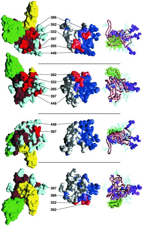
We have analyzed the unique epitope for the broadly neutralizing human monoclonal antibody (MAb) 2G12 on the gp120 surface glycoprotein of human immunodeficiency virus type 1 (HIV-1). Sequence analysis, focusing on the conservation of relevant residues across multiple HIV-1 isolates, refined the epitope that was defined previously by substitutional mutagenesis (A. Trkola, M. Purtscher, T. Muster, C. Ballaun, A. Buchacher, N. Sullivan, K. Srinivasan, J. Sodroski, J. P. Moore, and H. Katinger, J. Virol. 70:1100-1108, 1996). In a biochemical study, we digested recombinant gp120 with various glycosidase enzymes of known specificities and showed that the 2G12 epitope is lost when gp120 is treated with mannosidases. Computational analyses were used to position the epitope in the context of the virion-associated envelope glycoprotein complex, to determine the variability of the surrounding surface, and to calculate the surface accessibility of possible glycan- and polypeptide-epitope components. Together, these analyses suggest that the 2G12 epitope is centered on the high-mannose and/or hybrid glycans of residues 295, 332, and 392, with peripheral glycans from 386 and 448 on either flank. The epitope is mannose dependent and composed primarily of carbohydrate, with probably no direct involvement of the gp120 polypeptide surface. It resides on a face orthogonal to the CD4 binding face, on a surface proximal to, but distinct from, that implicated in coreceptor binding. Its conservation amidst an otherwise highly variable gp120 surface suggests a functional role for the 2G12 binding site, perhaps related to the mannose-dependent attachment of HIV-1 to DC-SIGN or related lectins that facilitate virus entry into susceptible target cells.


Figures

FIG. 1.
FIG. 1.
(A) Carbohydrates on gp120 and their contribution to the 2G12 epitope as identified by substitutional mutagenesis. The schematic of CHO-expressed IIIB and JR-FL gp120 indicates N-linked glycosylation sites. The composition of the carbohydrates in IIIB gp120 was experimentally determined (35); the carbohydrate designations in the schematic of JR-FL gp120 are based on that study, assuming that glycans are processed similarly on the two Env glycoproteins. Two sites in JR-FL gp120 that are not present in IIIB gp120 are designated as being of unknown carbohydrate composition. Arrows indicate sites that were shown to be important for 2G12 binding in a substitutional mutagenesis study. Note that the sites at 392 and 397 were only deleted in combination (69). (B) Specificities of glycosidases. The schematic is derived from reference . The cleavage sites of some of the endo- and exoglycosidases used in this study are indicated on the structures of the three classes of carbohydrates: complex, hybrid, and high mannose. Note that the number and characteristics of the sugar residues in the outer branches can vary. Complex glycans generally have two to four outer branches and may have a fucose residue attached to the inner N-acetylglucosamine. Asterisks indicate enzymatic cleavages that affect 2G12 binding (see Fig. 4). Abbreviations: GlcNAc, N-acetylglucosamine; Man, mannose; Gal, galactose; S.A., sialic acid.
FIG. 2.
FIG. 2.
The 2G12 epitope. Four different orientations of an HIV-1 gp120 monomer are shown in three different representations. The top panel shows gp120 as viewed from the target cell membrane, looking towards the virus. Each subsequent panel shows a view rotated by 90°, with the bottom panel showing core gp120 oriented such that the viral membrane would be positioned above it and the target cell would be below it. The leftmost column depicts the solvent-accessible surface of gp120, colored according to the functionality of the underlying atoms. Red, residues and associated glycans identified by mutagenesis as being part of the 2G12 epitope; cyan, carbohydrate; brown, remaining gp120 surface. Shown for reference are the solvent-accessible surfaces of CD4 (yellow; N-terminal two domains) and the human neutralizing antibody 17b (green; variable [Fv] portion), as they are oriented in the core gp120-CD4-17b ternary crystal structure (31, 32). The rightmost column depicts a carbon-alpha worm of gp120 (brown), the molecular surface of the V3 loop as modeled into the gp120 core context (33) (green), the atoms of neutral mutants for 2G12 binding identified previously (69) (purple), and the bonds of modeled carbohydrate (74) (cyan). The middle column depicts the variability of strains that 2G12 neutralizes efficiently, mapped onto the solvent-accessible gp120 core surface. Conserved residues are shown in white, variable residues are in blue, and the mutationally identified 2G12 epitope is in red, for substitutions that decrease 2G12 binding by at least 90%, or in purple for substitutions that decrease binding by 60 to 90%. Selected residues are labeled to aid in orientation.
FIG. 3.
FIG. 3.
Mobilities of glycosidase-treated gp120 on SDS-PAGE. CHO-expressed JR-FL gp120 was incubated with various endo- and exoglycosidases and then analyzed by SDS-PAGE and Western blotting. Control lanes included untreated and mock-treated gp120 (digestion buffer, no enzyme). Asterisks indicate enzymes that affect 2G12 binding (see Fig. 4).
FIG. 4.
FIG. 4.
Mannosidase treatment of gp120 inhibits 2G12 binding. (A) 2G12 binding of glycosidase-treated gp120. Aliquots of the same glycosidase-treated gp120 preparations analyzed by SDS-PAGE (Fig. 3) were tested for 2G12 reactivity in an ELISA. Bound gp120 was detected with either 2G12 (left panel) or serum from an HIV-1-infected individual (LSS; right panel). Modest variations in the ELISA signals derived from different enzymatic digests probably reflect small variations in the amounts of gp120 captured from the individual reaction buffers, and they are not considered experimentally significant. The results shown are representative of three independent experiments with similar outcomes, except where indicated in the text. (B) Deglycosylation of gp120 does not significantly affect IgG1b12 binding. The experiment was similar to that shown in panel A but compared the binding of the IgG1b12 and 2G12 MAbs. (C) Denaturation and reduction of gp120 significantly decreases 2G12 binding. Treatment of gp120 to denature and reduce it was performed as described in Materials and Methods. The binding of MAbs or HIV-1+ serum antibody (LSS) binding were then measured.
FIG. 5.
FIG. 5.
The 2G12 epitope in the context of the functional envelope trimer. Various representations of gp120 are displayed in the trimeric orientation that it mostly assumes in the functional, virion-associated Env complex. This orientation was determined by optimization of quantifiable surface parameters, as described previously (33). Three different views of the trimer are shown, each rotated by 90° about a horizontal axis. The top panel shows gp120 as viewed from the virus, the middle panel shows a side view with the virus membrane positioned above and the target cell below, and the bottom panel shows a view from the perspective of the target cell. (Note: the rightmost proteomer in the bottom panel corresponds in orientation to the top panel of gp120 monomers in Fig. 2). The left column depicts the solvent-accessible surface of gp120 colored according to functionality as follows: cyan, surface associated with carbohydrate; yellow, surface within 3 Å of CD4; green, surface associated with residues that are part of the CCR5-binding surface (64); brown, the remaining gp120 surface. The next column depicts a carbon-alpha worm trace of gp120 (brown), carbohydrate (cyan), and a carbon-alpha worm of CD4 (yellow). The third column depicts gp120 colored according to the sequence variability of the underlying residues, ranging from white (conserved) to dark blue (highly variable). The conservation scheme depicted here was described earlier in the structure analysis of core gp120 (31). Shown in red are the mannose residues of glycans 295, 332, and 392, which we have identified here as being critical for 2G12 binding; shown in olive-green are the mannose residues of 386 and 448, which also contribute to the epitope. The rightmost column depicts in purple the solvent-accessible surface associated with complex carbohydrates and, in green, the surface associated with main-chain atoms. Since main-chain atoms do not change upon amino acid variation, this portion is less subject to change upon side chain variation. Comparison of the leftmost and rightmost panels shows that much of the gp120 surface facing the cell is dominated by high-mannose and/or hybrid glycans. The figure was made using the program GRASP (53). (The left two columns were previously shown in reference and are reproduced here as a visual aid for orienting the other panels.)

References

    1. Agadjanyan, M., P. Luo, M. A. Westerink, L. A. Carey, W. Hutchins, Z. Steplewski, D. B. Weiner, and T. Kieber-Emmons. 1997. Peptide mimicry of carbohydrate epitopes on human immunodeficiency virus. Nat. Biotechnol. 15:547-551. - PubMed
    1. Baba, T. W., V. Liska, R. Hofmann-Lehmann, J. Vlasak, W. Xu, S. Ayehunie, L. A. Cavacini, M. R. Posner, H. Katinger, G. Stiegler, B. J. Bernacky, T. A. Rizvi, R. Schmidt, L. R. Hill, M. E. Keeling, Y. Lu, J. E. Wright, T. C. Chou, and R. M. Ruprecht. 2000. Human neutralizing monoclonal antibodies of the IgG1 subtype protect against mucosal simian-human immunodeficiency virus infection. Nat. Med. 6:200-206. - PubMed
    1. Baribaud, F., S. Pohlmann, and R. W. Doms. 2001. The role of DC-SIGN and DC-SIGNR in HIV and SIV attachment, infection, and transmission. Virology 286:1-6. - PubMed
    1. Bewley, C. A. 2001. Solution structure of a cyanovirin-N:Manα1-2Manα complex: structural basis for high-affinity carbohydrate-mediated binding to gp120. Structure 9:931-940. - PubMed
    1. Bewley, C. A., and S. Otero-Quinten. 2001. The potent anti-HIV protein cyanovirin-N contains two novel carbohydrate binding sites that selectively bind to Man8D1D3 and Man9 with nanomolar affinity: implications for binding to HIV envelope glycoprotein gp120. J. Am. Chem. Soc. 123:3892-3902. - PubMed

Publication types