. 2002 Jul;76(14):7306-21.
doi: 10.1128/jvi.76.14.7306-7321.2002.

The broadly neutralizing anti-human immunodeficiency virus type 1 antibody 2G12 recognizes a cluster of alpha1-->2 mannose residues on the outer face of gp120

Affiliations

The broadly neutralizing anti-human immunodeficiency virus type 1 antibody 2G12 recognizes a cluster of alpha1-->2 mannose residues on the outer face of gp120

Christopher N Scanlan et al. J Virol. 2002 Jul.

Abstract

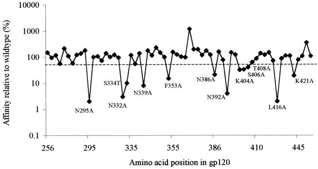
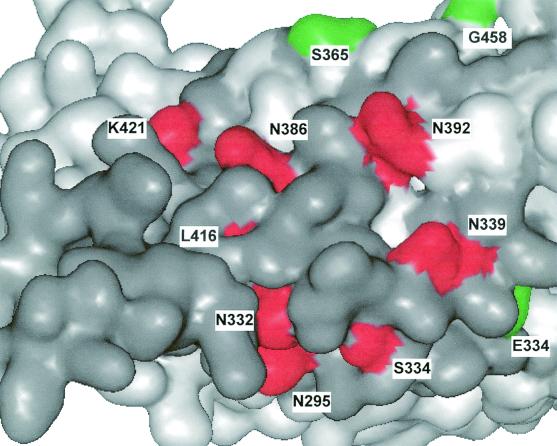
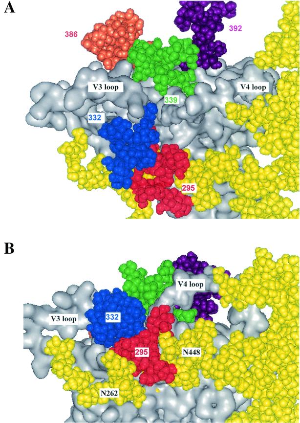
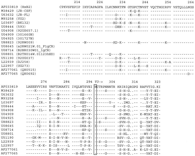
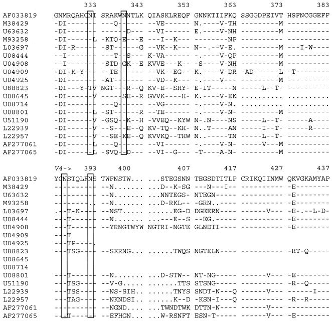
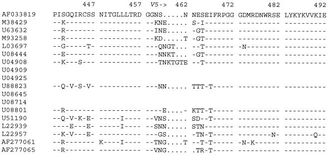
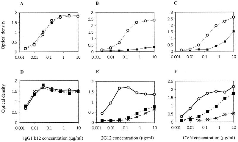
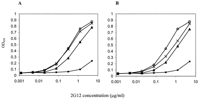
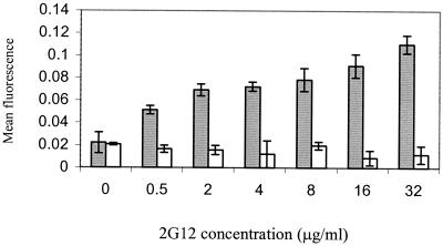
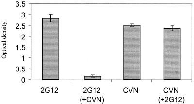
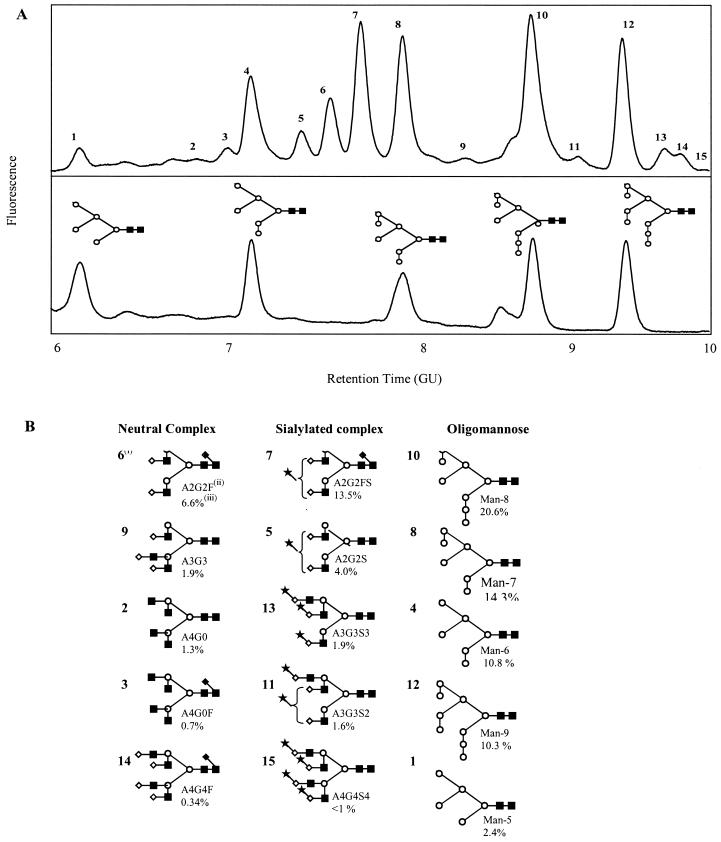
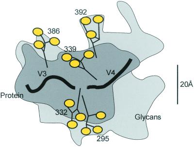
2G12 is a broadly neutralizing human monoclonal antibody against human immunodeficiency virus type-1 (HIV-1) that has previously been shown to bind to a carbohydrate-dependent epitope on gp120. Here, site-directed mutagenesis and carbohydrate analysis were used to define further the 2G12 epitope. Extensive alanine scanning mutagenesis showed that elimination of the N-linked carbohydrate attachment sequences associated with residues N295, N332, N339, N386, and N392 by N-->A substitution produced significant decreases in 2G12 binding affinity to gp120(JR-CSF). Further mutagenesis suggested that the glycans at N339 and N386 were not critical for 2G12 binding to gp120(JR-CSF). Comparison of the sequences of isolates neutralized by 2G12 was also consistent with a lesser role for glycans attached at these positions. The mutagenesis studies provided no convincing evidence for the involvement of gp120 amino acid side chains in 2G12 binding. Antibody binding was inhibited when gp120 was treated with Aspergillus saitoi mannosidase, Jack Bean mannosidase, or endoglycosidase H, indicating that Man(alpha)1-->2Man-linked sugars of oligomannose glycans on gp120 are required for 2G12 binding. Consistent with this finding, the binding of 2G12 to gp120 could be inhibited by monomeric mannose but not by galactose, glucose, or N-acetylglucosamine. The inability of 2G12 to bind to gp120 produced in the presence of the glucose analogue N-butyl-deoxynojirimycin similarly implicated Man(alpha)1-->2Man-linked sugars in 2G12 binding. Competition experiments between 2G12 and the lectin cyanovirin for binding to gp120 showed that 2G12 only interacts with a subset of available Man(alpha)1-->2Man-linked sugars. Consideration of all the data, together with inspection of a molecular model of gp120, suggests that the most likely epitope for 2G12 is formed from mannose residues contributed by the glycans attached to N295 and N332, with the other glycans playing an indirect role in maintaining epitope conformation.


Figures

FIG. 1.
FIG. 1.
Apparent affinity of 2G12 for alanine mutants of gp120JR-CSF relative to that of parent gp120JR-CSF. HxB2 sequence numbering is used (; Korber et al., HIV Sequence Database, 2001 [http://hiv-web.lanl.gov]). The substitutions that caused a more than 50% (dashed line) reduction in apparent affinity are labeled.
FIG. 2.
FIG. 2.
gp120 structure with amino acids colored to denote the effects of alanine substitutions on 2G12 affinity. The view shows the surface of the C4-V4 face of gp120. Coordinates were taken from the structure of the CD4-liganded core of gp120HxB2. Mutations which caused an increase in relative affinity are shown in green, those which did not cause a significant change in affinity are shown in dark grey, and those which caused a decrease in relative affinity are red. For clarity, the V4 loop has been omitted.
FIG. 3.
FIG. 3.
Location of gp120 N-linked glycans involved in 2G12 binding. The N-glycans which are likely to be primarily involved in 2G12 binding are shown in red (N-glycan of N295), blue (N-glycan of N332), and purple (N-glycan of N392). N-glycans which influence 2G12 binding but which are not directly involved in binding are shown in green (N-glycan of N339) and orange (N-glycan of N386). Other carbohydrate chains are shown in yellow. (A) Surface of the C4-V4 face of gp120 viewed from the perspective of the V4 loop. (B) Spatial location of the V3 and V4 loops, which are proposed to extend from the protein surface in the region of the 2G12 epitope. Glycans were modeled onto the core structure of gp120 according to highest population types and lineages, using mass spectrometry (70).
FIG. 4.
FIG. 4.
Sequence alignment of the C2-C5 region of gp120 from HIV-1 isolates neutralized by 2G12. Sequences were obtained from the GenBank database, aligned using ClustalW (http://www.ebi.ac.uk/clustalw/), and formatted for publication using SeqPublish (http://hiv-web.lanl.gov/content/hiv-db/SeqPublish/seqpublish.html). The names of isolates are shown in parentheses. Identical amino acid residues are indicated by dashes; for isolates with the GenBank accession numbers U04909, U04925, U08645, and U8714, only the C2, V3, and C3 regions have been fully sequenced. Rectangular boxes indicate glycosylation sites of N-glycans implicated in 2G12 binding.
FIG. 4.
FIG. 4.
Sequence alignment of the C2-C5 region of gp120 from HIV-1 isolates neutralized by 2G12. Sequences were obtained from the GenBank database, aligned using ClustalW (http://www.ebi.ac.uk/clustalw/), and formatted for publication using SeqPublish (http://hiv-web.lanl.gov/content/hiv-db/SeqPublish/seqpublish.html). The names of isolates are shown in parentheses. Identical amino acid residues are indicated by dashes; for isolates with the GenBank accession numbers U04909, U04925, U08645, and U8714, only the C2, V3, and C3 regions have been fully sequenced. Rectangular boxes indicate glycosylation sites of N-glycans implicated in 2G12 binding.
FIG. 5.
FIG. 5.
Binding of IgG1 b12, 2G12, and CVN to enzymatically treated gp120. Top panel: binding of IgG1 b12 (A), 2G12 (B), and CVN (C) to A. saitoi mannosidase-treated (▪) or untreated (○) gp120. Bottom panel: binding of IgG1 b12 (D), 2G12 (E), and CVN (F) to Jack Bean α-mannosidase-treated (×), endoH-treated (▪), or untreated (○) gp120.
FIG. 6.
FIG. 6.
Structure of Man9GlcNAc2. (A) Molecular model showing the Manα1→2Man-linked residues (red) and Manα1→2,3,6Man residues (blue) removed by A. saitoi mannosidase and Jack Bean mannosidase, respectively. (B) Chemical structure showing cleavage sites for the two mannosidases and endoH. The D1D3 isomer of Man8GlcNAc2 is derived by removing a single mannose from the D2 arm.
FIG. 7.
FIG. 7.
Monosaccharide inhibition of 2G12 binding to gp120. (A) Binding of 2G12 to gp120JR-FL in the presence of 0 (○), 5 (×), 50 (▴), or 500 (▪) mM mannose. (B) Binding of 2G12 to gp120JR-FL in the presence of 500 mM mannose (▪), galactose (▴), N-acetylglucosamine (×), or buffer (○).
FIG. 8.
FIG. 8.
2G12 binding to gp120 from cells grown in the presence of the glucosidase inhibitor NB-DNJ. Reactivity of 2G12 with recombinant gp120IIIB expressed in CHO cells, in the presence (open bars) or absence (solid bars) of 2 mM NB-DNJ, is shown.
FIG. 9.
FIG. 9.
Inhibition of 2G12 binding to gp120 by CVN. 2G12 and CVN reactivity with gp120JR-FL (1 μg/ml) alone or gp120 (1 μg/ml) preincubated with CVN (0.1 μg/ml) (+CVN) or with 2G12 (+2G12), respectively.
FIG. 10.
FIG. 10.
NP-HPLC of fluorescence-labeled gp120 N-linked glycans. (A) Charged, neutral, and oligomannose structures were released from gp120 by in-gel digestion using PNGase F and resolved by NP-HPLC (top panel). The retention time (GU value) for each peak was compared against a database of known structures. Preliminary assignments, made from the database, were confirmed by exoglycosidase digestions of the entire glycan pool using enzyme arrays. Following comprehensive digestion of complex glycan structures using a panel of exoglycosidases (see Materials and Methods), only oligomannose structures, which represent 58.4% of the total glycans, remained (bottom panel). (B) Symbolic representation of the most abundant sugars is shown together with the peak number (i), nomenclature (ii), and relative abundance (iii) in the glycan pool.
FIG. 11.
FIG. 11.
Schematic model of the approximate locations of the carbohydrate chains on gp120 implicated in 2G12 binding. The dark grey area represents the contour of the protein, and the light grey represents the contour of the glycans. The D1 terminal mannose structures of the glycans attached to asparagines at positions 332, 295, 339, 386, and 392 are shown in yellow. The 20 Å ruler represents the size of a typical antibody binding site.

References

    1. Baba, T. W., V. Liska, R. Hofmann-Lehmann, J. Vlasak, W. Xu, S. Ayehunie, L. A. Cavacini, M. R. Posner, H. Katinger, G. Stiegler, B. J. Bernacky, T. A. Rizvi, R. Schmidt, L. R. Hill, M. E. Keeling, Y. Lu, J. E. Wright, T. C. Chou, and R. M. Ruprecht. 2000. Human neutralizing monoclonal antibodies of the IgG1 subtype protect against mucosal simian-human immunodeficiency virus infection. Nat. Med. 6:200-206. - PubMed
    1. Bewley, C. A. 2001. Solution structure of a cyanovirin-N:Manα1-2Manα complex: structural basis for high-affinity carbohydrate-mediated binding to gp120. Structure 9:931-940. - PubMed
    1. Bewley, C. A., and S. Otero-Quinten. 2001. The potent anti-HIV protein cyanovirin-N contains two novel carbohydrate binding sites that selectively bind to Man8D1D3 and Man9 with nanomolar affinity: implications for binding to HIV envelope glycoprotein gp120. J. Am. Chem. Soc. 123:3892-3902. - PubMed
    1. Boyd, M. R., K. R. Gustafson, J. B. McMahon, R. H. Shoemaker, B. R. O'Keefe, T. Mori, R. J. Gulakowski, L. Wu, M. I. Rivera, C. M. Laurncot, M. J. Currens, J. H. Cardellina, R. W. Buckheit, P. L. Nara, L. K. Pannell, R. C. Sowder, and L. E. Henderson. 1997. Discovery of cyanovirin-N, a novel human immunodeficiency virus-inactivating protein that binds viral surface envelope glycoprotein gp120: potential applications to microbicide development. Antimicrob. Agents Chemother. 41:1521-1530. - PMC - PubMed
    1. Burton, D. R. 1997. A vaccine for HIV type 1: the antibody perspective. Proc. Natl. Acad. Sci. USA 94:10018-10023. - PMC - PubMed

Publication types

LinkOut - more resources