

Conditional mutation of the ErbB2 (HER2) receptor in cardiomyocytes leads to dilated cardiomyopathy
- PMID: 12072561
- PMCID: PMC124392
- DOI: 10.1073/pnas.122249299
Conditional mutation of the ErbB2 (HER2) receptor in cardiomyocytes leads to dilated cardiomyopathy
Abstract
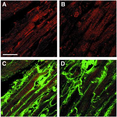
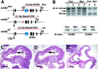
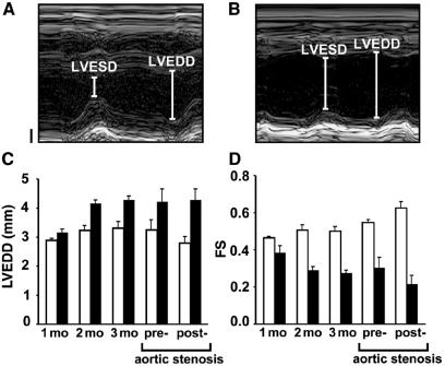
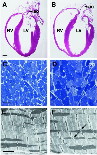
The ErbB2 (Her2) proto-oncogene encodes a receptor tyrosine kinase, which is frequently amplified and overexpressed in human tumors. ErbB2 provides the target for a novel and effective antibody-based therapy (Trastuzumab/Herceptin) used for the treatment of mammary carcinomas. However, cardiomyopathies develop in a proportion of patients treated with Trastuzumab, and the incidence of such complications is increased by combination with standard chemotherapy. Gene ablation studies have previously demonstrated that the ErbB2 receptor, together with its coreceptor ErbB4 and the ligand Neuregulin-1, are essential for normal development of the heart ventricle. We use here Cre-loxP technology to mutate ErbB2 specifically in ventricular cardiomyocytes. Conditional mutant mice develop a severe dilated cardiomyopathy, with signs of cardiac dysfunction generally appearing by the second postnatal month. We infer that signaling from the ErbB2 receptor, which is enriched in T-tubules in cardiomyocytes, is crucial for adult heart function. Conditional ErbB2 mutant mice provide a model of dilated cardiomyopathy. In particular, they will allow a rigorous assessment of the role of ErbB2 in the heart and provide insight into the molecular mechanisms that underlie the adverse effects of anti-ErbB2 antibodies.
Figures
Similar articles
-
ErbB2 is essential in the prevention of dilated cardiomyopathy.Nat Med. 2002 May;8(5):459-65. doi: 10.1038/nm0502-459. Nat Med. 2002. PMID: 11984589
-
ErbB2 pathways in heart and neural diseases.Trends Cardiovasc Med. 2003 Feb;13(2):80-6. doi: 10.1016/s1050-1738(02)00231-1. Trends Cardiovasc Med. 2003. PMID: 12586444 Review.
-
Neuregulin-1/erbB-activation improves cardiac function and survival in models of ischemic, dilated, and viral cardiomyopathy.J Am Coll Cardiol. 2006 Oct 3;48(7):1438-47. doi: 10.1016/j.jacc.2006.05.057. Epub 2006 Sep 14. J Am Coll Cardiol. 2006. PMID: 17010808
-
Essential roles of Her2/erbB2 in cardiac development and function.Recent Prog Horm Res. 2004;59:1-12. doi: 10.1210/rp.59.1.1. Recent Prog Horm Res. 2004. PMID: 14749494 Review.
-
Neuregulin receptors erbB2 and erbB4 in failing human myocardium -- depressed expression and attenuated activation.Basic Res Cardiol. 2005 May;100(3):240-9. doi: 10.1007/s00395-005-0514-4. Epub 2005 Feb 3. Basic Res Cardiol. 2005. PMID: 15685397
Cited by
-
Nrg1β Released in Remote Ischemic Preconditioning Improves Myocardial Perfusion and Decreases Ischemia/Reperfusion Injury via ErbB2-Mediated Rescue of Endothelial Nitric Oxide Synthase and Abrogation of Trx2 Autophagy.Arterioscler Thromb Vasc Biol. 2021 Aug;41(8):2293-2314. doi: 10.1161/ATVBAHA.121.315957. Epub 2021 May 27. Arterioscler Thromb Vasc Biol. 2021. PMID: 34039018 Free PMC article.
-
Ranolazine Attenuates Trastuzumab-Induced Heart Dysfunction by Modulating ROS Production.Front Physiol. 2018 Feb 6;9:38. doi: 10.3389/fphys.2018.00038. eCollection 2018. Front Physiol. 2018. PMID: 29467663 Free PMC article.
-
Cardiotoxicity of Epidermal Growth Factor Receptor 2-Targeted Drugs for Breast Cancer.Front Pharmacol. 2021 Nov 1;12:741451. doi: 10.3389/fphar.2021.741451. eCollection 2021. Front Pharmacol. 2021. PMID: 34790121 Free PMC article. Review.
-
Cardio-Oncology: mechanisms of cardiovascular toxicity.F1000Res. 2018 Jan 25;7:113. doi: 10.12688/f1000research.12598.1. eCollection 2018. F1000Res. 2018. PMID: 29399333 Free PMC article. Review.
-
Recent insights in the paracrine modulation of cardiomyocyte contractility by cardiac endothelial cells.Biomed Res Int. 2014;2014:923805. doi: 10.1155/2014/923805. Epub 2014 Mar 13. Biomed Res Int. 2014. PMID: 24745027 Free PMC article. Review.
References
-
- Seidman J G, Seidman C. Cell. 2001;104:557–567. - PubMed
-
- Towbin J A. Curr Opin Cell Biol. 1998;10:131–139. - PubMed
-
- Emanueli C, Maestri R, Corradi D, Marchione R, Minasi A, Tozzi M G, Salis M B, Straino S, Capogrossi M C, Olivetti G, Madeddu P. Circulation. 1999;100:2359–2365. - PubMed
-
- Nebigil C G, Hickel P, Messaddeq N, Vonesch J L, Douchet M P, Monassier L, Gyorgy K, Matz R, Andriantsitohaina R, Manivet P, et al. Circulation. 2001;103:2973–2979. - PubMed
-
- Kiriazis H, Kranias E G. Annu Rev Physiol. 2000;62:321–351. - PubMed
Publication types
MeSH terms
Substances
LinkOut - more resources
Full Text Sources
Other Literature Sources
Molecular Biology Databases
Research Materials
Miscellaneous
