. 2002 Jun 25;99(13):8506-11.
doi: 10.1073/pnas.132142799. Epub 2002 Jun 18.

All intermediates of the arsenate reductase mechanism, including an intramolecular dynamic disulfide cascade

Affiliations

All intermediates of the arsenate reductase mechanism, including an intramolecular dynamic disulfide cascade

Joris Messens et al. Proc Natl Acad Sci U S A. .

Abstract

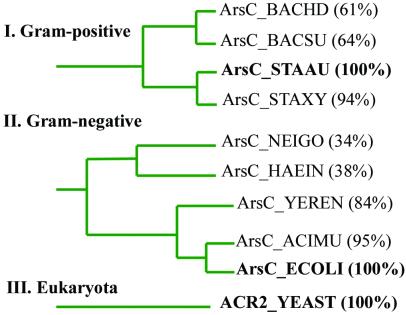
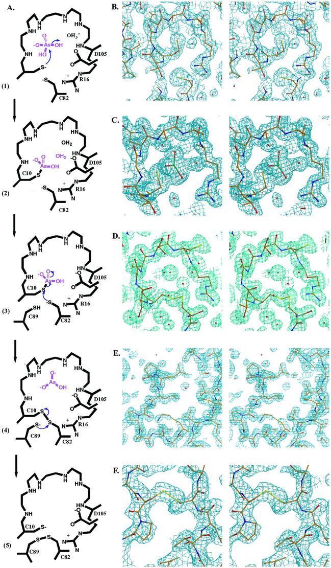
The mechanism of pI258 arsenate reductase (ArsC) catalyzed arsenate reduction, involving its P-loop structural motif and three redox active cysteines, has been unraveled. All essential intermediates are visualized with x-ray crystallography, and NMR is used to map dynamic regions in a key disulfide intermediate. Steady-state kinetics of ArsC mutants gives a view of the crucial residues for catalysis. ArsC combines a phosphatase-like nucleophilic displacement reaction with a unique intramolecular disulfide bond cascade. Within this cascade, the formation of a disulfide bond triggers a reversible "conformational switch" that transfers the oxidative equivalents to the surface of the protein, while releasing the reduced substrate.


Figures

Figure 1
Figure 1
ArsC families from Gram-positive bacteria (I), Gram-negative bacteria (II), and eukaryota (III). Within each family, the sequence identity of ArsC proteins is shown behind the SWISS-PROT database name. ArsC_STAAU (S. aureus) (bold face) represents the Gram-positive family (Bacillus halodurans, B. subtilis, Staphylococcus xylosus), ArsC_ECOLI (E. coli) (bold face) represents the Gram-negative family (Neisseria gonorrhoeae, Haemophilus influenzae, Yersinia enterocolitica, Acidiphilium multivorum), and ACR 2 (S. cerevisiae) (bold face) represents the only known member of the eukaryotic family. The blast network service of the Swiss Institute of Bioinformatics was used to construct the phylogenetic tree from the percentage of sequence identity to the family representative. The interfamilial sequence identity is lower than 20%.
Figure 2
Figure 2
Ribbon diagram of the overall structure of reduced ArsC wild type visualized from two different positions. The P-loop CX5R motif (red), the catalytic key residues in ball-and-stick representation, and the flexible short α-helix region (yellow) are shown.
Figure 3
Figure 3
(A) Scheme of the reaction mechanism of pI258 ArsC. (1) The nucleophilic attack of the thiol of Cys-10; (2) the formation of a covalent Cys-10-HAsOformula image intermediate; (3) the nucleophilic attack of the thiol of Cys-82 with arsenite release; (4) the formation of a Cys-10–Cys-82 intermediate and the nucleophilic attack of the thiol of Cys-89; (5) the formation of a Cys-82–Cys-89 disulfide. (B–F) A stereo view of the 2FoFc electron density maps contoured at 1.0 σ placed next to its corresponding reaction step in A. (B) The P-loop (residues 10–17) in the structure of reduced wild-type ArsC with Cys-10 in the center of the image. The P-loop is fully structured, despite the absence of bound oxyanion (2.0 Å). (C) In the structure of C15A ArsC-HAsOformula image, an arsenic is covalently bound on Cys-10, surrounded by three oxygens in a plane and a water molecule opposite the sulfur of Cys-10 (1.4 Å). (D) Oxidized ArsC C89L with the intermediate Cys-10–Cys-82 disulfide bond (1.6 Å). (E) A view on the flexible looped-out region of oxidized ArsC C89L, where Cys-89 has left the hydrophobic core and is replaced by Leu-92 upon Cys-10–Cys-82 formation. The electron density in this highly flexible region is not so well defined. (F) A view on the surface of oxidized ArsC C10SC15A (6) with the Cys-82–Cys-89 disulfide bond.
Figure 4
Figure 4
The movement of the “conformational switch” in the flexible segment (residues 80–98) trapped in four different ArsC crystals. Starting with a helix in the reduced ArsC wild type (blue), via oxidized ArsC C89L (Cys-10–Cys-82) in the first (yellow) and in the second (red) molecule in the asymmetric unit to finally looping out to form the C82-C89 disulfide (green). The two arrows indicate the movement of L92 and C89.

References

    1. Broer S, Ji G, Broer A, Silver S. J Bacteriol. 1993;175:3480–3485. - PMC - PubMed
    1. Xu C, Zhou T, Kuroda M, Rosen B P. J Biochem. 1998;123:16–23. - PubMed
    1. Rosen B P, Dey S, Dou D, Ji G, Kaur P, Ksenzenko M Y, Silver S, Wu J. Ann NY Acad Sci. 1992;671:257–272. - PubMed
    1. Ji G, Silver S. Proc Natl Acad Sci USA. 1992;89:9474–9478. - PMC - PubMed
    1. Mukhopadhyay R, Rosen B P. FEMS Microbiol Lett. 1998;168:127–136. - PubMed

Publication types

LinkOut - more resources