

Interrupted aortic arch in an adult single-stage extra-anatomic repair
- PMID: 12075868
- PMCID: PMC116738
Interrupted aortic arch in an adult single-stage extra-anatomic repair
Abstract
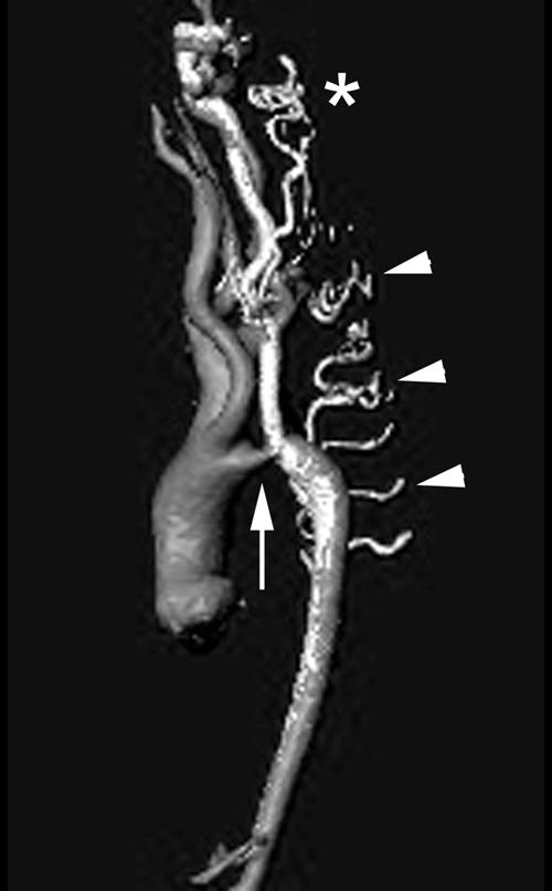
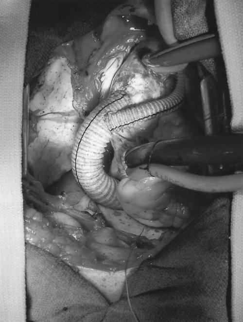
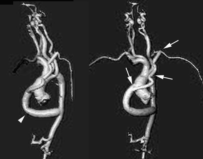
Interrupted aortic arch is a rare congenital malformation of the aortic arch that occurs in 3 per million live births. Defined as a loss of luminal continuity between the ascending and descending portions of the aorta, this anomaly entails a very poor prognosis without surgical treatment. To our knowledge, the world medical literature contains only 12 reports of isolated interrupted aortic arch diagnosed in adults. Nine of these patients underwent successful surgical repair, but 1 died during the early postoperative period. We describe a 10th successful surgical repair, which involved a 42-year-old woman who had an asymptomatic type B interrupted aortic arch (characterized by interruption between the left subclavian and left carotid arteries). We performed a single-stage extra-anatomic repair by placing a 16-mm extra-anatomic Dacron graft between the ascending and descending portions of the thoracic aorta and by interposing a 7-mm extra-anatomic Dacron graft between the 16-mm graft and the left subclavian artery. The patient recovered uneventfully and continued to do well 6 months later.
Figures
References
-
- Canova CR, Carrel T, Dubach P, Turina M, Reinhart WH. Interrupted aortic arch: fortuitous diagnosis in a 72-year-old female patient with severe aortic insufficiency [in German]. Schweiz Med Wochenschr 1995;125(1–2):26–30. - PubMed
-
- Backer CL, Mavroudis C. Congenital Heart Surgery Nomenclature and Database Project: patent ductus arteriosus, coarctation of the aorta, interrupted aortic arch. Ann Thorac Surg 2000;69(4 Suppl):S298–307. - PubMed
-
- Steidele RJ. Samml Chir u Med Beob (Vienna) 1778;2:114.
-
- Celoria GC, Patton RB. Congenital absence of the aortic arch. Am Heart J 1959;58:407–13. - PubMed
-
- Prasad SV, Gupta SK, Reddy KN, Murthy JS, Gupta SR, Somnath HS. Isolated interrupted aortic arch in adult. Indian Heart J 1988;40:108–12. - PubMed
Publication types
MeSH terms
LinkOut - more resources
Full Text Sources