A role for humoral mechanisms in the pathogenesis of Devic's neuromyelitis optica
- PMID: 12076996
- PMCID: PMC5444467
- DOI: 10.1093/brain/awf151
A role for humoral mechanisms in the pathogenesis of Devic's neuromyelitis optica
Abstract
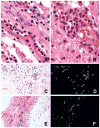
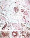
Devic's disease [neuromyelitis optica (NMO)] is an idiopathic inflammatory demyelinating disease of the CNS, characterized by attacks of optic neuritis and myelitis. The mechanisms that result in selective localization of inflammatory demyelinating lesions to the optic nerves and spinal cord are unknown. Serological and clinical evidence of B cell autoimmunity has been observed in a high proportion of patients with NMO. The purpose of this study was to investigate the importance of humoral mechanisms, including complement activation, in producing the necrotizing demyelination seen in the spinal cord and optic nerves. Eighty-two lesions were examined from nine autopsy cases of clinically confirmed Devic's disease. Demyelinating activity in the lesions was immunocytochemically classified as early active (21 lesions), late active (18 lesions), inactive (35 lesions) or remyelinating (eight lesions) by examining the antigenic profile of myelin degradation products within macrophages. The pathology of the lesions was analysed using a broad spectrum of immunological and neurobiological markers, and lesions were defined on the basis of myelin protein loss, the geography and extension of plaques, the patterns of oligodendrocyte destruction and the immunopathological evidence of complement activation. The pathology was identical in all nine patients. Extensive demyelination was present across multiple spinal cord levels, associated with cavitation, necrosis and acute axonal pathology (spheroids), in both grey and white matter. There was a pronounced loss of oligodendrocytes within the lesions. The inflammatory infiltrates in active lesions were characterized by extensive macrophage infiltration associated with large numbers of perivascular granulocytes and eosinophils and rare CD3(+) and CD8(+) T cells. There was a pronounced perivascular deposition of immunoglobulins (mainly IgM) and complement C9neo antigen in active lesions associated with prominent vascular fibrosis and hyalinization in both active and inactive lesions. The extent of complement activation, eosinophilic infiltration and vascular fibrosis observed in the Devic NMO cases is more prominent compared with that in prototypic multiple sclerosis, and supports a role for humoral immunity in the pathogenesis of NMO. Based on this study, future therapeutic strategies designed to limit the deleterious effects of complement activation, eosinophil degranulation and neutrophil/macrophage/microglial activation are worthy of further investigation.
Figures


Comment in
-
Devic's disease: bridging the gap between laboratory and clinic.Brain. 2002 Jul;125(Pt 7):1425-7. doi: 10.1093/brain/awf147. Brain. 2002. PMID: 12076994 No abstract available.
-
Glatiramer acetate treatment in Devic's neuromyelitis optica.Brain. 2003 Jun;126(Pt 6):1E; author reply 1E-a. doi: 10.1093/brain/awg140. Brain. 2003. PMID: 12764069 No abstract available.
References
-
- April RS, Vansonnenberg E. A case of neuromyelitis optica (Devic’s syndrome) in systemic lupus erythematosus: clinico-pathologic report and review of the literature. Neurology. 1976;26:1066–70. - PubMed
-
- Asghar SS, Pasch MC. Complement as a promiscuous signal transduction device. [Review] Lab Invest. 1998;78:1203–25. - PubMed
-
- Biliciler S, Uygucgil H, Saip S, Altintas A, Soysal T, Ozdemir SE, et al. Plasmapheresis in multiple sclerosis patients with different indications [abstract] Mult Scler. 2001;7(Suppl 1):S64.
-
- Bonnet F, Mercie P, Morlat P, Hocke C, Vergnes C, Ellie E, et al. Devic’s neuromyelitis optica during pregnancy in a patient with systemic lupus erythematosus. Lupus. 1999;8:244–7. - PubMed
-
- Breitschopf H, Suchanek G, Gould RM, Colman DR, Lassmann H. In situ hybridization with digoxigenin-labeled probes: sensitive and reliable detection method applied to myelinating rat brain. Acta Neuropathol (Berl) 1992;84:581–7. - PubMed
Publication types
MeSH terms
Substances
Grants and funding
LinkOut - more resources
Full Text Sources
Other Literature Sources
Research Materials

