. 2002 Jun 15;22(12):4767-75.
doi: 10.1523/JNEUROSCI.22-12-04767.2002.

ATP as a putative sensory mediator: activation of intrinsic sensory neurons of the myenteric plexus via P2X receptors

Affiliations

ATP as a putative sensory mediator: activation of intrinsic sensory neurons of the myenteric plexus via P2X receptors

Paul P Bertrand et al. J Neurosci. .

Abstract

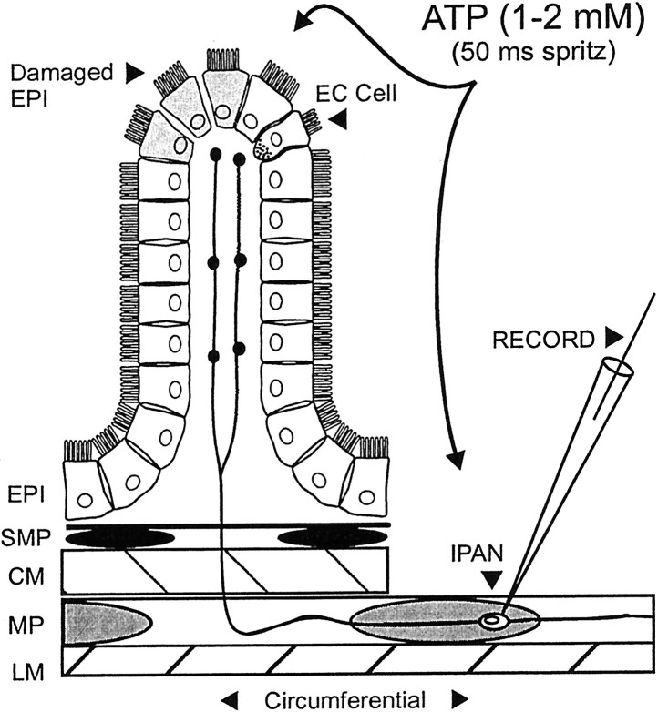
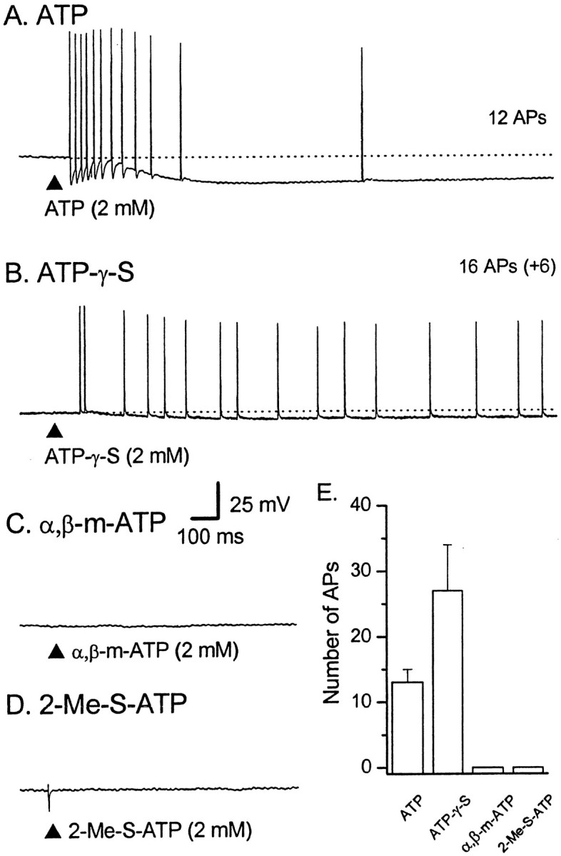
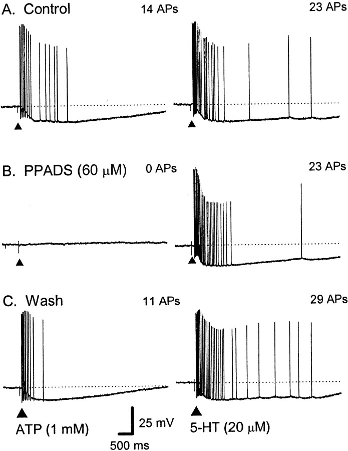
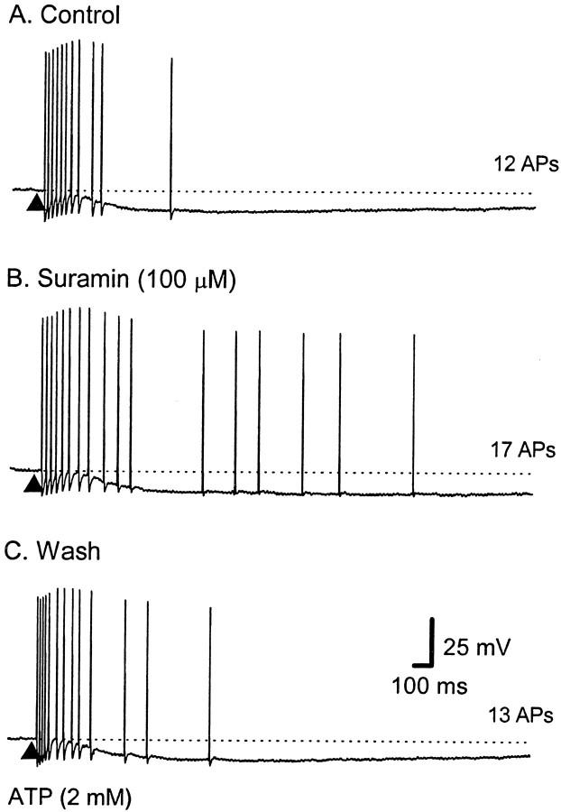
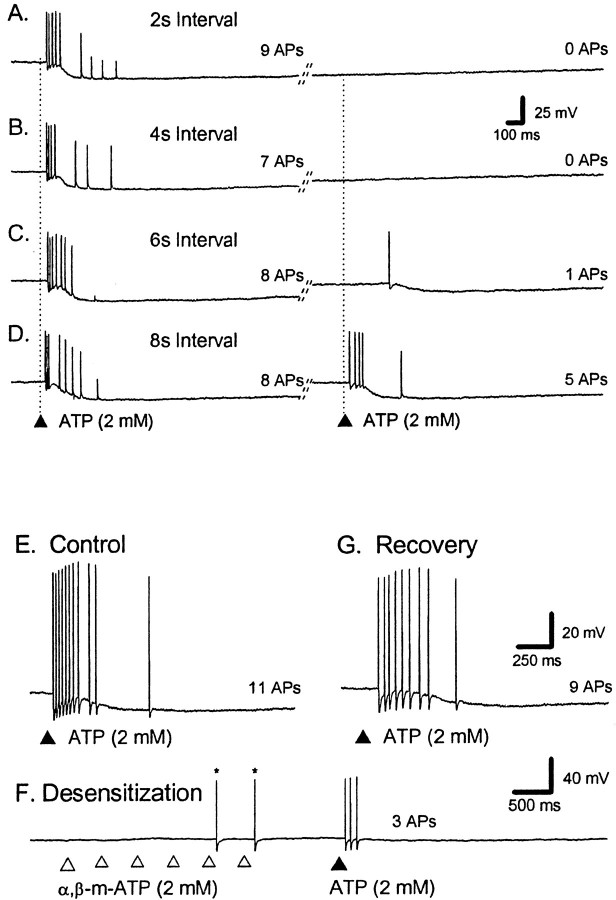
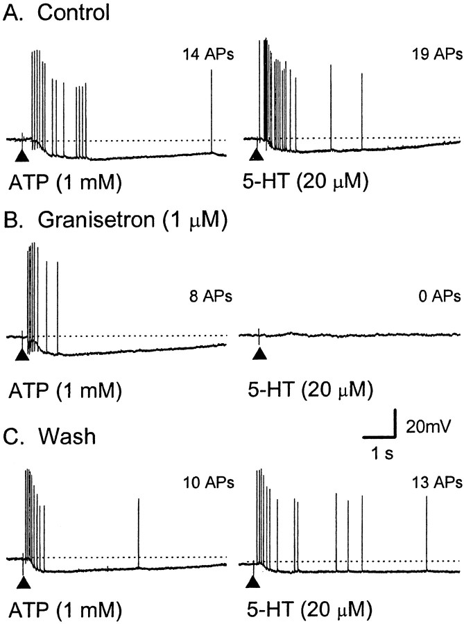
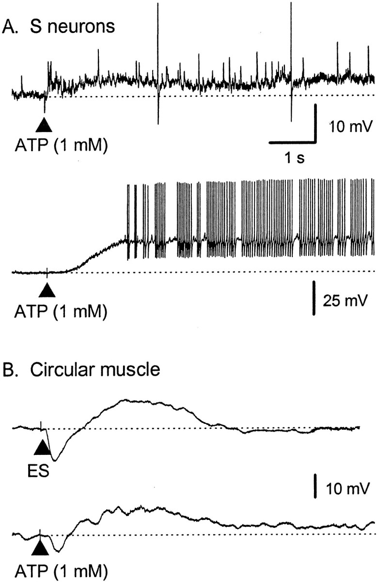
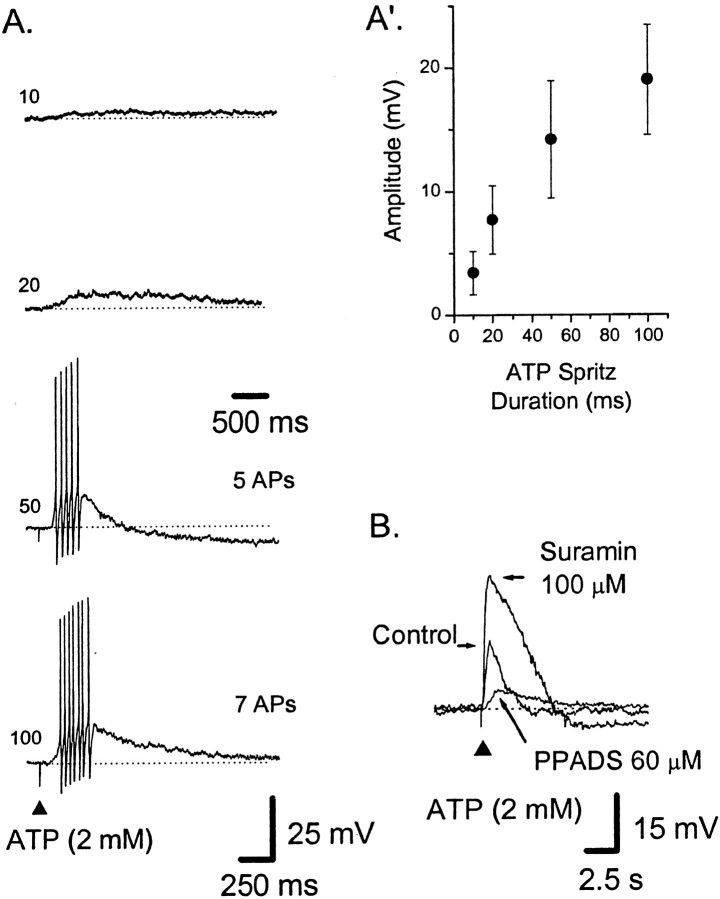
The mucosal terminals of sensory neurons intrinsic to the wall of the intestine are sensitive to the chemical environment within the lumen. Lumenal stimuli probably release sensory mediators from the mucosal epithelium, which then activate the nerve terminals indirectly. Here, we tested the idea that ATP activates intrinsic sensory nerve terminals in a way consistent with its being a sensory mediator. We made intracellular recordings from intrinsic sensory neurons located in the myenteric plexus [identified as AH neurons, which are neurons with a long-lasting afterhyperpolarization following the action potential (AP)], located within 1 mm of intact mucosa. Focal electrical stimulation of the mucosa was used to locate and map regions innervated by each neuron. Application of ATP (1-2 mm in the pressure pipette) to these regions elicited trains of APs that originated at the sensory terminals. ATP-gamma-S produced a similar response, but alpha,beta-methylene ATP and 2-methylthio-ATP were only weakly active. The P2 receptor antagonist pyridoxalphosphate-6-azophenyl-2',5'-disulphonic acid (PPADS) (60 microm in the bath) abolished the APs evoked by ATP and ATP-gamma-S but spared similar responses evoked by 5-hydroxytryptamine (5-HT). Another P2 receptor antagonist suramin (100 microm in the bath) did not significantly change the number of APs evoked by ATP. Either ATP or alpha,beta-methylene ATP desensitized the ATP-evoked APs; 50% recovery occurred after approximately 5 sec. The number of APs evoked by ATP was reduced, but not abolished, by the selective 5-HT3 receptor antagonist granisetron (1 microm in the bath). ATP was applied to the cell bodies of sensory neurons to investigate whether the cell bodies express the same P2X receptor as the terminals. ATP evoked a fast depolarization associated with a reduction in input resistance and a reversal potential of -11 mV. This depolarization was potentiated by suramin and blocked by PPADS. We conclude that activation of an atypical excitatory P2X receptor by ATP triggers AP generation in the mucosal processes of the sensory neurons; endogenous 5-HT release may also contribute to activation of the nerve terminals. A similar P2X receptor exists on the cell body of the sensory neuron. Together, these data are consistent with a role for ATP as a sensory mediator in gastrointestinal chemosensory transduction.


Figures

Fig. 1.
Fig. 1.
Illustration of the experimental arrangement and the relation of the epithelium and the AH–sensory nerve terminals. A side view of the preparation used in the present study. From thebottom: LM, the longitudinal muscle;MP, myenteric plexus; CM, circular muscle; SMP, submucosal plexus; EPI, epithelium. Note that the circular muscle, submucosal plexus, and epithelium have been dissected away from the right half of the preparation to allow an intracellular recording electrode (RECORD) to impale myenteric AH neurons [sensory neurons and intrinsic primary afferent neurons (IPAN, at the open circle)]. When the cell body of the AH neuron is close enough to the intact mucosa (<1 mm), there is a good chance that one or more of its projections is intact and innervates the mucosa. ATP and other agonists were applied to the mucosa and to the cell body of AH neurons via short-duration pressure ejection. The serotonin-containing enterochromaffin cells (EC Cell) are evenly spaced among the enterocytes. They represent ∼1% of the total population of endothelial cells. The mucosal epithelium is likely to be damaged when maintained in vitro (Damaged EPI).
Fig. 2.
Fig. 2.
Responses in AH–sensory neurons to mucosal application of ATP, ATP-γ-S, α,β-me-ATP or 2-Me-S-ATP. Representative voltage traces from four different AH neurons during application of ATP or ATP analogs to the mucosa; dotted lines indicate RMP. Calibration in Capplies to all traces. The total number of APs is shown to the right of each trace.A, Brief application (100 msec; at the filled triangle) of ATP (2 mm) elicited a train of APs that showed a slowing in frequency during the 1.1 sec duration of the discharge; the average instantaneous frequency (ƒINT) was 29 Hz [resting membrane potential (RMP) of −67 mV]. B, ATP-γ-S (2 mm) elicited a 3.0-sec-long train of 22 APs (16 APs shown) that fired at a slow, relatively constant frequency (fINT of 12 Hz; RMP of −82 mV).C, α,β-methylene ATP (2 mm) failed to elicited any APs in the majority of cases (RMP of −68 mV).D, 2-Me-S-ATP (2 mm) also failed to elicit any APs under control conditions (n = 3).E, Histogram showing the average number of APs evoked by agonists for two to three repetitions per cell. ATP,n = 18; ATP-γ-S, n = 7; α,β-methylene ATP, n = 7; 2-Me-S-ATP,n = 3.
Fig. 3.
Fig. 3.
Effect of the P2 receptor antagonist PPADS on the train of APs evoked by ATP and 5-HT. Representative voltage traces from a single AH neuron; dotted lines indicate RMP. Calibration in C applies to all traces.Left, A, ATP (2 mm, 100 msec; applied at the filled triangle) elicited a 1.8 sec duration train of 14 APs at an average instantaneous frequency (fINT) of 13 Hz.B, The response was blocked by PPADS (60 μm). C, Twenty-five minutes after washout of PPADS, APs have reappeared, and the response is shorter in duration and of a higher frequency; fINT of 26 Hz (RMP, control, −63 mV; PPADS, −46 mV; recovery, −59 mV).Right, A, 5-HT (20 μm, 100 msec; applied at the filled triangle) elicited a train of 23 APs; fINT of 24 Hz. B, The response was not affected during superfusion with PPADS (23 APs;fINT of 22 Hz) and remained stable during washout of PPADS (C, 29 APs;fINT of 22 Hz) (RMP, control, −64 mV; PPADS, −48 mV; wash, −56 mV).
Fig. 4.
Fig. 4.
Effect of the P2 receptor antagonist suramin on the train of APs evoked by ATP. Representative voltage traces from a single AH neuron; the dotted lines indicate RMP. Calibration in C applies to all traces. The total number of APs is shown to the right of eachtrace. A, ATP (2 mm, 100 msec; applied at the filled triangle) elicited a train of 12 APs at an average instantaneous frequency (fINT) of 30 Hz for a duration of 0.8 sec. B, The response was not reduced by suramin (100 μm) (17 APs; ƒINT of 16 Hz; duration of 2.1 sec). C, Thirty minutes after washout, the response is shorter and of a higher frequency than in suramin (13 APs;fINT of 30 Hz; duration of 1.0 sec) (RMP, control, −69 mV; suramin, −70 mV; recovery, −65 mV).
Fig. 5.
Fig. 5.
ATP can desensitize the train of APs evoked by ATP. A–D, Representative voltage traces from a single AH neuron; the dotted lines indicate RMP. Calibration in B applies to all traces. ATP was applied twice to the mucosa at the vertical lines with a 2, 4, 6, or 8 sec interval (A–D, respectively); the total number of APs is shown to theright of each trace. The diagonal lines indicate a break in the trace of a variable length of time. In control (A–D), ATP evokes a burst of APs consisting of between seven and nine APs. Subsequent APs evoked by the second application of ATP are depressed when the interval is shorter than 8 sec. Note that the long AHP after AP generation does not prevent incoming APs from the distal processes from evoking PPPs.E–G, Calibration in G also applies to E. The total number of ATP-evoked APs is shown to the right of each trace.E, In control, ATP evoked a burst of APs consisting of between seven and nine APs (3 repetitions). F, α,β-me-ATP was then applied six times in rapid succession to the same region of mucosa, and ATP was applied immediately after.G, Two minutes later, the ATP response was fully recovered.
Fig. 6.
Fig. 6.
Effect of granisetron on trains of APs evoked by ATP and 5-HT. Representative voltage traces from a single AH neuron; the dotted lines indicate RMP. Calibration inC applies to all traces. The total number of APs is shown to the right of eachtrace. Left, A, ATP (1 mm, 100 msec; applied at the filled triangle) elicited a train of 10 APs at an average instantaneous frequency (fINT) of 12 Hz for a duration of 2.1 sec. B, Granisetron (1 μm) caused a significant reduction in the number of APs (8 APs; fINT of 13 Hz; duration of 0.6 sec; RMP of −77 mV). C, The train of APs recovered after washout of granisetron (9 APs;fINT of 11 Hz; duration of 0.8 sec).Right, A, 5-HT (20 μm, 100 msec; applied at the filled triangle) elicited a train (15 APs; fINT of 11 Hz; duration of 3.9 sec). B, Granisetron (1 μm) fully blocked this response. C, Twenty-five minutes after washout of granisetron, the number of APs had increased (8 APs;fINT of 10 Hz; duration of 3.1 sec). Note that the long AHP after AP generation in C does not prevent incoming APs from the distal processes from evoking PPPs.
Fig. 7.
Fig. 7.
Effect of ATP applied to the mucosa on myenteric S neurons and circular muscle. ATP (1 mm) was applied to mucosa directly circumferential from the impalement. Calibrations inA applies to all traces.A, Representative voltage traces from two S neurons; thedotted lines indicate RMP. Top, ATP evoked a burst of fast EPSPs, two of which reach threshold for AP generation. Bottom, ATP evoked a slow EPSP-like depolarization that evoked many APs. B, Voltage traces from a single circular muscle cell. Top, Electrical stimulation of the mucosa, directly circumferential to the recording site, evoked a biphasic inhibitory junction potential (RMP of −48 mV).Bottom, Application of ATP to the same site on the mucosa evoked a biphasic inhibitory event, similar to that evoked electrically (RMP of −55 mV). ES, Electrical stimulation.
Fig. 8.
Fig. 8.
Effect of ATP applied to the cell body of myenteric AH–sensory neurons. Representative voltage traces from two AH neurons (A, B); the dotted lines indicate RMP. A, ATP was applied to mucosa with an increasing duration in milliseconds (numbers to the left of the trace). Thetop calibration bar applies to the top two traces; the bottom calibration applies to alltraces. A′, Summary data from three AH neurons in which ATP evoked depolarizations that did not generate APs.B, ATP was applied to the cell body under control conditions and during superfusion with PPADS (60 μm), which blocked, and suramin (100 μm), which potentiated, the depolarization.

References

    1. Barajas-Lopez C, Huizinga JD, Collins SM, Gerzanich V, Espinosa-Luna R, Peres AL. P2X-purinoceptors of myenteric neurones from the guinea-pig ileum and their unusual pharmacological properties. Br J Pharmacol. 1996;119:1541–1548. - PMC - PubMed
    1. Bertrand PP, Galligan JJ. Alfaxalone, pentobarbital and diazepam potentiate γ-aminobutyric acid-induced depolarizations in single myenteric neurons of guinea pig intestine. J Pharmacol Exp Ther. 1992;262:677–682. - PubMed
    1. Bertrand PP, Kunze WAA, Bornstein JC, Furness JB, Smith ML. Analysis of the responses of myenteric neurons in the small intestine to chemical stimulation of the mucosa. Am J Physiol. 1997;273:G422–G435. - PubMed
    1. Bertrand PP, Kunze WA, Bornstein JC, Furness JB. Electrical mapping of the projections of intrinsic primary afferent neurons to the mucosa of the guinea-pig small intestine. Neurogastroenterol Motil. 1998;10:533–541. - PubMed
    1. Bertrand PP, Kunze WAA, Furness JB, Bornstein JC. The terminals of myenteric intrinsic primary afferent neurons of the guinea-pig ileum are excited by 5-hydroxytryptamine acting at 5-hydroxytryptamine-3 receptors. Neuroscience. 2000;101:459–469. - PubMed

Publication types

MeSH terms

LinkOut - more resources