. 2002 Jun 15;22(12):4814-24.
doi: 10.1523/JNEUROSCI.22-12-04814.2002.

P2X receptor trafficking in neurons is subunit specific

Affiliations

P2X receptor trafficking in neurons is subunit specific

Laura K Bobanovic et al. J Neurosci. .

Abstract

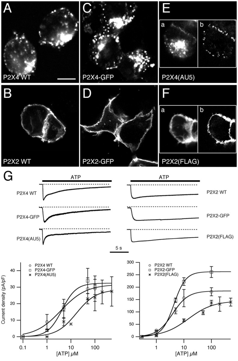
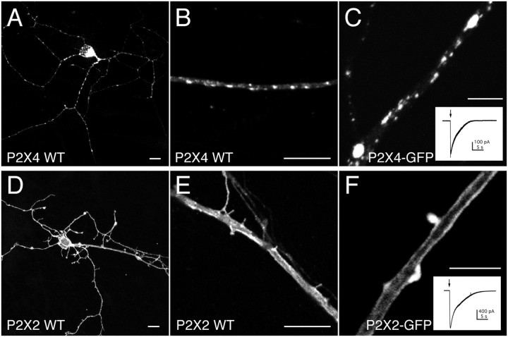
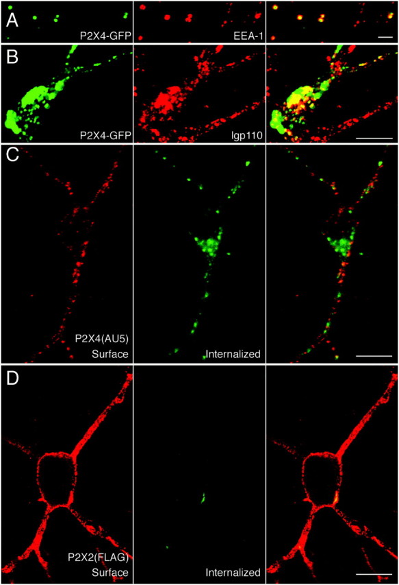
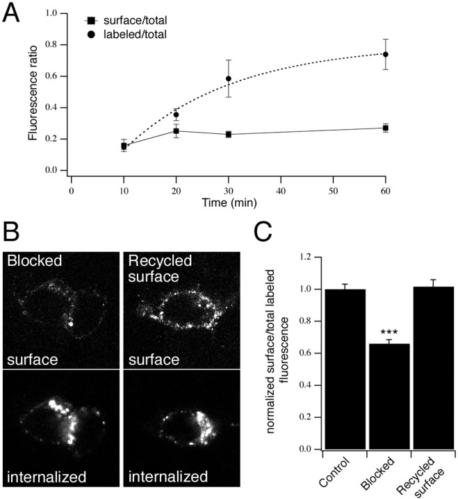
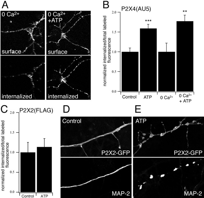
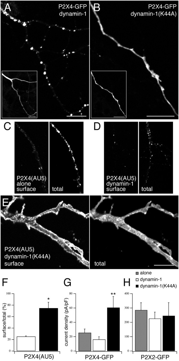
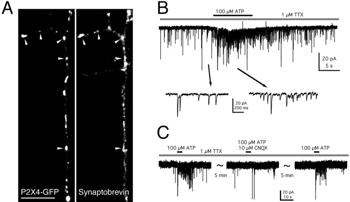
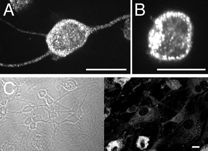
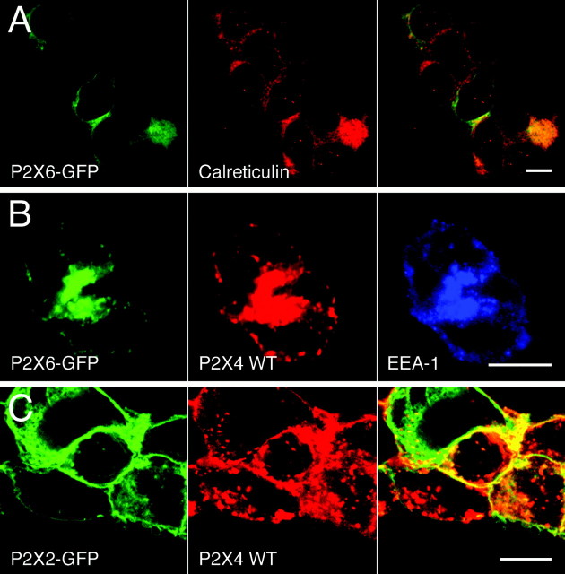
P2X receptors within the CNS mediate excitatory synaptic transmission and also act presynaptically to modulate neurotransmitter release. We have studied the targeting and trafficking of P2X4 and P2X2 receptors heterologously expressed in cultured olfactory bulb neurons. Homomeric P2X4 receptors had a punctate distribution, and many of the puncta colocalized with early endosomes. In contrast, P2X2 receptors were primarily localized at the plasma membrane. By antibody-labeling of surface receptors in living neurons, we showed that P2X4 receptors undergo rapid constitutive internalization and subsequent reinsertion into the plasma membrane, whereas P2X2 receptors were not regulated in such a way. The internalization of P2X4 receptors was dynamin-dependent, and the binding of ATP enhanced the basal rate of retrieval in a Ca2+-independent manner. The presence of the P2X4 subunit in a P2X4/6 heteromer governed the trafficking properties of the receptor. P2X receptors acted presynaptically to enhance the release of glutamate, suggesting that the regulated cycling of P2X4-containing receptors might provide a mechanism for modulation of synaptic transmission.


Figures

Fig. 1.
Fig. 1.
Subcellular distribution and functional properties of P2X constructs in HEK293 cells. A–F, The subcellular distribution of P2X4 WT (A), P2X4–GFP (C), P2X4(AU5) (E), P2X2 WT (B), P2X2-GFP (D), and P2X2(FLAG) (F) receptors in HEK293 cells. Cells expressing untagged receptors were stained using anti-P2X4/Cy3 or anti-P2X2/Cy3. Cells expressing P2X4(AU5) were stained using anti-AU5/Cy3 before (E, left panel) and anti-P2X4/FITC after permeabilization. Cells expressing P2X2(FLAG) were stained using anti-FLAG/Cy3 before (F, left panel) and anti-P2X2/FITC after permeabilization. Scale bar, 10 μm. G, Concentration–effect curves for ATP and representative traces showing ATP-evoked whole-cell currents for P2X4 (left) and P2X2 (right) constructs used in this study. ATP-evoked peak currents shown are normalized to compare the time course of desensitization. Calculated EC50values were 3.8, 4.2, and 17 μm for P2X4 WT, P2X4–GFP, and P2X4(AU5), respectively. Calculated EC50 values for P2X2 WT, P2X2–GFP, and P2X2(FLAG) were 3.9, 4.4, and 14.6 μm, respectively (n = 3–7 cells for each concentration).
Fig. 2.
Fig. 2.
Subcellular distribution of heterologously expressed P2X receptors in neurons. Confocal images of heterologously expressed P2X4 WT (A, B), P2X4–GFP (C), P2X2 WT (D, E), and P2X2–GFP (F) receptors in mature OB neurons.Insets,C, F, Example traces of whole-cell currents evoked by a 0.5 sec application of ATP (100 μm). Cells expressing untagged receptors were stained using anti-P2X4/Cy3 and anti-P2X2/Cy3, respectively. Scale bars: A, B, D,E, 10 μm; C, F, 5 μm.
Fig. 3.
Fig. 3.
P2X4, but not P2X2, receptors are constitutively internalized. In distal processes, P2X4–GFP puncta colocalize with anti-EEA1/Cy3 staining (A), and costaining with anti-lgp110/Cy3 shows that large accumulations of P2X4–GFP in the cell soma are within lysosomes (B). Live-labeling of P2X4(AU5) (C) and P2X2(FLAG) (D) receptors for 30 min at 37°C with anti-AU5 and anti-FLAG, respectively. In C and D, cell surface (left) and internalized (middle) receptors were visualized using Cy3-conjugated and FITC-conjugated secondary antibodies, before and after permeabilization, respectively. Overlaid images are shown on the right panels. Scale bars: A, 2 μm;B–D, 10 μm.
Fig. 4.
Fig. 4.
Cycling of P2X4(AU5) receptors to and from the surface. A, Time course of P2X4(AU5) constitutive internalization in neurons. Transfected neurons were labeled with anti-AU5 for different lengths of time at 37°C. Cells were stained using secondary antibody before (surface/total) or after (labeled/total) permeabilization and then stained for total P2X4 receptor. Fluorescence ratios are shown plotted against antibody incubation time. The time course of labeling is described by a single exponential (τ = 21 min;n = 4–7 neurons for each time point).B, C, Internalized P2X4(AU5) receptors recycle back to the plasma membrane. Representative confocal images used for analysis show an increase in P2X4(AU5) receptors recycled to the cell surface (B). Histogram (C) shows the surface fluorescence as a fraction of total (surface plus internalized) fluorescence, normalized toControl cells that were fixed immediately after a 30 min incubation with anti-AU5. Blocked represents cells that were fixed immediately after incubating cells with a nonfluorescent secondary antibody at 4°C. Recycled surface represents cells that were returned to 37°C for 15 min after blocking. Surface and internalized receptors were detected before and after permeabilization using Cy3- and FITC-conjugated secondary antibodies, respectively (n = 14–22 neurons for each condition).
Fig. 5.
Fig. 5.
The effect of ATP on trafficking of P2X receptors in neurons. A–C, ATP-dependent P2X4(AU5) and P2X2(FLAG) receptor internalization was measured by live-labeling neurons for 30 min at 37°C with anti-AU5 and anti-FLAG, respectively. Cell surface and internalized receptors were stained after 15 min incubation in control or Ca2+-free solution with or without ATP (100 μm) (A). B, C, Quantification of P2X4(AU5) and P2X2(FLAG) receptor internalization. Histograms show the internalized fluorescence as a fraction of total labeled (surface plus internalized) fluorescence, normalized to control cells (n = 7–29 neurons for each condition). ATP-dependent (100 μm) internalization of P2X4(AU5) receptors is independent of extracellular Ca2+(B). D, E, Neurons expressing P2X2-GFP receptors were stained with anti-MAP-2. Application of ATP (100 μm) causes dendritic injury in P2X2-GFP transfected neurons.
Fig. 6.
Fig. 6.
Trafficking of P2X4 is dynamin-dependent.A, B, Confocal images of P2X4-GFP coexpressed with either wild-type (A) or dominant-negative (B) dynamin-1.Inset images show detection of the coexpressed dynamin-1 constructs using anti-HA/Cy3. C–E, Confocal images to show neurons expressing P2X4(AU5) alone (C) or with either wild-type dynamin-1 (D) or dynamin-1(K44A) (E) were immunostained for cell surface receptors (anti-AU5/Cy3 before permeabilization) and total receptor (anti-P2X4/FITC after permeabilization). Scale bars, 10 μm.F, Quantification of surface versus total receptor in neurons coexpressing P2X4(AU5) and either dynamin-1 or dynamin-1(K44A) (n = 3 neurons for both condition).G, H, Histograms of the mean whole-cell current densities after application of ATP (100 μm) in neurons expressing either P2X4–GFP (G) or P2X2–GFP (H) alone (shaded) or coexpressed with either dynamin-1 (white bars) or dynamin-1(K44A) (black bars) (n = 5–15 neurons for each condition).
Fig. 7.
Fig. 7.
Presynaptic function of P2X4 receptors.A, Confocal image to show the colocalization of P2X4–GFP puncta (left panel) with anti-synaptobrevin/Cy-3 immunofluorescence (right panel). Arrowheads indicate areas of colocalization. Scale bar, 10 μm. B, Increase in the frequency of mEPSCs recorded from an untransfected neuron, after application of ATP to the process of a neighboring transfected neuron, in the presence of TTX (1 μm). The holding potential was −40 mV. C, The ATP-mediated increase in the frequency of mEPSCs was reversibly blocked by CNQX (10 μm).
Fig. 8.
Fig. 8.
Endogenous P2X4 receptors have a punctate distribution. A, B, Subcellular distribution of endogenous P2X4 in freshly plated OB neurons (4 hrin vitro). C, P2X4 immunoreactivity in OB neurons was downregulated after 7 DIV, whereas microglia remained P2X4-positive. Cells were stained using anti-P2X4/FITC. Scale bars, 10 μm.
Fig. 9.
Fig. 9.
P2X4 determines heteromeric P2X4/6 receptor trafficking. A, Confocal image to show the colocalization between P2X6–GFP and the ER marker calreticulin.B, Coexpression of P2X6–GFP with P2X4 WT and their colocalization with the early endosomal marker EEA-1. C, Coexpression of P2X2–GFP and P2X4 WT in HEK293 cells. Cells were stained using anti-calreticulin/Cy3 (A) or anti-P2X4/Cy5 and anti-EEA-1/Cy3 (B) and anti-P2X4/TRITC (C). Right panelsshow overlaid images (A, C). Scale bars, 10 μm.

Similar articles

Cited by

References

    1. Beattie EC, Carroll RC, Yu X, Morishita W, Yasuda H, von Zastrow M, Malenka RC. Regulation of AMPA receptor endocytosis by a signaling mechanism shared with LTD. Nat Neurosci. 2000;3:1291–1300. - PubMed
    1. Burrone J, Murthy VN. Synaptic plasticity: rush hour traffic in the AMPA lanes. Curr Biol. 2001;11:R274–R277. - PubMed
    1. Carroll RC, Beattie EC, Xia HH, Luscher C, Altschuler Y, Nicoll RA, Malenka RC, von Zastrow M. Dynamin-dependent endocytosis of ionotropic glutamate receptors. Proc Natl Acad Sci USA. 1999;96:14112–14117. - PMC - PubMed
    1. Carroll RC, Beattie EC, von Zastrow M, Malenka RC. Role of AMPA receptor endocytosis in synaptic plasticity. Nat Rev Neurosci. 2001;2:315–324. - PubMed
    1. Collo G, North RA, Kawashima E, Merlo-Pich E, Neidhart S, Surprenant A, Buell G. Cloning of P2X5 and P2X6 receptors and the distribution and properties of an extended family of ATP-gated ion channels. J Neurosci. 1996;16:2495–2507. - PMC - PubMed

Publication types

MeSH terms

LinkOut - more resources