Comparative Study
. 2002 Jun 15;22(12):4850-9.
doi: 10.1523/JNEUROSCI.22-12-04850.2002.

Control and plasticity of intercellular calcium waves in astrocytes: a modeling approach

Affiliations
Comparative Study

Control and plasticity of intercellular calcium waves in astrocytes: a modeling approach

Thomas Höfer et al. J Neurosci. .

Abstract

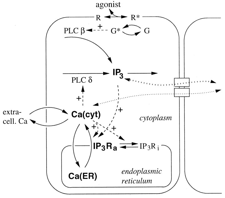
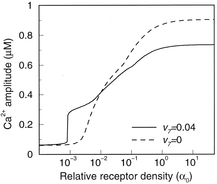
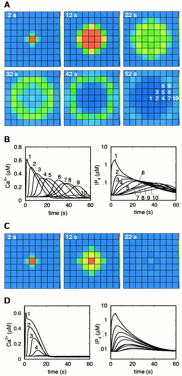
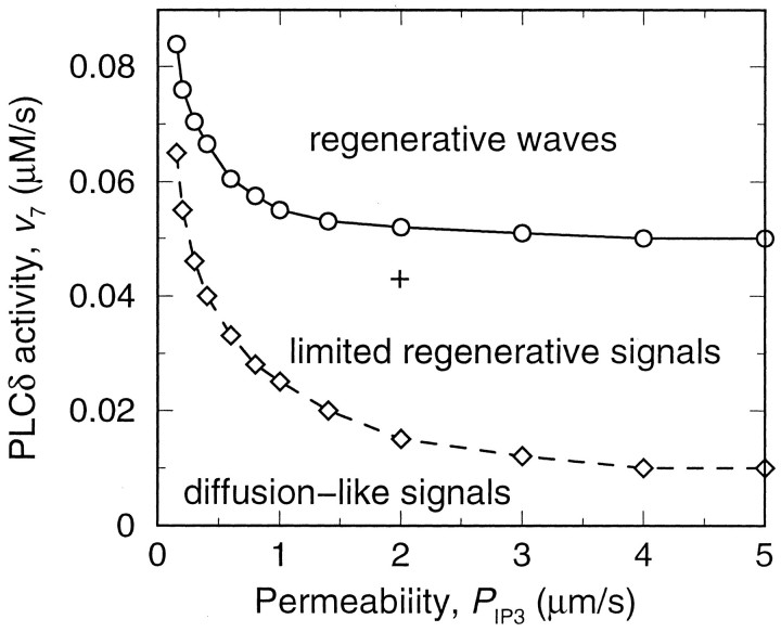
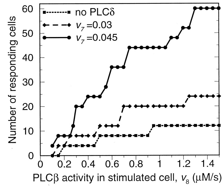
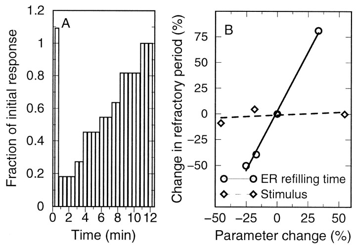
Intercellular Ca2+ waves in astrocytes are thought to serve as a pathway of long-range signaling. The waves can propagate by the diffusion of molecules through gap junctions and across the extracellular space. In rat striatal astrocytes, the gap-junctional route was shown to be dominant. To analyze the interplay of the processes involved in wave propagation, a mathematical model of this system has been developed. The kinetic description of Ca2+ signaling within a single cell accounts for inositol 1,4,5-trisphosphate (IP3) generation, including its activation by cytoplasmic Ca2+, IP3-induced Ca2+ liberation from intracellular stores and various other Ca2+ transports, and cytoplasmic diffusion of IP3 and Ca2+. When cells are coupled by gap junction channels in a two-dimensional array, IP3 generation in one cell triggers Ca2+ waves propagating across some tens of cells. The spatial range of wave propagation is limited, yet depends sensitively on the Ca2+-mediated regeneration of the IP3 signal. Accordingly, the term "limited regenerative signaling" is proposed. The gap-junctional permeability for IP3 is the crucial permissive factor for wave propagation, and heterogeneity of gap-junctional coupling yields preferential pathways of wave propagation. Processes involved in both signal initiation (activation of IP3 production caused by receptor agonist) and regeneration (activation of IP3 production by Ca2+, loading of the Ca2+ stores) are found to exert the main control on the wave range. The refractory period of signaling strongly depends on the refilling kinetics of the Ca2+ stores. Thus the model identifies multiple steps that may be involved in the regulation of this intercellular signaling pathway.


Figures

Fig. 1.
Fig. 1.
Scheme of the Ca2+ and IP3 dynamics included in the model. Solid,dashed, and dotted arrows indicate reaction/transport steps, regulatory interactions, and molecular diffusion, respectively. The bold quantities indicate the model variables. R, Agonist receptor; G, G-protein (active forms are denoted by asterisks);IP3Ri, inactive conformation of the IP3 receptor. For further abbreviations and explanation see Mathematical Model.
Fig. 2.
Fig. 2.
Agonist response in the model of a single cell. Activation level was varied by changing the relative receptor density at saturating agonist dose. This translates into different activities of PLCβ (cf. Eq. 9). Shown are the Ca2+ responses in the presence (solid line) and absence (dashed line) of PLCδ. The duration of activation was 4 sec.
Fig. 3.
Fig. 3.
Intercellular Ca2+ waves in the model. A, [Ca2+] isignal for the reference parameter set (Table 1). B, Time courses of [Ca2+]i and IP3 in the cells as labeled in the last frame ofA. C, [Ca2+]i signal in the absence of Ca2+ activation of IP3 production by PLCδ, under otherwise identical conditions. D, Time courses of [Ca2+]i and IP3corresponding to C. The central area of a simulated field of 19 × 19 cells is shown. The stimulus was an elevation of the PLCβ activity v8 in the central cell to 1 μm/sec for 4 sec.
Fig. 4.
Fig. 4.
Types of intercellular signals in the model as a function of the gap junction permeability for IP3 and the maximal activity of PLCδ. According to the spatial range of propagation, three qualitatively different behaviors are observed: diffusion-like signals, the range of which equals the range obtained by IP3 diffusion from the stimulated cell alone; limited regenerative signals, for which the spatial range is increased by IP3 regeneration yet remains finite; and regenerative waves without an intrinsic limit to their propagation. The + indicates the parameter values used for Figure 3, A andB. All parameters except PIP3and v7 are as in Figure 3. The upper boundary (solid line) is obtained by applying a small stimulus to one cell and evaluating whether it triggers a limited signal or develops into a wave with constant concentration profile. The lower boundary (dashed line) gives the parameter values at which the wave range is increased above the range achieved by pure IP3 diffusion from the stimulated cell, for the same stimulus as in Figure 3.
Fig. 5.
Fig. 5.
Dependence of ICW range on the model parameters.A, Maximal activity of PLCδ; B, activity of SERCA pump; C, IP3 permeability of the gap junctions; D, comparison of the effects of the three parameters. The reference conditions (0% parameter change) are as for Figure 3, A and B. All parameters that are not shown and the stimulation conditions are as in Figure 3.
Fig. 6.
Fig. 6.
Dependence of ICW range on the strength of the stimulus, as measured by the PLCβ rate in the stimulated cell.
Fig. 7.
Fig. 7.
Effect of heterogeneous distribution of gap junction channels between cells on ICWs. A, Histogram of experimental data from double patch-clamp recordings of gap junction conductance in striatal astrocyte pairs (n = 24) [experiments as described in Venance et al. (1995)].B, ICW obtained for a random distribution of intercellular conductances drawn from the probability distribution obtained as a smooth fit to the data in A.
Fig. 8.
Fig. 8.
Refractory period of the ICWs. A, For the parameters and stimulation conditions of Figure 3,A and B, a second stimulus of equal magnitude was applied to the same cell after the time indicated, and the fraction of the wave range compared with the range of the initial signal (44 responding cells) was measured. When the second stimulus was applied within 30 sec after the initial stimulus, a slight potentiation of the response is seen; for longer intervals, the system is refractory. B, Effect of Ca2+ store refilling time and stimulus strength on the refractory period. The reference conditions (0% parameter change) are as for Figure 3,A and B.

Similar articles

Cited by

References

    1. Araque A, Parpura V, Sanzgiri RP, Haydon PG. Tripartite synapses: glia, the unacknowledged partner. Trends Neurosci. 1999;22:208–215. - PubMed
    1. Atri A, Ammundson J, Clapham D, Sneyd J. A single-pool model for intracellular calcium oscillations and waves in the Xenopus laevis oocyte. Biophys J. 1993;65:1727–1739. - PMC - PubMed
    1. Bezprozvanny I, Watras J, Ehrlich BE. Bell-shaped calcium response curves of Ins(1, 4, 5)P3 and calcium gated channels from endoplasmic reticulum of the cerebellum. Nature. 1991;351:751–754. - PubMed
    1. Blomstrand F, Giaume C, Hansson E, Ronnback L. Distinct pharmacological properties of ET-1 and ET-3 on astroglial gap junctions and Ca2+ signaling. Am J Physiol. 1999;277:C616–627. - PubMed
    1. Braet K, Paemeleire K, D'Herde K, Sanderson MJ, Leybaert L. Astrocyte-endothelial cell calcium signals conveyed by two signalling pathways. Eur J Neurosci. 2001;13:79–91. - PubMed

Publication types

Substances

LinkOut - more resources