. 2002 Jun 15;22(12):4869-77.
doi: 10.1523/JNEUROSCI.22-12-04869.2002.

Cyclin-dependent kinase 5 phosphorylates disabled 1 independently of Reelin signaling

Affiliations

Cyclin-dependent kinase 5 phosphorylates disabled 1 independently of Reelin signaling

Lakhu Keshvara et al. J Neurosci. .

Abstract

Two major signaling pathways that control neuronal positioning during brain development have been uncovered as a result of genetic and biochemical studies on neurological mouse mutants. Mice deficient in Reelin, Disabled 1 (Dab1), or both the very low-density lipoprotein receptor (VLDLR) and the apolipoprotein E receptor 2 (ApoER2) exhibit identical neuroanatomic defects in laminar structures throughout the brain. These proteins function as components of the Reelin signaling pathway. Reelin is a secreted glycoprotein that binds to VLDLR and ApoER2, inducing tyrosine phosphorylation of Dab1, an intracellular adapter protein. Neuronal migration is also regulated by cyclin-dependent kinase 5 (Cdk5) and its activating subunits p35 and p39. Mice deficient in Cdk5, p35, or both p35 and p39 exhibit lamination defects that are similar but not identical to those observed in mice with a defect in the Reelin signaling pathway. Cdk5 phosphorylates proteins that maintain cytoskeletal structures and promote cell motility. To explore the possibility that Cdk5 influences the Reelin pathway, we sought to determine whether Dab1 is a substrate for Cdk5. Here we show that Cdk5 phosphorylates Dab1 on serine 491 in vitro and in vivo, independently of Reelin signaling. We also show that ectopic neurons in Cdk5-deficient mice exhibit reduced levels of Reelin signaling during later stages of cortical development, although Cdk5 is not required for Reelin-induced tyrosine phosphorylation of Dab1. Although the functional significance of Dab1 serine phosphorylation is unclear, our results suggest that there is biochemical cross-talk between two signaling pathways that control cell positioning.


Figures
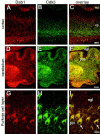
Fig. 1.
Fig. 1.
Dab1 and Cdk5 display distinct but overlapping expression patterns in the cerebral cortex and cerebellum at postnatal day 0. A, Dab1 (red) is present in the cell bodies and dendritic processes of neurons in the cerebral cortex.B, Cdk5 (green) is detected in postmitotic neurons in the cortical plate and in the thalamocortical fibers. C, Double staining shows that Dab1 and Cdk5 expression overlap in postmitotic neurons in the cerebral cortex.D, In the cerebellum, Dab1 is expressed in Purkinje cells as they migrate to form the Purkinje cell layer.E, Cdk5 is expressed in Purkinje cells, but it is also present in cerebellar nuclei and in axon tracts in the cerebellum.F, Double staining shows that Dab1 and Cdk5 overlap in the Purkinje cells of the developing cerebellum (yellow). Scale bar (in F), ∼100 μm. G, Dab1 is present in Purkinje cell bodies and dendrites in the postnatal day 7 cerebellum. H, Cdk5 is also present in Purkinje cell bodies and dendrites.I, Double staining in the same tissue section shows the overlap of Dab1 and Cdk5 expression in Purkinje cells.Arrows point out individual Purkinje cells in the overlay image. Scale bar (in I), ∼10 μm.mz, Marginal zone; cp, cortical plate;wm, white matter; egl, external germinal layer; pcl, Purkinje cell layer.
Fig. 2.
Fig. 2.
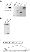
Cdk5–p35 immunoprecipitated from brain phosphorylates Dab1 in vitro. A, The Cdk5–p35 complex was immunoprecipitated from detergent lysates of either Cdk5−/− orCdk5+/+ P0 brains. The immunoprecipitates were then used as source of kinase activity to phosphorylate either full-length Dab1-His or GST fusion proteins containing Dab1 domains (GST-PTB, residues 1–179; GST-Mid, residues 180–399; GST-CT, residues 400–555) in the presence of [γ-32P]ATP in vitro. Proteins were separated by SDS-PAGE, transferred to nitrocellulose, and located by autoradiography. B, In vitrophosphorylated GST-CT and Dab1-His (from A) were trypsinized off the membrane, and tryptic fragments were resolved by alkaline 40% polyacrylamide gel electrophoresis. Autoradiography revealed two major phosphopeptides (phosphopeptides 1and 2). C, Schematic representation of Dab1 domains. The C-terminal region of Dab1 (CT) gives rise to two phosphopeptides as a result of phosphorylation by Cdk5–p35. Two serines, serine 491 and serine 515, contain the consensus sequence for Cdk5 phosphorylation, including proline in +1 position and lysine residue in +3 position.
Fig. 3.
Fig. 3.
Characterization of anti-PSer491 phosphopeptide-specific antibodies. A, HEK293T cells were transfected with either Dab1-HA alone (−) or Dab1-HA plus both Cdk5 and p35 (+). Lysates from the transfected cells were separated by SDS-PAGE, transferred to nitrocellulose, and probed with antibodies raised against a phosphopeptide encompassing PSer491. To confirm the presence of Dab1, the membrane was stripped and reprobed with anti-Dab1 antibodies. B, Wild-type Dab1-HA (Wt) or mutant forms of Dab1 carrying alanine substitutions at residues 491 (491A), 515 (515A), or both 491 and 515 (AA) were transiently expressed in HEK293T cells along with Cdk5 and p35. The site-specificity of the antibodies was assessed by Western blotting of the resulting lysates.
Fig. 4.
Fig. 4.
Cdk5 is required for in vivophosphorylation of serine 491. Brain lysates prepared from E16.5 (A) or P0 (B)Cdk5−/−,Cdk5−/+, orCdk5+/+ embryos were immunoprecipitated with anti-Dab1 antibodies. The immunoprecipitates were separated by SDS-PAGE and analyzed by Western blotting using anti-PSer491, anti-PTyr198, 4G10, or anti-Dab1 antibodies. Western blots with anti-Cdk5 and anti-actin antibodies were performed on total lysates.
Fig. 5.
Fig. 5.
Dab1 is elevated in ectopic neurons in the cerebral cortex and cerebellum. Immunohistochemistry was performed on cortical and cerebellar tissue sections from E18.5Cdk5+/+ (A,B, E, F) andCdk5−/− (C,D, G, H) mice.A, Cresyl violet staining ofCdk5+/+ mice was used to visualize the normal arrangement of neurons in the cerebral cortex. B, In Cdk5+/+ mice, Dab1 is present in the cell bodies and within the dendrites of cortical plate (cp) neurons. Low or no immunostaining was observed in the subplate (sp) and intermediate zone (iz). D, In theCdk5−/− cerebral cortex, a thin cortical plate (cp) forms above a subplate (sp), and neurons accumulate beneath the subplate in an ectopic cortical plate (e-cp). C, Dab1 levels appear elevated in neurons that occupy the ectopic cortical plate (e-cp), located beneath the subplate.E, Cresyl violet staining of the cerebellum inCdk5+/+ mice shows the location of the Purkinje cell layer (pcl) and the external germinal layer (egl). F, Dab1 is present at detectable levels in Purkinje cells aligned in the Purkinje cell layer (arrows in E andF). H, In theCdk5−/− cerebellum, the ectopic Purkinje cells (e-pc) fail to form a Purkinje cell layer and instead accumulate close to the ventricle as shown by cresyl violet staining. G, Dab1 is dramatically elevated in the ectopic Purkinje cells (arrowheads). Digital images were captured at identical settings in B and Cand in F and G. Scale bar (inH), ∼100 μm. mz, Marginal zone; vz, ventricular zone.
Fig. 6.
Fig. 6.
Cdk5 is not required for Reelin-induced tyrosine phosphorylation of Dab1. Neurons isolated from E16.5Cdk5−/− orCdk5+/+ embryos were suspended in DMEM alone (Con) or Reelin-enriched DMEM (Reln). Treated neurons were then lysed and immunoprecipitated with anti-Dab1 antibodies. The immunoprecipitates were separated by SDS-PAGE and transferred to nitrocellulose, and Western blots were performed using 4G10 anti-phosphotyrosine (PTyr) or anti-Dab1 (Dab1) antibodies. Lysates were also probed with anti-NSE antibodies to confirm equal sample loading.
Fig. 7.
Fig. 7.
Phosphorylation of Dab1 on serine 491 does not require Reelin signaling. A, Brain lysates prepared from E14.5 reeler (rl/rl) or wild-type (wt) littermates were immunoprecipitated with anti-Dab1 antibodies. The immunoprecipitates were then separated by SDS-PAGE, transferred to nitrocellulose, and probed with anti-Dab1, anti-phosphotyrosine, or anti-PSer491 antibodies. B, E16.5 reeler neurons were either left untreated (Con) or treated with Reelin (Reln), and phosphorylation was determined by anti-phosphotyrosine and anti-PSer491 antibodies as before.
Fig. 8.
Fig. 8.
Changes in Dab1 serine and tyrosine phosphorylation during development. Brain lysates were prepared from E15.5, P0, P3, P8, P16, or P42 wild-type mice. Anti-Dab1 immunoprecipitates from these lysates were separated by SDS-PAGE and transferred to nitrocellulose. The membrane was sequentially probed with anti-phosphotyrosine, anti-PSer491, and anti-Dab1 antibodies.

Similar articles

Cited by

References

    1. Borrell V, Del Rio JA, Alcantara S, Derer M, Martinez A, D'Arcangelo G, Nakajima K, Mikoshiba K, Derer P, Curran T, Soriano E. Reelin regulates the development and synaptogenesis of the layer-specific entorhino-hippocampal connections. J Neurosci. 1999;19:1345–1358. - PMC - PubMed
    1. Chae T, Kwon YT, Bronson R, Dikkes P LiE, Tsai LH. Mice lacking p35, a neuronal specific activator of Cdk5, display cortical lamination defects, seizures, and adult lethality. Neuron. 1997;18:29–42. - PubMed
    1. D'Arcangelo G, Curran T. Reeler: new tales on an old mutant mouse. BioEssays. 1998;20:235–244. - PubMed
    1. D'Arcangelo G, Miao GG, Chen SC, Soares HD, Morgan JI, Curran T. A protein related to extracellular matrix proteins deleted in the mouse mutant reeler. Nature. 1995;374:719–723. - PubMed
    1. D'Arcangelo G, Homayouni R, Keshvara L, Rice DS, Sheldon M, Curran T. Reelin is a ligand for lipoprotein receptors. Neuron. 1999;24:471–479. - PubMed

Publication types

MeSH terms