. 2002 Jun 25;99(13):8554-61.
doi: 10.1073/pnas.082253699. Epub 2002 Jun 19.

Zinc finger as distance determinant in the flexible linker of intron endonuclease I-TevI

Affiliations

Zinc finger as distance determinant in the flexible linker of intron endonuclease I-TevI

Amy B Dean et al. Proc Natl Acad Sci U S A. .

Abstract

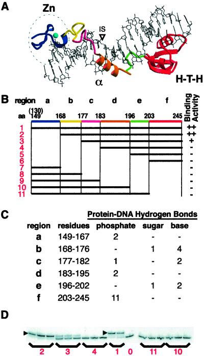
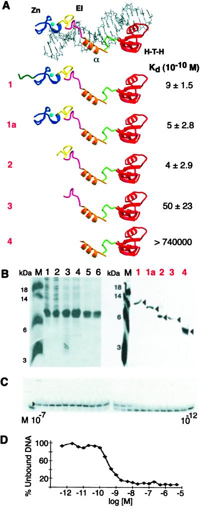
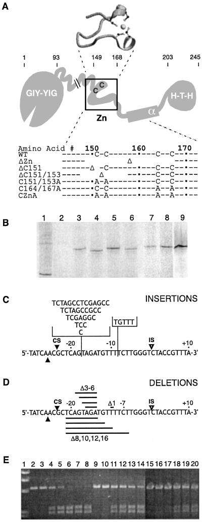
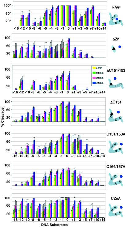
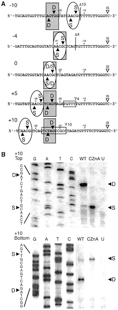
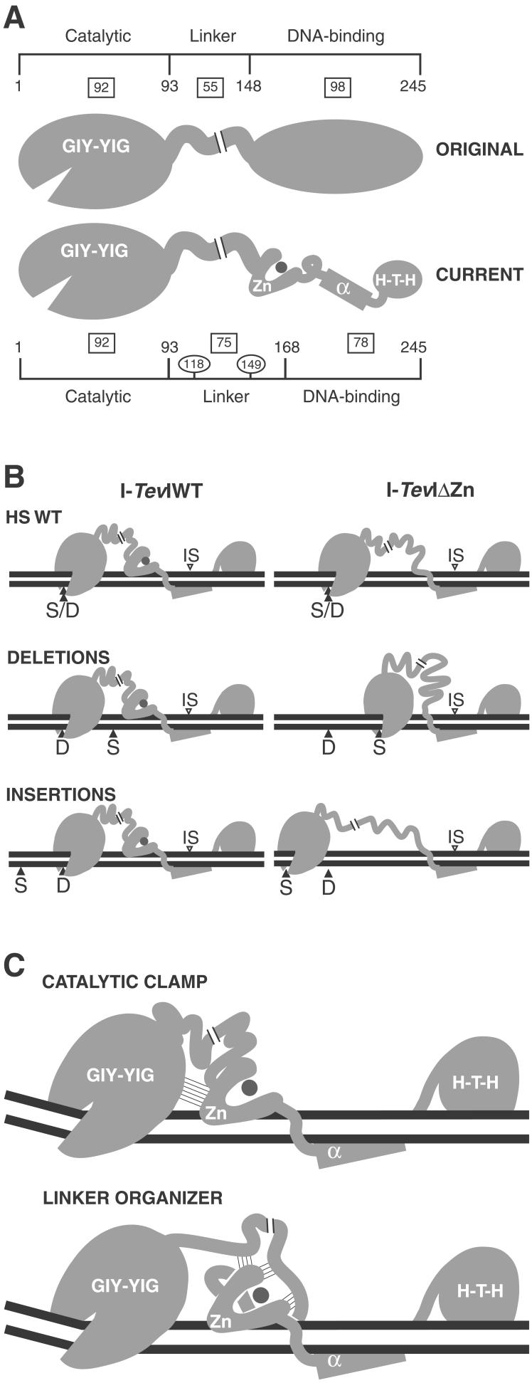
I-TevI, the phage T4 td intron-encoded endonuclease, recognizes a lengthy DNA target and initiates intron mobility by introducing a double-strand break in the homing site. The enzyme uses both sequence and distance determinants to cleave the DNA 23-25 bp upstream of the intron insertion site. I-TevI consists of an N-terminal catalytic domain and a C-terminal DNA-binding domain separated by a long, flexible linker. The DNA-binding domain consists of three subdomains: a zinc finger, a minor-groove binding alpha-helix, and a helix-turn-helix. In this study, a mutational analysis was undertaken to assess the roles of these subdomains in substrate binding and cleavage. Surprisingly, the zinc finger is not required for DNA binding or catalysis. Rather, the zinc finger is a component of the linker and directs the catalytic domain to cleave the homing site at a fixed distance from the intron insertion site. When the cleavage site (CS) is shifted outside a given range, wild-type I-TevI defaults to the fixed distance, whereas zinc-finger mutants have lost the distance determinant and search out the displaced cleavage sequences. Although counterintuitive, a protein containing a 19-aa deletion of the zinc finger can extend further than can wild-type I-TevI to cleave a distant CS sequence, and a Cys-to-Ala mutant of the ligands for zinc, nominally a longer protein, can retract to cleave at a closer CS sequence. Models are presented for the novel function of the zinc finger, as a molecular constraint, whereby intramolecular protein-protein interactions position the catalytic domain by "catalytic clamp" and/or "linker-organizer" mechanisms.


Figures

Figure 1
Figure 1
Binding activity of deletion derivatives. (A) The crystal structure of the I-TevI DNA-binding domain bound to substrate (12). Three subdomains are: Zn, zinc finger; α, α-helix; H-T-H, helix–turn–helix. The colored elements correspond to segments a–f of equivalent color in B. (B) Deletion derivatives. The horizontal black bars show the portion of the binding domain that is expressed in each construct, numbered 1–11. Protein 1 starts at residue 130 but the visible crystal structure begins at residue 149. Region a, zinc finger; b and c, extended region I; d, α-helix; e, extended region II; and f, H-T-H. Qualitative binding activities with cell lysates are shown on the right. ++, strong binding; +, weak binding; −, no detectable binding. (C) Protein hydrogen bond contacts to DNA organized by protein region and DNA contact. (D) EMSAs for representative constructs. ▸ = shifted complex. Numbers correspond to constructs in B. Lane 0 = DNA only.
Figure 2
Figure 2
Kd determination for I-TevI subdomain derivatives. (A) Summary of deletion derivatives and Kds. Protein–DNA complex labeled as in Fig. 1A. EI, elongated region I. Data from 3–5 experiments were used to derive the Kds. (B) Purification of I-TevI deletion derivatives. (Left) Purification of construct 2. M, molecular mass markers; lane 1, crude extract; lane 2, soluble lysate; lane 3, 0.2% polyethyleneimine supernatant; lane 4, 30–60% saturated (NH4)2SO4 pellet and heparin column load; lanes 5 and 6, heparin column fractions. (Right) Purified protein samples that were used for the Kd determinations of constructs 1, 1a, 2, 3, and 4. (C) EMSA with construct 1a. I-TevI derivatives were bound to 100-bp DNA (3.4 × 10⁻¹² M) containing the homing site. The binding reaction mixtures contained the protein (1a) at concentrations ranging from 1.0 × 10⁻⁷ M to 1.0 × 10⁻¹² M. Protein concentration ranges were as follows for the remaining proteins: 1, 2.5 × 10⁻⁷ M to 2.5 × 10⁻¹¹ M; 2, 2.0 × 10⁻⁶ M to 2.0 × 10⁻¹³ M; and 3, 5.0 × 10⁻⁶ M to 5.0 × 10⁻¹³ M. Protein 4 showed no evidence of binding to homing site DNA at 7.4 × 10⁻⁵ M. (D) Bjerrum plot of the binding experiment depicted in C.
Figure 3
Figure 3
Endonuclease and substrate variants. (A) Schematic representation of full-length I-TevI variants. The structure of the zinc finger as seen in the cocrystal structure (12) is shown above the schematic. C and A indicate relevant cysteines and alanines; Δ indicates deletion; spaces indicate the extent of deletion; and ⋅ indicates every 10th amino acid. The regions of I-TevI are: GIY-YIG, catalytic domain; Zn, zinc finger; α, α-helix; H-T-H, helix–turn–helix. (B) In vitro translation of I-TevI zinc-finger deletion variants. Wheat germ extract and 35S-labeled methionine were used for translations with RNA runoff transcript (10). Lane 1, bovine mosaic virus translation product as a positive control; lane 2, unprogrammed extract as a negative control; lane 3, wild-type I-TevI; lane 4, ΔZn; lane 5, ΔC151; lane 6, ΔC151/153; lane 7, C151/153A; lane 8, C164/167A; and lane 9, CZnA. (C) Schematic of the homing-site insertion variants. The locations for insertions of base pairs (+1, +3, +5, +7, +10, +14) are indicated on the top strand with the vertical line. The horizontal brackets contain the inserted sequences in each case. IS and open arrowheads indicate the IS while CS and closed arrowheads indicate the CSs on each strand. (D) Schematic of the homing-site deletion variants. The deletions of base pairs (−1, −3, −4, −5, −6, −8, −10, −12, −16) are indicated on the bottom strand by the horizontal black bands. (E) Representative cleavage data with cleavage products resolved on an agarose gel. Lane 1, molecular weight markers; lanes 2–8, wild-type I-TevI on wild-type DNA; lanes 9–14, CZnA mutant on +3 DNA; and lanes 15–20, CZnA mutant on +7 DNA. Lanes 2, 9, and 15 contain DNA only; lanes 3, 10, and 16 contain DNA with unprogrammed lysates. Incubation times were 1, 2, 4, 7, and 10 min for lanes 4–8, and 5, 10, 20 and 30 min for lanes 11–14 and 17–20, respectively.
Figure 4
Figure 4
Summary of cleavage data of zinc-finger mutants. Each graph represents the compiled data for one protein on the different DNA homing sites. The homing sites are ordered from deletions to wild type to insertions from left to right [−16, −12, −10, −8, −6, −5, −4, −3, −1, 0 (wild type), +1, +3, +5, +7, +10, and +14]. The average percent cleavage at four time points is presented for each protein/DNA pair (5, 10, 20, and 30 min). *, The % cleavage for wild-type I-TevI was derived at a 3-fold lower protein concentration than was used for the mutants. The data for which 100% cleavage is seen with no error bars indicate that the DNA was completely cleaved under the conditions of this experiment; cleavage activity is in reality higher. Accurate relative activities of the mutants compared with wild-type I-TevI on wild-type homing-site DNA are given in the text. The mutant proteins were all run at equivalent concentrations and can be compared with one another directly. Error bars represent the SD in the data from 2–5 cleavage assays. The schematic on the right corresponds to the boxed portion of I-TevI in Fig. 3A. The zinc ion (dot) is shown either coordinated (wild-type I-TevI) or not (mutants). The triangles correspond to deletions.
Figure 5
Figure 5
CS mapping for I-TevI variants. (A) CSs indicated on each of the DNA sequences. Vertical lines indicate the site of the 10- and 4-bp deletions. The horizontal boxes indicate the 5- and 10-bp insertions. Gray rectangles indicate cleavage by wild-type I-TevI, and ovals indicate cleavage by ΔZn and CZnA. (B) Top and bottom strand CS mapping for wild-type I-TevI and CZnA on the +10 homing site variant (Fig. 3). G, A, T, and C indicate the bases represented in the sequencing ladders. WT, cleavage by wild-type I-TevI for 5 min; CZnA, cleavage by CZnA for 20 min; U, unprogrammed extract; S, cleavage at the wild-type cleavage sequence; D, cleavage at the wild-type cleavage distance from the IS. Other labels are as in Fig. 3.
Figure 6
Figure 6
Models for the role of the zinc finger in I-TevI. The regions of I-TevI were: GIY-YIG, catalytic domain; Zn, zinc finger; α, α-helix; H-T-H, helix–turn–helix. (A) Domains of I-TevI. Original and current models are shown. Boxes indicate the number of residues in each segment of the protein. Residue numbers indicate domain boundaries. Numbers in ovals demarcate the three segments of the linker. (B) Distance-constraining activity of the zinc finger. At large deletions and insertions, wild-type I-TevI defaults to cleave at wild-type distance (Left), whereas ΔZn cleaves predominantly at native cleavage sequence (Right). Labels are as in Fig. 5. (C) Models for directing catalytic domain to a fixed distance. Thin lines depict hypothetical interactions. The catalytic-clamp model shows interactions between the zinc finger and the catalytic domain, whereas the linker-organizer model shows the zinc finger interacting with the linker, to position the catalytic center of I-TevI at the CS. In both models the zinc finger is proposed to be anchored on the DNA, and in both cases, protein–protein interactions limit flexibility and therefore promote distance-specific cleavage by the sequence-tolerant catalytic domain.

Similar articles

Cited by

References

    1. Lambowitz A M, Belfort M. Annu Rev Biochem. 1993;62:587–622. - PubMed
    1. Belfort M, Roberts R J. Nucleic Acids Res. 1997;25:3379–3388. - PMC - PubMed
    1. Lambowitz A M, Caprara M G, Zimmerly S, Perlman P S. In: The RNA World. Gesteland R F, Cech T R, Atkins J F, editors. Plainview, NY: Cold Spring Harbor Lab. Press; 1999. pp. 451–485.
    1. Gimble F S. FEMS Microbiol Lett. 2000;185:99–107. - PubMed
    1. Chevalier B S, Stoddard B L. Nucleic Acid Res. 2001;29:3757–3774. - PMC - PubMed

Publication types

LinkOut - more resources