. 2002 Jun 25;99(13):8512-7.
doi: 10.1073/pnas.132274899. Epub 2002 Jun 19.

Identification of a gene encoding MGAT1, a monoacylglycerol acyltransferase

Affiliations

Identification of a gene encoding MGAT1, a monoacylglycerol acyltransferase

Chi-Liang Eric Yen et al. Proc Natl Acad Sci U S A. .

Abstract

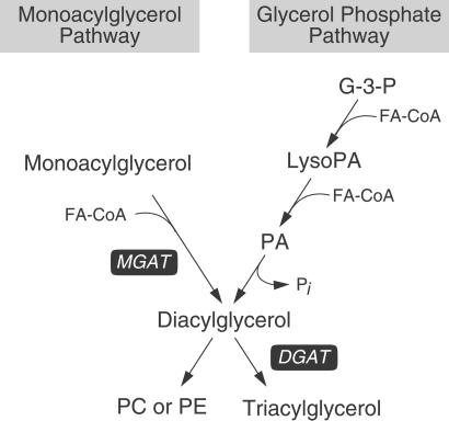
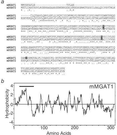
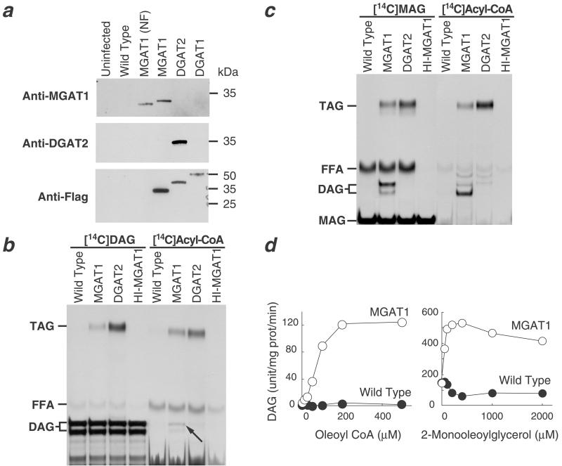
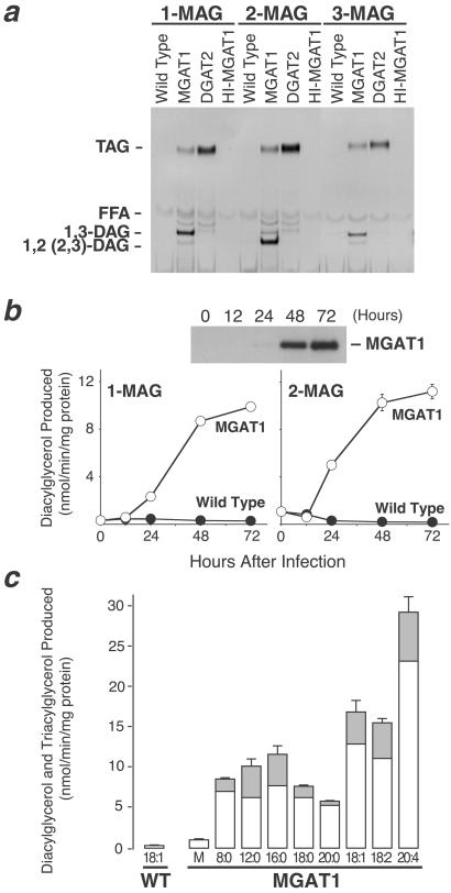
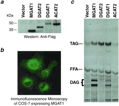
Acyl-CoA:monoacylglycerol acyltransferase (MGAT) catalyzes the synthesis of diacylglycerol, the precursor of physiologically important lipids such as triacylglycerol and phospholipids. In the intestine, MGAT plays a major role in the absorption of dietary fat because resynthesis of triacylglycerol is required for the assembly of lipoproteins that transport absorbed fat to other tissues. MGAT activity has also been reported in mammalian liver and white adipose tissue. However, MGAT has never been purified to homogeneity from mammalian tissues, and its gene has not been cloned. We identified a gene that encodes an MGAT (MGAT1) in mice. This gene has sequence homology with members of a recently identified diacylglycerol acyltransferase gene family. Expression of the MGAT1 cDNA in insect cells markedly increased MGAT activity in cell membranes. In addition, MGAT activity was proportional to the level of MGAT1 protein expressed, and the amount of diacylglycerol produced depended on the concentration of either of its substrates, oleoyl-CoA or monooleoylglycerol. In mice, MGAT1 expression and MGAT activity were detected in the stomach, kidney, white and brown adipose tissue, and liver. However, MGAT1 was not expressed in the small intestine, implying the existence of a second MGAT gene. The identification of the MGAT1 gene should greatly facilitate research on the identification of the intestinal MGAT gene and on the function of MGAT enzymes in mammalian glycerolipid metabolism.


Figures

Figure 1
Figure 1
The two major pathways for synthesizing diacylglycerol. In the monoacylglycerol pathway, MGAT produces diacylglycerol, the precursor of triacylglycerol and certain phospholipids, by covalently joining a fatty acyl moiety to monoacylglycerol. In the glycerol phosphate pathway, diacylglycerol is derived from the dephosphorylation of phosphatidic acid (PA), which is produced by sequential acylation of glycerol 3-phosphate (G-3-P). FA-CoA, fatty acyl-CoA; LysoPA, lysophosphatidic acid; Pi, inorganic phosphate; PC, phosphatidylcholine; PE, phosphatidylethanolamine.
Figure 2
Figure 2
Mouse MGAT1 protein sequence analysis. (a) Alignment of predicted mouse MGAT1 (mMGAT1) amino acid sequences with mouse DGAT2 (mDGAT2). Amino acid residues identical for both MGAT1 and DGAT2 are indicated with an asterisk; conservation of strong groups is indicated with a colon; conservation of weak groups is indicated with a period. Two putative N-linked glycosylation sites and one potential tyrosine phosphorylation site are also indicated. (b) Hydrophobicity plot of mMGAT1 as assessed by Kyte–Doolittle analysis (20). The bold line indicates a predicted transmembrane domain (http://www.cbs.dtu.dk/services/TMHMM/).
Figure 3
Figure 3
Expression of mouse MGAT1 cDNA in Sf9 insect cells. (a) Immunoblots of insect cell membranes. Expression of MGAT1 and DGAT2 was verified by immunoblotting with specific antibodies or an anti-FLAG antibody. Membrane proteins (5 μg) were analyzed from uninfected Sf9 cells, cells infected with wild-type virus, non-FLAG-tagged MGAT1 (MGAT1-NF), FLAG-tagged-MGAT1 (MGAT1), FLAG-tagged-DGAT2 (DGAT2), or FLAG-tagged-DGAT1 (DGAT1) recombinant baculoviruses. (b) Weak DGAT activity conferred by expression of MGAT1. DGAT activity was detected by incorporation of [14C]dioleoylglycerol ([14C]DAG) or [14C]oleoyl-CoA ([14C]Acyl-CoA) into triacylglycerol. The arrow indicates the incorporation of [14C]oleoyl-CoA into diacylglycerol by membranes expressing MGAT1. In this TLC system, the three stereoisomers of diacylglycerol migrate as a doublet, in which the upper species is sn-1,3-diacylglycerol and the lower species is a combination of sn-1,2- and sn-2,3-diacylglycerol. FFA, free fatty acid; HI-MGAT1, heat-inactivated MGAT1; TAG, triacylglycerol. (c) Strong MGAT activity conferred by expression of MGAT1. MGAT activity was detected by using either sn-1-[14C]monooleoylglycerol ([14C]MAG) or [14C]oleoyl-CoA ([14C]acyl-CoA) as the radioactive substrate. (d) Dependence of MGAT activity in membranes expressing MGAT1 on substrate concentrations. MGAT activity was assessed by measuring diacylglycerol mass in membranes after the assay. Values are the mean ± SD of four measurements.
Figure 4
Figure 4
Substrate specificities of MGAT1. (a) Ability of MGAT1 expressed in insect cells to acylate each of the three monoacylglycerol stereoisomers [sn-1-monooleoylglycerol (1-MAG), sn-2-monooleoylglycerol (2-MAG), or sn-3-monostearoylglycerol (3-MAG)]. FFA, free fatty acid. (b) Proportional increases in MGAT1 protein and MGAT activity in insect cell membranes. The specific activities of MGAT1 with sn-1-monooleoylglycerol (1-MAG) and sn-2-monooleoylglycerol (2-MAG) substrates are similar. Values are the mean ± SD of three measurements. (c) Preference of MGAT1 for unsaturated fatty acyl-CoAs. sn-2-[3H]monooleoylglycerol was used as the acyl acceptor for different fatty acyl-CoAs (50 μM each). M, malonyl-CoA; 8:0, n-octanoyl-CoA; 12:0, lauroyl-CoA; 16:0, palmitoyl-CoA; 18:0, stearoyl-CoA; 20:0, arachidoyl-CoA; 18:1, oleoyl-CoA; 18:2, linoleoyl-CoA, and 20:4, arachidonoyl-CoA. White and gray bars represent radioactivity recovered from incorporation of the label into diacylglycerol and triacylglycerol, respectively. Because sn-2-[3H]monooleoylglycerol was used the labeled substrate, incorporation of radioactivity into diacylglycerol and triacylglycerol represents total MGAT activity. Values are the mean ± SD of four measurements and are representative of two independent experiments.
Figure 5
Figure 5
Expression of MGAT1 in mammalian cells. (a) Immunoblotting of FLAG-tagged proteins to verify expression of MGAT1 and control proteins. Cells were transfected with vector only or with FLAG-tagged versions of MGAT1, DGAT2, DGAT1, or ACAT2. (b) Immunocytochemistry of COS-7 cells expressing FLAG-tagged MGAT1. An anti-FLAG antibody and FITC-conjugated secondary antibody were used. (c) Acyltransferase activities in COS-7 cell membranes. [14C]oleoyl-CoA was used for enzyme assays.
Figure 6
Figure 6
Tissue expression pattern of mouse MGAT1. (a) MGAT1 mRNA expression was detected by Northern analysis with 20 μg of total RNA. The same blots were probed for DGAT2 as a control for the presence of mRNA. (b) MGAT1 mRNA expression in liver was detected by Northern analysis with 2 μg (Left) or 5 μg (Right) poly(A)+ RNA. Sm, small; Sk, skeletal; WAT, white adipose tissue; BAT, brown adipose tissue. (c) MGAT activity in mouse tissues. Values are the mean ± SD of four measurements.

Similar articles

Cited by

References

    1. Ron D, Kazanietz M G. FASEB J. 1999;13:1658–1676. - PubMed
    1. Lehner R, Kuksis A. Prog Lipid Res. 1996;35:169–201. - PubMed
    1. Bell R M, Coleman R A. Annu Rev Biochem. 1980;49:459–487. - PubMed
    1. Coleman R A, Haynes E B. J Biol Chem. 1984;259:8934–8938. - PubMed
    1. Mostafa N, Bhat B G, Coleman R A. Lipids. 1994;29:785–791. - PubMed

Publication types