. 2002 Jul;22(14):5089-99.
doi: 10.1128/MCB.22.14.5089-5099.2002.

The Dorsal Rel homology domain plays an active role in transcriptional regulation

Affiliations

The Dorsal Rel homology domain plays an active role in transcriptional regulation

Songtao Jia et al. Mol Cell Biol. 2002 Jul.

Abstract

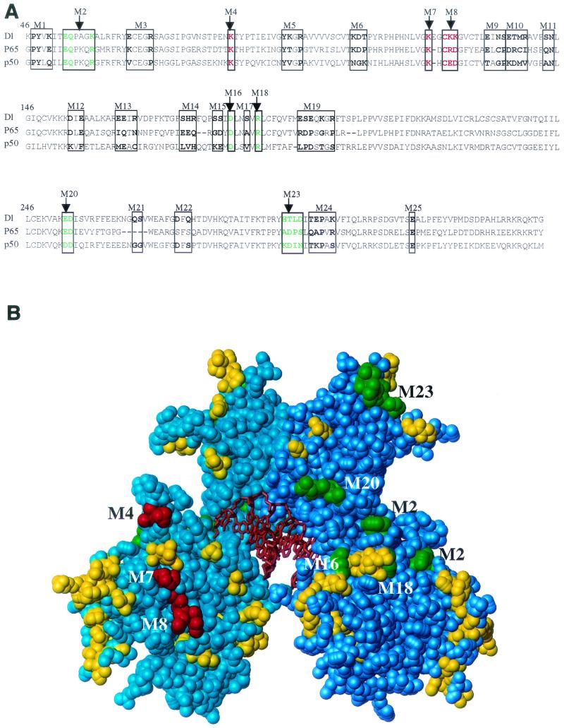
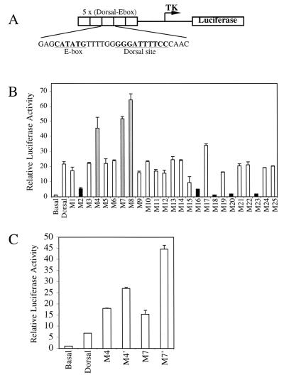
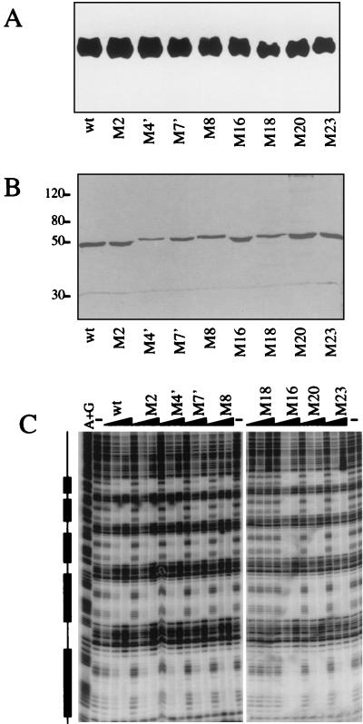
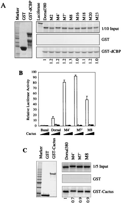
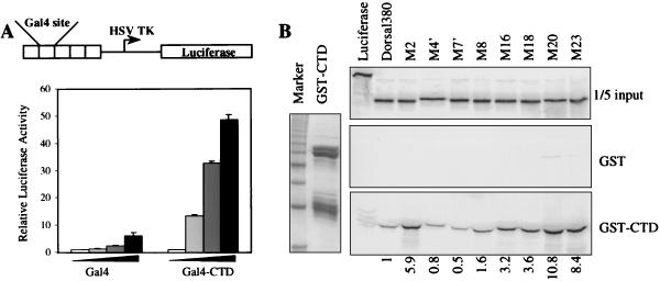
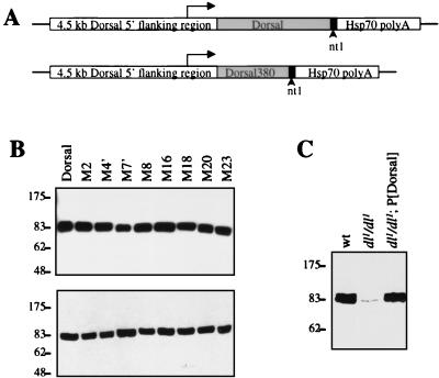
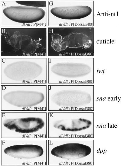
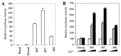
The Dorsal morphogen directs formation of the Drosophila dorsoventral axis by both activating and repressing transcription. It contains an N-terminal Rel homology domain (RHD), which is responsible for DNA binding and regulated nuclear import, and a C-terminal domain (CTD) that contains activation and repression motifs. To determine if the RHD has a direct role in transcriptional control, we analyzed a series of RHD mutations in S2 cells and embryos. Two classes of mutations (termed class I and class II mutations) that alter activation without affecting DNA binding or nuclear import were identified. The two classes appear to define distinct protein interaction surfaces on opposite faces of the RHD. Class I mutations enhance an apparently inhibitory interaction between the RHD and the CTD and eliminate both activation and repression by Dorsal. In contrast, class II mutations result in increased activation in S2 cells but severely decreased activation in embryos and have little effect on repression. Analysis of the cuticles of class II mutant embryos suggests that, in the absence of Dorsal-mediated activation, Dorsal-mediated repression is not sufficient to pattern the embryo. These results provide some of the first evidence that the RHD plays an active role in transcriptional regulation in intact multicellular organisms.


Figures

FIG. 1.
FIG. 1.
Mutagenesis of the Dorsal RHD. (A) Sequence alignment of the Dorsal RHD with those of p65 and p50. The amino acids that were mutated are indicated in bold and boxed. Amino acids changed in class I and class II mutants are colored green and red, respectively. (B) Space-filling model of the RHD. The RHD homodimer structure is based on the coordinates determined for p50 (Protein Data Bank identification no. 1NF-K). The two polypeptide chains are shown in cyan and blue, and the DNA is shown in orange. The amino acids that were altered in the mutants are shaded green (class I mutations), red (class II mutations), and yellow (phenotypically silent mutations).
FIG. 2.
FIG. 2.
Cotransfection assays reveal two classes of mutants. (A) Structure of the DE5 reporter. (B) Activation of the DE5 reporter by Dorsal mutants. S2 cells were transfected with 60 ng of a plasmid encoding the wild-type or indicated mutant forms of Dorsal together with 20 ng of a plasmid encoding Twist, 5 μg of the DE5 reporter, and 0.1 μg of a plasmid containing the Renilla luciferase gene under the control of the herpes simplex virus thymidine kinase (TK) promoter. Cell extracts were prepared and assayed for luciferase activity 2 days posttransfection. Firefly luciferase activities were first divided by the Renilla luciferase activities to normalize for variations in transfection efficiency. The normalized values were then divided by the basal value (no activators) to obtain the relative luciferase activity. Results shown are the averages of duplicate assays, and error bars show the standard deviation. Three mutants (class II mutants M4, M7, and M8; gray bars) were found to give significantly better activation than wild-type Dorsal, while five mutants (class I mutants M2, M16, M18, M20, and M23; black bars) yielded significantly reduced Dorsal-mediated activation. (C) Effect of replacing the alanine substitutions in the class II mutants with glutamic acid substitutions. The mutants were analyzed as described for panel B.
FIG. 3.
FIG. 3.
Effects of the mutations on protein stability and DNA-binding activity. (A) Mutations do not affect levels of nuclear Dorsal protein. S2 cells (50 ml) were transiently transfected with 2 μg of wild-type (wt) Dorsal380 or the indicated mutant forms of Dorsal380. Cells were harvested 2 days posttransfection, and nuclear extracts were prepared. The proteins were resolved by SDS-10% PAGE, transferred to a polyvinylidene difluoride membrane, and probed with anti-Dorsal antibody. (B) Purification of recombinant Dorsal mutants. Flag-tagged Dorsal380 or the indicated Dorsal380 mutants were immunopurified to homogeneity from Sf9 cells infected with recombinant baculoviruses. Proteins were resolved by SDS-10% PAGE and silver stained. (C) With one exception, the mutations do not alter DNA-binding activity. The purified proteins shown in panel B were assayed for binding to the DE5 enhancer by a DNase I footprinting assay. The boxes to the left indicate the Dorsal binding sites. Each protein was assayed at 2, 10, and 50 ng. Lane A+G, A+G chemical sequencing ladder; lane −, no-protein control.
FIG. 4.
FIG. 4.
Mutations do not affect interactions of Dorsal with known partner proteins. (A) Mutations do not affect binding of Dorsal to the coactivator dCBP. Left, purified GST and GST-dCBP(781-1159) were resolved by SDS-PAGE and visualized by staining with Coomassie blue. Right, in vitro-translated and 35S-labeled luciferase, Dorsal380 (a C-terminal truncation of Dorsal containing the intact RHD), and the indicated Dorsal380 mutants were incubated with equal amounts of GST or GST-dCBP immobilized on glutathione beads and washed extensively. The eluted proteins were resolved by SDS-PAGE and imaged by autoradiography. One-tenth of the input protein amount is shown for comparison. (B) Class II mutants are still responsive to Cactus in vivo. S2 cells was cotransfected with 60 ng of a plasmid encoding full-length Dorsal or the indicated Dorsal mutants together with 20 ng of a plasmid encoding Twist, 5 μg of the DE5 reporter, and 0 ng (white bars), 100 ng (gray bars), or 500 ng (black bars) of a plasmid encoding Cactus. Luciferase activity was determined as described in the legend to Fig. 1A. (C) Class II mutants bind Cactus in vitro. Left, GST and GST-Cactus purified from E. coli and stained with Coomassie blue. Right, In vitro-translated and 35S-labeled Dorsal380 or the indicated Dorsal380 mutants were incubated with either GST or GST-Cactus immobilized on glutathione beads and washed extensively. The eluted proteins were resolved by SDS-PAGE and imaged by autoradiography. One-fifth of the input protein amount is shown for comparison. Quantification of the interaction is shown below the lanes, with wild-type binding arbitrarily set to 1.
FIG. 5.
FIG. 5.
Interaction between RHD and CTD. (A) Dorsal CTD can activate transcription. Top, schematic diagram of the G5 luciferase reporter. The firefly luciferase gene is under the control of five copies of a module containing Gal4 binding sites. These are inserted immediately upstream of the herpes simplex virus thymidine kinase (HSV TK) core promoter. Bottom, S2 cells were transfected with 5 μg of the G5 luciferase reporter and 0.1 μg of a plasmid containing the Renilla luciferase gene under the control of the herpes simplex virus thymidine kinase promoter, together with 0 ng, 150 ng, 500 ng, or 2 μg of pPac-Gal4 or pPac-Gal4-CTD. (B) The RHD binds the CTD. Left, purified GST-CTD. Right, in vitro-translated and 35S-labeled Dorsal380 or the indicated Dorsal380 mutants were incubated with either GST or GST-Dorsal-CTD immobilized on glutathione beads and washed extensively. The eluted proteins were resolved by SDS-PAGE and imaged by autoradiography. One-fifth of the input protein amount is shown for comparison. Quantification of the interaction is shown below the lanes, with wild-type binding arbitrarily set to 1.
FIG. 6.
FIG. 6.
Transformation of germ line with P-elements encoding Dorsal mutants. (A) Structure of expression vectors. A 4.5-kb region from the endogenous dl locus was used to direct the expression of full-length Dorsal, full-length Dorsal mutants, and Dorsal380. The 19-amino-acid nt1 epitope was appended to the C terminus of the encoded proteins. The constructs also contained the hsp70 polyadenylation signal. (B) Analysis of the expression level of dl transgenes. Forty embryos from mothers bearing transgenes encoding the indicated nt1-tagged forms of Dorsal were homogenized in SDS-PAGE loading buffer, resolved by SDS-PAGE, transferred to a polyvinylidene difluoride membrane, and probed with anti-nt1 antibody. (C) Comparison of the expression levels of endogenous and transgenic Dorsal proteins. Embryos with the indicated maternal genotypes were analyzed as in panel B except with anti-Dorsal antibody.
FIG. 7.
FIG. 7.
Class I mutations abolish Dorsal function in the embryo. (A, F, K, P, and U) Whole-mount anti-nt1 antibody staining of embryos laid by mothers with the indicated genotypes. (B, G, L, Q, and V) Cuticle preparations of embryos laid by mothers with the indicated genotypes. (C, H, M, R, and W) Whole-mount in situ hybridization with digoxigenin-labeled antisense riboprobe against the twi mRNA to embryos laid by mothers with the indicated genotypes. (D, I, N, S, and X) Whole-mount in situ hybridization with digoxigenin-labeled antisense riboprobe against the sna mRNA to embryos laid by mothers with the indicated genotypes. (E, J, O, T, and Y) Whole-mount in situ hybridization with digoxigenin-labeled antisense riboprobe against the dpp mRNA to embryos laid by mothers with the indicated genotypes. Embryos are oriented with the anterior to the left and the dorsal side up. Arrows in panels M, N, R, and S indicate the positions in the embryos where twi and sna expression is reduced.
FIG. 8.
FIG. 8.
Class II mutants and Dorsal380 show reduced activation in vivo. (A and G) Whole-mount anti-nt1 antibody staining of embryos laid by mothers with the indicated genotypes. (B and H) Cuticle preparations of embryos laid by mothers with the indicated genotypes. Filzkörper are indicated by arrows. (C and I) Whole-mount in situ hybridization with digoxigenin-labeled antisense riboprobe against the twi mRNA to embryos laid by mothers with the indicated genotypes. (D, E, J, and K) Whole-mount in situ hybridization with digoxigenin-labeled antisense riboprobe against the sna mRNA to embryos laid by mothers with the indicated genotypes. (F and L) Whole-mount in situ hybridization with digoxigenin-labeled antisense riboprobe against the dpp mRNA to embryos laid by mothers with the indicated genotypes. All in situ hybridizations show blastoderm stage embryos except panels D and J, which show germ band-elongated embryos.
FIG. 9.
FIG. 9.
Comparison of unintegrated and integrated templates. (A) Class II mutants can activate transcription of unintegrated templates in the absence of Twist. S2 cells were cotransfected with 5 μg of DE5 reporter together with 1 μg of either wild-type full-length Dorsal or class II mutants. (B) Class II mutants can activate transcription of integrated templates. The DE5 reporter was stably integrated into the genome of S2 cells. Full-length Dorsal or the indicated Dorsal mutants under the control of the metallothionein promoter were also integrated into the genome. Luciferase activity was measured 2 days after induction with CuSO4. For each mutant, luciferase activity in the absence of CuSO4 was set to 100. White bars, no CuSO4; gray bars, 100 μM CuSO4; black bars, 500 μM CuSO4.

Similar articles

Cited by

References

    1. Akimaru, H., D. X. Hou, and S. Ishii. 1997. Drosophila CBP is required for dorsal-dependent twist gene expression. Nat. Genet. 17:211-214. - PubMed
    1. Anderson, K. V., and C. Nusslein-Volhard. 1986. Dorsal-group genes of Drosophila, p. 177-194. In J. Gall (ed.), Gametogenesis and the early embryo. A. R. Liss, New York, N.Y.
    1. Bhaskar, V., S. A. Valentine, and A. J. Courey. 2000. A functional interaction between dorsal and components of the Smt3 conjugation machinery. J. Biol. Chem. 275:4033-4040. - PubMed
    1. Biehs, B., V. Francois, and E. Bier. 1996. The Drosophila short gastrulation gene prevents Dpp from autoactivating and suppressing neurogenesis in the neuroectoderm. Genes Dev. 10:2922-2934. - PubMed
    1. Blair, W. S., H. P. Bogerd, S. J. Madore, and B. R. Cullen. 1994. Mutational analysis of the transcription activation domain of RelA: identification of a highly synergistic minimal acidic activation module. Mol. Cell. Biol. 14:7226-7234. - PMC - PubMed

Publication types

MeSH terms

LinkOut - more resources