. 2002 Jul;22(14):5141-56.
doi: 10.1128/MCB.22.14.5141-5156.2002.

Mammalian PRP4 kinase copurifies and interacts with components of both the U5 snRNP and the N-CoR deacetylase complexes

Affiliations

Mammalian PRP4 kinase copurifies and interacts with components of both the U5 snRNP and the N-CoR deacetylase complexes

Graham Dellaire et al. Mol Cell Biol. 2002 Jul.

Abstract

A growing body of evidence supports the coordination of pre-mRNA processing and transcriptional regulation. We demonstrate here that mammalian PRP4 kinase (PRP4K) is associated with complexes involved in both of these processes. PRP4K is implicated in pre-mRNA splicing as the homologue of the Schizosaccharomyces pombe pre-mRNA splicing kinase Prp4p, and it is enriched in SC35-containing nuclear splicing speckles. RNA interference of Caenorhabditis elegans PRP4K indicates that it is essential in metazoans. In support of a role for PRP4K in pre-mRNA splicing, we identified PRP6, SWAP, and pinin as interacting proteins and demonstrated that PRP4K is a U5 snRNP-associated kinase. In addition, BRG1 and N-CoR, components of nuclear hormone coactivator and corepressor complexes, also interact with PRP4K. PRP4K coimmunoprecipitates with N-CoR, BRG1, pinin, and PRP6, and we present data suggesting that PRP6 and BRG1 are substrates of this kinase. Lastly, PRP4K, BRG1, and PRP6 can be purified as components of the N-CoR-2 complex, and affinity-purified PRP4K/N-CoR complexes exhibit deacetylase activity. We suggest that PRP4K is an essential kinase that, in association with the both U5 snRNP and N-CoR deacetylase complexes, demonstrates a possible coordination of pre-mRNA splicing with chromatin remodeling events involved in transcriptional regulation.


Figures
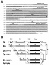
FIG. 1.
FIG. 1.
Primary structure of murine PRP4K and phylogenic comparison of PRP4K homologues. (A) Amino acid sequence and domain structure of murine PRP4K. The mouse and human proteins are 1,007 aa in length and share 95% identity and 97% similarity (accession numbers HsPRP4K, AF283465, MmPRP4K, and AF283466). Conserved domains (KKHK box, RS-like domain, MI and MII) and the kinase domain of MmPRP4K are shaded in gray. Unlike the canonical RS domain of splicing proteins such as SC35, the RS-like domain of PRP4K is extended and shares similarities with RS-like domains of the U1 70K and SRp75 splicing factors. Bipartite nuclear localization signals are boxed. Peptides MRC1 and MRC2, used to produce antibodies, are underlined, and the arrow indicates the site of gene-trap integration in cell line CT143 (56). (B) Domain structure and phylogenic comparison of S. pombe Prp4 (Sp Prp4p) and PRP4K homologues in humans (Hs), mice (Mm), D. melanogaster (Dm), C. elegans (Ce), and A. thaliana (At). The amino acid position of each domain is shown above the human sequence only. The position of each conserved domain within these homologues is shown relative to HsPRP4K. The length of each protein is shown at the right, and the percentage identity to human PRP4K is shown above each domain. The evolutionary relationship between these proteins is depicted as a phylogeny tree at the far right.
FIG. 2.
FIG. 2.
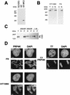
Characterization of PRP4K by Western blot analysis and indirect immunofluorescence. (A) Specificity of the anti-PRP4K antibody MRC2. Western blot analysis shows that affinity purified anti-PRP4K antibody MRC2 can detect the MRC2 epitope fused to GST (GST-MRC2) but does not cross-react with GST alone or with total bacterial protein extract. (B) Western blot analysis with the anti-PRP4K antibody MRC2 on total (T), cytoplasmic (C), or nuclear (N) extracts prepared from the human HT1080 or murine F9 embryocarcinoma cells indicates that PRP4K is a nuclear protein. (c) Salt extraction of PRP4K from human HT1080 nuclei was performed. Western blot analysis with the MRC2 antibody of soluble (S) and pellet (P) fractions of nuclei extracted with 200 mM, 400 mM, or 1 M KCl was then carried out. Total nuclei are shown for comparison (T). Three bands were detected: band A, 167 kDa; band B, 152 kDa; and band C, 147 kDa. Identical results were obtained with both the MRC1 and the H143 anti-PRP4K antibodies (data not shown). (D) Subcellular localization of PRP4K in murine F9 and human HT1080 cells by indirect immunofluorescence. Immunofluorescence of PRP4K (FITC) is shown in cells costained with DAPI to visualize the DNA. For each cell type, nuclei are shown in the first row of images, followed by mitotic chromosomes shown in the second row. In interphase cells, PRP4K is nucleoplasmic but enriched in foci that do not correspond to regions of concentrated DNA (DAPI). At mitosis, some PRP4K is also localized to mitotic chromosomes. Immunodetection of T7-tagged recombinant PRP4K (T7-PRP4K) in HT1080 cells produces a localization pattern similar to that of endogenous PRP4K in interphase cells and on mitotic chromosomes. Scale bars, 5 μm.
FIG. 3.
FIG. 3.
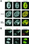
PRP4K is concentrated in SC35-containing splicing speckles. (A) Coimmunofluorescence with antibodies detecting PRP4K (H143) (FITC) and SC35 (Texas red) in human HT1080 cells alone, in the presence of actinomycin D (+Act D), or subjected to heat shock. PRP4K is nucleoplasmic but also concentrates in speckles that colocalize with SC35 in both transcriptionally active and transcriptionally inactive cells (i.e., heat shock and actinomycin D treated). (B) The RS-like domain of PRP4K is sufficient for localization to splicing speckles. HT1080 cells were transfected with either GFP-tagged murine PRP4K (GFP-PRP4K) or the RS domain of PRP4K fused to GFP (GFP-RS). Localization of GFP-tagged proteins was assessed by immunofluorescence in combination with the detection of endogenous SC35 (Texas red). GFP-tagged PRP4K forms distinct and large nuclear foci that colocalize with SC35. Scale bars, 5 μm.
FIG. 4.
FIG. 4.
Yeast two-hybrid analysis of human PRP4K. Yeast two-hybrid analysis of the N terminus or the C-terminal kinase domain of human PRP4K was carried out by using an adult human brain library. The region of PRP4K used in each screen is represented above each table. The identity of each protein and the number and relative strength of each interaction (β-galactosidase activity) is shown in the table. Each interacting protein is depicted graphically to the right, with the minimal PRP4K-interacting region underlined.
FIG. 5.
FIG. 5.
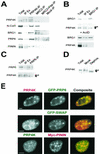
In vivo interactions of PRP4K. (A to C) Co-IP of PRP4K with pinin, N-CoR, BRG1, and PRP6. Complexes containing PRP6, BRG1, or PRP4K were immunoprecipitated from HeLa nuclear extracts with antibodies against each protein coupled to protein A- or protein G-agarose. Total, total nuclei; IP, IP of the indicated protein; IP Ex, IP extract. (A) Western blot analysis with antibodies recognizing each protein shows that N-CoR, BRG1, PRP6, and pinin coimmunoprecipitated with PRP4K by using anti-PRP4K immune serum against the MRC1 and two epitopes (PRP4K-IP) or the affinity-purified anti-PRP4K antibody MRC2 (MRC2-IP). Control IPs were carried out with MRC2 antibody preblocked with the MRC2 peptide (MRC2+Pep-IP) and with rabbit anti-sheep IgG (Mock-IP). (B) Western blot analysis of proteins immunoprecipitated with BRG1 by using anti-BRG1 antibodies (BRG1-IP). Only the fastest-migrating form of PRP4K coimmunoprecipitated with BRG1. In the presence of actinomycin D (+ActD), PRP4K fails to coimmunoprecipitate with BRG1, suggesting that the interaction between these proteins is transcription dependent. Arrows indicate two prominent forms of PRP4K; the slower-migrating form is phosphorylated (P). (C) Western blot analysis of proteins immunoprecipitated with PRP6 with anti-PRP6 antibodies (PRP6-IP). Only the fastest-migrating form of PRP4K coimmunoprecipitated with PRP6. (D) Dephosphorylation of HeLa nuclear extracts by using CIAP. Two major forms of PRP4K are indicated by the arrows; P indicates the hyperphosphorylated and slower-migrating form. Mock dephosphorylation results are shown for comparison. These results indicate that BRG1 and PRP6 interact primarily with the hypophosphorylated form of PRP4K. (E) Colocalization of PRP4K with GFP-tagged human PRP6 (GFP-PRP6), GFP-tagged murine SWAP (GFP-SWAP), or myc-tagged pinin (Myc-PININ) in HeLa cells. Endogenous PRP4K was detected with either Texas red-labeled (for GFP-PRP6 and GFP-SWAP transfections) or FITC-labeled secondary antibodies (for myc-pinin transfections). Myc-tagged pinin (Myc-PININ) was detected with Texas red-labeled secondary antibodies. Scale bars, 5 μm.
FIG. 6.
FIG. 6.
In vitro kinase activity of PRP4K in immunoprecipitates. IPs with antibodies against PRP4K (MRC2 antibody [PRP4K-IP]), BRG1 (BRG1-IP), or PRP6 (PRP6-IP) were subjected to an in vitro kinase assay and resolved by SDS-PAGE, and phosphorylated proteins were visualized by autoradiography (Kinase Assay). Control reactions were carried out with either peptide-blocked MRC2 antibody (Pep. Block) or rabbit anti-sheep IgG (Mock). Western blot analysis of the PRP4K-IP complex was also carried out. Bands corresponding to known proteins are designated with arrows (arrow A, BRG1; arrow B, PRP4K; arrow C, PRP6). A contaminating band (arrow D) is present in the peptide-blocked kinase assay, and a strong band of ca. 54 kDa (arrow “?”) may correspond to phosphorylated IgG. In the PRP4K-IP, the strongly labeled band (arrow B) probably corresponds to autophosphorylated PRP4K.
FIG. 7.
FIG. 7.
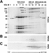
PRP4K specifically cosediments with the 20S U5-snRNP during glycerol gradient centrifugation. (A) Proteins from each fraction of the glycerol gradient were separated by SDS-10% PAGE and visualized by Coomassie blue staining (Protein). Fractions that cosediment with the 12S U1/U2 snRNP, 20S U5-snRNP, and the U4/U6.U5 snRNP were determined by the position of the corresponding snRNAs (data not shown) as indicated above the panels. The positions of PRP4K, PRP6, the U1-specific U1-70K protein, the U5 snRNP-specific 200K and 220K proteins, and the U4/U6 snRNP-specific 90K, 61K, and 60K proteins are arrowed. (B and C) Western blot analysis of fractions with antibodies to PRP4K (B) or PRP6 (C).
FIG. 8.
FIG. 8.
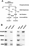
Detection of the presence of PRP4K, BRG1, and PRP6 in the N-CoR-1 and -2 complexes. (A) Schematic representation of the purification of the N-CoR-1 and N-CoR-2 complexes (57). (B) Western blot analysis of the N-CoR-1 and N-CoR-2 complexes after elution from the N-CoR affinity column with antibodies against N-CoR, PRP4K, BRG1, and PRP6. The control lane is an aliquot from the mock affinity purification, and the input lane is the pooled S300 fractions containing N-CoR prior to immunoaffinity chromatography. Whereas BRG1 and PRP6 are components of both the N-CoR-1 and the N-CoR-2 complexes, PRP4K is primarily a component of the N-CoR-2 complex.
FIG. 9.
FIG. 9.
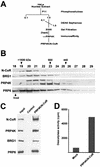
Affinity purification of the PRP4K/N-CoR deacetylase complex. (A) Purification scheme for the isolation of complexes enriched in both N-CoR and PRP4K. At each stage, N-CoR- and PRP4K-containing fractions were determined by Western blot analysis and pooled before we continued with the next chromatography step. Affinity-purified PRP4K/N-CoR complex was then subjected to both Western blot analysis and deacetylase assays. (B) S300 gel filtration profile of N-CoR, BRG1, PRP4K, and PRP6. Fractions corresponding to the protein peak from the S300 gel filtration column (fractions 18 to 29) were analyzed by Western blotting for the presence of each protein. Approximate molecular masses of the various complexes eluting from the S300 column are indicated in kilodaltons above the corresponding fraction numbers. N-CoR, BRG1, PRP4K, and PRP6 coelute from the gel filtration column in a large-molecular-mass complex of between 660 and 1,500 kDa. (C) Western blot analysis of the PRP4K/N-CoR complex. The PRP4K/N-CoR complex was affinity purified from the S300 fractions 20 to 22 by using an anti-PRP4K column. Western blot analysis indicates the PRP4K/N-CoR complex contains N-CoR, PRP4K, BRG1, and PRP6. (D) Deacetylase activity of the PRP4K/N-CoR complex. Deacetylase activity of affinity-purified PRP4K/N-CoR complex was monitored by the release of [H3]acetate from in vitro-tritiated histones in the presence of immunoprecipitated complex with anti-PRP4K antibody coupled to protein G-Sepharose. The activity is presented as the total counts per minute, and a mock deacetylase assay (Mock) was carried out by using mock-affinity-purified PRP4K/N-CoR complex immunoprecipitated with protein G-Sepharose alone. The PRP4K/N-CoR complex exhibits strong deacetylase activity compared to the nonspecific activity found associated with the mock-affinity-purified complex.

Similar articles

Cited by

References

    1. Aasland, R., A. F. Stewart, and T. Gibson. 1996. The SANT domain: a putative DNA-binding domain in the SWI-SNF and ADA complexes, the transcriptional co-repressor N-CoR and TFIIIB. Trends Biochem. Sci. 21:87-88. - PubMed
    1. Alahari, S. K., H. Schmidt, and N. F. Kaufer. 1993. The fission yeast prp4+ gene involved in pre-mRNA splicing codes for a predicted serine/threonine kinase and is essential for growth. Nucleic Acids Res. 21:4079-4083. - PMC - PubMed
    1. Andrews, N. C., and D. V. Faller. 1991. A rapid micropreparation technique for extraction of DNA-binding proteins from limiting numbers of mammalian cells. Nucleic Acids Res. 19:2499.. - PMC - PubMed
    1. Bauren, G., and L. Wieslander. 1994. Splicing of Balbiani ring 1 gene pre-mRNA occurs simultaneously with transcription. Cell 76:183-192. - PubMed
    1. Brandner, J. M., S. Reidenbach, C. Kuhn, and W. W. Franke. 1998. Identification and characterization of a novel kind of nuclear protein occurring free in the nucleoplasm and in ribonucleoprotein structures of the “speckle” type. Eur. J. Cell Biol. 75:295-308. - PubMed

Publication types

MeSH terms

Substances

Associated data