. 2002 Jul;22(14):5182-93.
doi: 10.1128/MCB.22.14.5182-5193.2002.

Drosophila Mi-2 negatively regulates dDREF by inhibiting its DNA-binding activity

Affiliations

Drosophila Mi-2 negatively regulates dDREF by inhibiting its DNA-binding activity

Fumiko Hirose et al. Mol Cell Biol. 2002 Jul.

Abstract

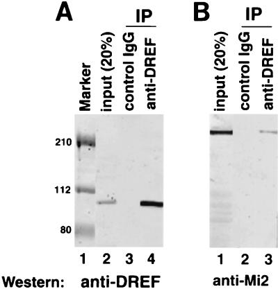
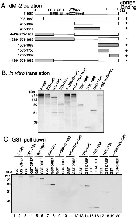
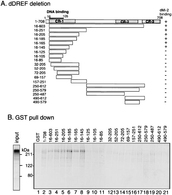
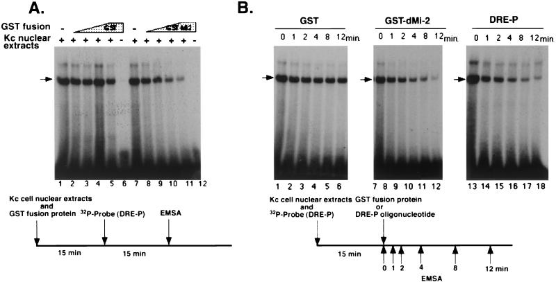
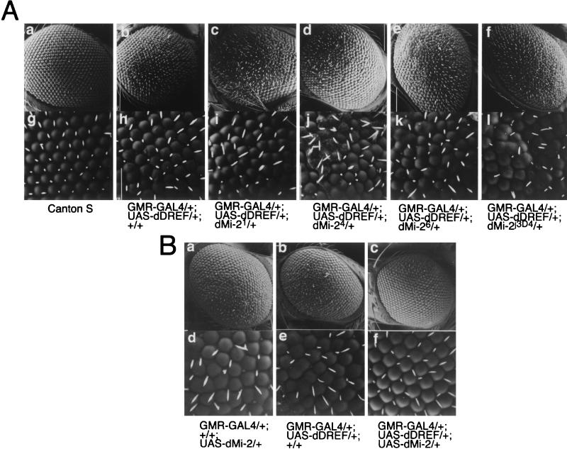
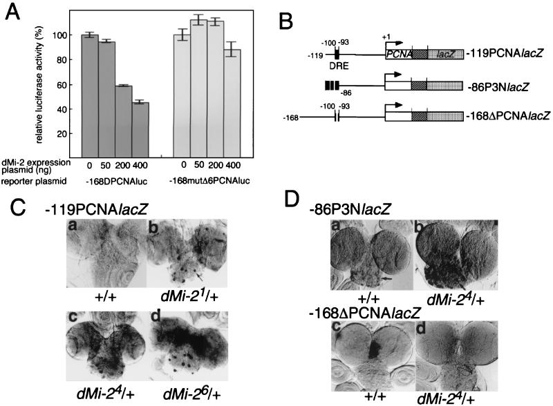
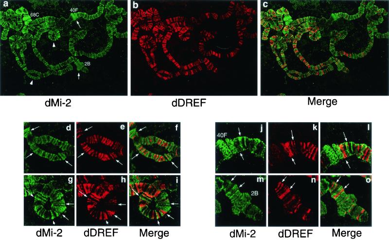
Drosophila melanogaster DNA replication-related element (DRE) factor (dDREF) is a transcriptional regulatory factor required for the expression of genes carrying the 5'-TATCGATA DRE. dDREF has been reported to bind to a sequence in the chromatin boundary element, and thus, dDREF may play a part in regulating insulator activity. To generate further insights into dDREF function, we carried out a Saccharomyces cerevisiae two-hybrid screening with DREF polypeptide as bait and identified Mi-2 as a DREF-interacting protein. Biochemical analyses revealed that the C-terminal region of Drosophila Mi-2 (dMi-2) specifically binds to the DNA-binding domain of dDREF. Electrophoretic mobility shift assays showed that dMi-2 thereby inhibits the DNA-binding activity of dDREF. Ectopic expression of dDREF and dMi-2 in eye imaginal discs resulted in severe and mild rough-eye phenotypes, respectively, whereas flies simultaneously expressing both proteins exhibited almost-normal eye phenotypes. Half-dose reduction of the dMi-2 gene enhanced the DREF-induced rough-eye phenotype. Immunostaining of polytene chromosomes of salivary glands showed that dDREF and dMi-2 bind in mutually exclusive ways. These lines of evidence define a novel function of dMi-2 in the negative regulation of dDREF by its DNA-binding activity. Finally, we postulated that dDREF and dMi-2 may demonstrate reciprocal regulation of their functions.


Figures

FIG. 1.
FIG. 1.
dDREF and dMi-2 interact in vivo. Ten microliters of supernatant of a hybridoma producing anti-dDREF monoclonal antibody no. 1 were added to 300 μl of crude nuclear extract from Drosophila embryos (approximately 150 μg of protein) and incubated for 3 h at 4°C. Ten microliters of protein G-Sepharose beads (Roche) were then added to the mixture, followed by incubation for 1 h. After extensive washing, proteins bound to the beads were separated by SDS-PAGE, transferred to a Hybond membrane, and probed with (A) anti-dDREF antibody or (B) anti-dMi-2 antibody. (A) Lane 1, prestained molecular size markers; lane 2, 20% of input protein used for immunoprecipitation (IP); lane 3, immunoprecipitation with normal rabbit IgG as a negative control; lane 4, immunoprecipitation with the anti-DREF antibody. (B) Lane 1, 20% of input protein used for immunoprecipitation; lane 2, immunoprecipitation without normal rabbit IgG as a negative control; lane 3, immunoprecipitation with anti-DREF antibody.
FIG. 2.
FIG. 2.
Mapping of the dMi-2 domain interacting with dDREF by GST pull-down assay. (A) Schematic representation of deletions for the dMi-2 polypeptide. (B) In vitro translation products of deleted dMi-2 forms used for the GST pull-down assay. (C) GST pull-down assay using GST-dDREF and in vitro-translated 35S-dMi-2. Odd lanes, GSTs were immobilized on glutathione-Sepharose beads and incubated with either 35S-dMi-2 polypeptide as indicated; even lanes, GST-dDREF (aa 1 to 708) was immobilized on glutathione-Sepharose beads and incubated with either 35S-dMi-2 polypeptide as indicated. Protein complexes were washed and resolved by SDS-PAGE, and bound proteins were detected by autoradiography.
FIG. 3.
FIG. 3.
Mapping of the dDREF domain interacting with dMi-2 by GST pull-down assay. (A) Schematic representation of deletions for the dDREF polypeptide. These deletion polypeptides were expressed as fusion proteins with GST. (B) GST pull-down assay results with GST-dDREF and in vitro-translated 35S-dMi-2. GST (lane 1) or GST-dDREF polypeptides (lanes 2 through 21) were immobilized on glutathione-Sepharose beads and incubated with either 35S-dMi-2 polypeptide (aa residues 4 through 1982). Protein complexes were washed and resolved by SDS-PAGE, and bound dMi-2 was detected by autoradiography. The input gel on the left of the panel shows an aliquot of 35S-dMi-2 polypeptide used for each binding reaction.
FIG. 4.
FIG. 4.
dMi-2 inhibits DNA-binding activity of dDREF. (A) Nuclear extracts from Kc cells were preincubated with E. coli extracts containing GST (lanes 2 through 5) or GST-dMi-2 fusion protein (lanes 8 through 11), and then 32P-labeled DRE-P oligonucleotides were added. After an additional 15-min incubation, complexes of DRE/dDREF were analyzed by EMSA. (B) Nuclear extracts from Kc cells were incubated with 32P-labeled DRE-P oligonucleotides for 15 min; then E. coli extracts containing GST (lanes 1 through 6), E.coli extracts containing GST-dMi-2 fusion protein (lanes 7 through 12), or a 200-fold molar excess of unlabeled DRE-P oligonucleotide as a competitor (lanes 13 through 18) were added to reactions. Aliquots of reaction mixtures were subjected to electrophoresis at the indicated time points after addition of E. coli extracts or competitor oligonucleotide.
FIG. 5.
FIG. 5.
dMi-2 genetically interacts with dDREF. (A) Half-dose reduction of the dMi-2 gene enhances the dDREF rough-eye phenotype. All alleles of dMi-2 are balanced with TM6C or TM6B balancer chromosomes. Female flies expressing dDREF (GMR-GAL4/GMR-GAL4, UAS-dDREF/UAS-dDREF, +/+) were crossed with males carrying dMi-21, dMi-24, dMi-26, and dMi-2j3D4 alleles. F1 progeny developed at 25°C without a balancer chromosome were used for analysis of eye phenotype by scanning electron microscopy. (a and g) Wild-type eye; (b and h) GMR-GAL4/+, UAS-dDREF/+, and +/+; (c and i) GMR-GAL4/+, UAS-dDREF/+, and dMi-21/+; (d and j) GMR-GAL4/+, UAS-dDREF/+, and dMi-24/+; (e and k) GMR-GAL4/+, UAS-dDREF/+, and dMi-26/+; (f and l) GMR-GAL4/+, UAS-dDREF/+, and dMi-2j3D4/+. (B) Expression of dMi-2 suppresses the dDREF-induced rough-eye phenotype. Scanning electron micrographs of adult compound eyes. (a and d) GMR-GAL4/+, +/+, and UAS-HA-dMi-2/+; (b and e) GMR-GAL4/+, UAS-Flag-dDREF/+, and +/+; (c and f) GMR-GAL4/+, UAS-Flag-dDREF/+, and UAS-HA-dMi-2/+. Note that the transgenic fly simultaneously expressing dDREF and dMi-2 in eye imaginal discs exhibits a normal eye morphology.
FIG. 6.
FIG. 6.
dMi-2 negatively regulates the PCNA gene promoter. (A) Effect of cotransfecting dMi-2-expressing plasmids on luciferase activity directed by the PCNA gene promoter. Schneider cells (1.5 × 10⁵/well) were cotransfected with 50 ng of reporter plasmid (either −168DPCNAluc or −168mutΔ6PCNAluc), 50 to 400 ng of pUAS-HA-dMi-2 and 100 ng of pAct-GAL4 as effector plasmids, and 1 ng of pRL-actin5C as an internal control plasmid. The total amount of DNA in the transfection mixture was adjusted to 1 μg by the addition of pGEM3. Cells were harvested 48 h after transfection. The luciferase assay was carried out by means of the Dual-Glo Luciferase Reporter Assay System (Promega). Firefly luciferase activity was normalized to Renilla luciferase activity. Averaged values obtained from three independent transfections are shown. (B) Constructs of PCNA-lacZ fusion genes used to establish the transgenic lines analyzed in panels C and D. (C) Half-dose reduction of dMi-2 activates the PCNA gene promoter depending on the presence of DRE. Detection of expression of β-galactosidase in brain lobes from transgenic flies carrying the PCNA-lacZ reporter gene. Male transgenic flies carrying −119PCNAlacZ on the second chromosome were crossed with females with dMi-21, dMi-24, and dMi-26 alleles balanced with the TM6C chromosome. Brain lobes were dissected from the third-instar larvae of F1 progeny, fixed, and immunostained with anti-β-galactosidase antibody. (D) Male transgenic flies carrying either −86P3NlacZ or −168ΔPCNAlacZ reporter genes on the second chromosome were crossed with females with the dMi-24 allele balanced with the TM6C chromosome. Brain lobes were dissected from the third-instar larvae of F1 progeny, fixed, and immunostained with anti-β-galactosidase antibody.
FIG. 7.
FIG. 7.
Immunostaining of polytene chromosomes with anti-dMi-2 and anti-dDREF antibodies. (a through c) Polytene chromosomes from the third-instar larvae of wild-type flies were spread and double-stained with rabbit anti-dMi-2 antiserum (green) and mouse anti-dDREF monoclonal antibodies (red). Arrows indicate puffs. Arrowheads indicate regions shown as enlarged images in panels d through o. (d through i) Enlarged images of the localization patterns of dMi-2 and dDREF. Note that they bind DNA in a mutually exclusive manner. Typical features of localization of the two proteins are highlighted by arrows. (j through o) Enlarged images of the localization patterns of dMi-2 and dDREF in puffed regions.

Similar articles

Cited by

References

    1. Aubry, F., M.-G. Mattei, and F. Galibert. 1998. Identification of a human 17p-located cDNA encoding a protein of the Snf2-like helicase family. Eur. J. Biochem. 254:558-564. - PubMed
    1. Brand, A. H., and N. Perrimon. 1993. Targeted gene expression as a means of altering cell fates and generating dominant phenotypes. Development 118:401-415. - PubMed
    1. Brehm, A., G. Langst, J. Kehle, C. R. Clapier, A. Imhof, A. Eberharter, J. Muller, and P. B. Becker. 2000. dMi-2 and ISWI chromatin remodelling factors have distinct nucleosome binding and mobilization properties. EMBO J. 19:4332-4341. - PMC - PubMed
    1. Duronio, R. J., P. H. O'Farrell, J. E. Xie, A. Brook, and N. Dyson. 1995. The transcription factor E2F is required for S phase during Drosophila embryogenesis. Genes Dev. 9:1445-1455. - PubMed
    1. Feng, Q., and Y. Zhang. 2001. The MeCP1 complex represses transcription through preferential binding, remodeling, and deacetylating methylated nucleosome. Genes Dev. 15:827-832. - PMC - PubMed

Publication types

MeSH terms

LinkOut - more resources