

doi: 10.1126/science.1070790.
Regulation of the different chromatin states of autosomes and X chromosomes in the germ line of C. elegans
Affiliations
- PMID: 12077420
- PMCID: PMC2435369
- DOI: 10.1126/science.1070790
Item in Clipboard
Regulation of the different chromatin states of autosomes and X chromosomes in the germ line of C. elegans
Science.
.
Abstract
The Maternal-Effect Sterile (MES) proteins are essential for germline viability in Caenorhabditis elegans. Here, we report that MES-4, a SET-domain protein, binds to the autosomes but not to the X chromosomes. MES-2, MES-3, and MES-6 are required to exclude MES-4 and markers of active chromatin from the X chromosomes. These findings strengthen the emerging view that in the C. elegans germ line, the X chromosomes differ in chromatin state from the autosomes and are generally silenced. We propose that all four MES proteins participate in X-chromosome silencing, and that the role of MES-4 is to exclude repressors from the autosomes, thus enabling efficient repression of the Xs.
Figures

MES-4 in the germ line and in embryos and larvae. Samples were stained with either DAPI (4′,6-diamidino-2-phenylindole) or antibodies to chromatin, and with anti−MES-4 (10). (A) Adult hermaphrodite gonad. (Upper panel) DAPI stain. (Lower panel) Anti−MES-4 stain. Asterisk marks the distal end, o marks oocytes. Bar, 50 μm. (B) One-cell embryo in metaphase. (C) Six-cell, (D) ~80-cell, and (E) ~180-cell embryo. (F) L1 larva. Arrowhead points to the germline cells Z2 and Z3. Bar, 10 μm (B) to (F).
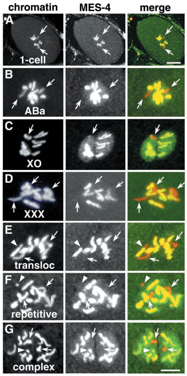
MES-4 associates with autosomes and complex extrachromosomal arrays, but not with X chromosomes and repetitive arrays. Embryos were stained with either anti-chromatin (A to E) or ethidium bromide (F and G) (red in merge), and with anti−MES-4 (green) (10). Arrows point to chromatin lacking MES-4. (A) One-cell embryo at pronuclear meeting. Bar, 10 μm. (B) Nucleus in the ABa blastomere of a four-cell embryo. (C) Presumptive XO one-cell embryo generated by mating fer-1 females with wild-type males. (D) Presumptive XXX one-cell embryo from a him-8 hermaphrodite. (E) Embryo carrying mnDp10, a fusion of chromosome I and a duplicated portion of the X (25) (arrowhead marks the I:X boundary). The X-autosome translocation mnT12 gave similar results (11). (F) Embryo carrying a repetitive extrachromosomal array (arrowhead) that contains many copies of GFP::let-858 and rol-6 (21). A repetitive GFP::cam-1 array gave similar results (11). (G) Embryo carrying a complex extrachromosomal array (arrowhead) that contains genomic DNA and few copies of GFP::let-858 and rol-6 (21). A complex GFP::tbg-1 array gave similar results (11). Bar, 5 μm (B) to (G).

MES-4, acetyl (Lys12) H4, methyl (Lys9) H3, and active RNA polymerase II in the germ lines of wild-type and mes mutant worms. Adult germ lines were stained with combinations of DAPI or anti-chromatin (red in merge), anti−MES-4 (green), anti-acetyl (Lys12) H4 (green), anti-dimethyl (Lys9) H3 (green), and the H5 antibody to phosphorylated RNA polymerase II (green) (10). Arrows point to the presumptive X chromosomes. (A) mes + hermaphrodites. (Upper panels) Mitotic nuclei in a glp-4 germ line, which contains few germ nuclei in prophase (26). (Lower panels) Oocyte chromosomes in wild type. (B) M+Z− mes hermaphrodites. (Upper panels) Distal mitotic nuclei in a mes-6(bn64) gonad. (Lower panels) mes-2(bn11) oocyte. (C) Pachytene nuclei in wild-type (upper two rows), M− Z− mes-6(bn64) (third row), and M−Z− mes-3(bn88) (bottom row) hermaphrodites. (D) Transition-zone nuclei in wild-type (upper panels), M+Z− mes-3(bn35) (middle panels), and M−Z+ mes-6(bn64)/+ (bottom panels) hermaphrodites. (E) Pachytene nuclei in wild-type (upper panels), M−Z− mes-6(bn66)him-8 (middle panels), and M− Z− mes-4(bn67); him-8 (bottom panels) males. Bars [(C), rows 2 and 4], 2 μm. All other bars, 5 μm. (F) Schematic time line of marks on the autosomes (As) and Xs during germline development (mitotic, transition zone, pachytene, diplotene, diakinesis) in wild-type and M+Z− mes hermaphrodites. Derived from (3) and this paper.
References
Publication types
MeSH terms
Substances
Grants and funding
LinkOut - more resources
Full Text Sources
Other Literature Sources
Molecular Biology Databases
Miscellaneous
